Imagine the frustration: a patient undergoes successful cataract surgery, their world once again vibrant and clear after years of gradual dimming. Then, months or even years later, the familiar fog begins to creep back. Blurred vision, glare from oncoming headlights, and a frustrating dimness cast a shadow over their regained sight. Fear sets in—have the cataracts returned? For nearly 20-40% of post-cataract surgery patients, this is a disheartening reality. However, the solution is not a repeat of the complex surgery that first restored their vision. Instead, it is an elegant, non-invasive, and remarkably swift procedure performed in an ophthalmologist’s office: the YAG laser capsulotomy.
This article serves as the definitive guide to understanding this common ophthalmic procedure, not just from a clinical perspective but through the critical lens of medical coding and reimbursement. At the heart of this process is a five-digit code that, when used correctly, seamlessly translates clinical work into financial stability for a practice: CPT Code 66821. We will embark on a detailed journey, exploring the intricate anatomy that leads to the problem, the brilliant physics of the laser solution, the meticulous steps of the procedure itself, and the absolute necessity of precise documentation and coding. For ophthalmologists, practice managers, and medical coders alike, mastering the nuances of 66821 is essential for ensuring patient access to this sight-restoring treatment and for the operational health of the practice itself.

2. Understanding the Foundation: The Anatomy of the Crystalline Lens and Cataract Surgery
To truly appreciate what a YAG capsulotomy fixes, one must first understand the original structure and what occurs during cataract surgery.
The Natural Lens: A Marvel of Biological Optics
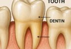
The natural crystalline lens is a transparent, biconvex structure located behind the iris and the pupil. Its primary function is to focus light onto the retina, working in concert with the cornea to provide a clear image. It is encapsulated by a thin, elastic, clear membrane called the lens capsule. The lens itself is made up of precisely arranged proteins and water, and its transparency is maintained by the unique structure and arrangement of these proteins.
Cataract Formation: The Clouding of a Lens
A cataract is the opacification of this naturally clear lens. It is not a “film” over the eye but a physical clouding within the lens itself. This process is most commonly age-related but can be accelerated by factors like diabetes, trauma, prolonged steroid use, and UV light exposure. As the lens becomes cloudy, it scatters and blocks light from reaching the retina, leading to the classic symptoms of blurred vision, glare, halos around lights, fading colors, and eventually, functional blindness if left untreated.
Modern Cataract Surgery: Phacoemulsification and IOL Implantation
Modern cataract surgery is a miracle of microsurgery. The standard technique is phacoemulsification (phaco). In this procedure:
- The surgeon creates tiny corneal incisions (often 2.2 mm or smaller).
- A continuous circular tear (capsulorhexis) is made in the front (anterior) portion of the lens capsule.
- An ultrasonic probe is inserted to emulsify (break up) the cloudy natural lens into microscopic pieces.
- These pieces are suctioned out of the eye, leaving the clear, empty capsular bag intact.
- An artificial intraocular lens (IOL), made of silicone or acrylic, is folded, inserted through the tiny incision, and unfolded into the vacant capsular bag, where it remains permanently.
The Capsule’s Crucial Role: A Natural Pocket for the IOL
The lens capsule is the hero of this story. Surgeons deliberately leave this “bag” or “pocket” in place because it provides the ideal, natural scaffolding to secure and center the new IOL. Its integrity is paramount during surgery. However, the survival of these capsule cells after surgery is also what leads to the most common long-term complication.
3. The Problem: Posterior Capsular Opacification (PCO) – The “After-Cataract”
What is PCO? Pathophysiology and Cellular Mechanisms
Posterior Capsular Opacification (PCO) is not the return of the cataract. It is a separate process of wound healing and cellular proliferation. During surgery, not all lens epithelial cells (LECs) are removed. Residual LECs, which originally resided on the anterior capsule, survive the procedure. In the weeks, months, or years following surgery, these cells undergo:
- Proliferation: They multiply and migrate across the posterior capsule (the back wall of the bag).
- Metaplasia: They transform from their normal, transparent form into large, swollen, blister-like cells (called “Elschnig pearls”) or into fibrous, collagen-producing cells.
This migration and transformation cause the once-clear posterior capsule to become cloudy, wrinkled, and opaque, effectively acting like a frosted glass window behind the new, clear IOL. This scatters light and blurs vision, mimicking the original cataract symptoms.
Epidemiology: How Common is PCO?
PCO is the most frequent long-term complication of modern cataract surgery. Its incidence varies widely based on:
- Patient age: Younger patients have more robust cell proliferation and thus a much higher risk.
- IOL material and design: Newer IOLs with square-edged optics and hydrophobic materials significantly reduce PCO rates.
- Surgical technique: A well-performed capsulorhexis that completely overlaps the IOL optic and thorough cortical cleanup reduce risk.
Studies show Nd:YAG capsulotomy rates can range from under 10% to over 50% at five years post-surgery, making it a very common procedure in any comprehensive ophthalmology practice.
Clinical Presentation: Symptoms and Patient Complaints
Patients present with a gradual, often frustrating, decline in vision. Common complaints include:
- “My vision is getting blurry again, like it did before my cataract surgery.”
- “I’m seeing a lot of glare and starbursts from car headlights at night.”
- “My vision seems hazy or foggy.”
- “My glasses prescription seems to have changed, but new glasses don’t help.”
It is crucial to differentiate these symptoms from those caused by other conditions, such as dry eye or retinal issues.
Diagnosing PCO: Slit-Lamp Biomicroscopy and Differential Diagnosis
The diagnosis is confirmed clinically with a slit-lamp biomicroscope. The ophthalmologist will see a hazy, often pearly or fibrous membrane behind the IOL. Retinal visualization may be difficult due to the opacity. It is critical to rule out other causes of vision loss post-cataract surgery, such as:
- Cystoid Macular Edema (CME)
- Refractive error (need for updated glasses prescription)
- Pre-existing retinal pathology (e.g., Age-Related Macular Degeneration)
- Corneal edema or other corneal issues
- IOL decentration or dislocation
The definitive diagnostic test is the improvement of subjective vision and objective visual acuity when the patient looks through a pinhole. If vision improves significantly with a pinhole (which eliminates the scatter from the cloudy capsule), it is highly suggestive of PCO.
4. The Solution: YAG Laser Capsulotomy – Principles and Physics
What is a YAG Laser? Understanding Nd:YAG Technology
The YAG laser used for this procedure is a Neodymium-doped Yttrium Aluminum Garnet (Nd:YAG) laser. It is a solid-state laser that produces a beam of light in the near-infrared spectrum (1064 nm wavelength). This specific wavelength is largely invisible and passes freely through the clear structures of the eye—the cornea, aqueous humor, IOL, and vitreous—without being absorbed by them. It is only absorbed at the point of focus, where its energy is concentrated.
The Photodisruption Phenomenon: Creating a Plasma Spark
The Nd:YAG laser does not work by burning or cutting tissue through thermal energy. Instead, it operates through a process called photodisruption or optical breakdown. The laser emits an extremely short, high-energy pulse (measured in nanoseconds). When this pulse is focused precisely on the opaque posterior capsule, the high electric field at the focal point ionizes the molecules, tearing electrons from their atoms and creating a microscopic plasma spark. This spark rapidly expands, creating a shockwave and a cavitation bubble that mechanically disrupts and tears the tissue. It is this mechanical disruption that creates a clean opening in the capsule without generating significant heat that could damage surrounding structures like the IOL or retina.
From Cloudy to Clear: The Goal of the Procedure
The goal is to create a central opening, or window, in the opacified posterior capsule. This opening allows light to pass unimpeded through the clear IOL and the new opening, onto the retina, restoring clear vision instantly. The procedure does not remove the opacification; it simply pushes it aside out of the visual axis. The opening is permanent.
5. A Deep Dive into CPT Code 66821: The Language of Reimbursement
Code Definition and Official CPT® Descriptor
The American Medical Association’s (AMA) Current Procedural Terminology (CPT®) code set is the universal language for describing medical, surgical, and diagnostic services. For a YAG capsulotomy, the designated code is:
CPT 66821: Discission of secondary membranous cataract (opacified posterior lens capsule and/or anterior hyaloid); laser surgery (e.g., YAG laser) (1 or more stages)
This descriptor is critical to understand:
- “Discission”: An old surgical term meaning “to cut.” This covers the laser’s action.
- “Secondary membranous cataract”: This is the formal term for PCO.
- “(opacified posterior lens capsule…”: This specifies the exact anatomical target.
- “laser surgery (e.g., YAG laser)”: Specifies the technology used.
- “(1 or more stages)”: Indicates that the code is reported only once per eye, regardless of how many laser applications or “bursts” are required to complete the capsulotomy.
Global Period: Understanding the 10-Day Postoperative Timeline
CPT 66821 is assigned a 10-day global surgical period. This means the reimbursement for this code is intended to cover:
- The procedure itself.
- Any related postoperative care provided by the surgeon for the 10 days immediately following the procedure.
This has significant billing implications:
- If the patient returns for a follow-up visit within those 10 days for a reason related to the YAG procedure (e.g., checking intraocular pressure), it is not separately billable. It is bundled into the payment for 66821.
- An evaluation and management (E/M) service for an unrelated problem (e.g., a sudden eye infection) during the global period may be billed with a modifier -24 (Unrelated Evaluation and Management Service by the Same Physician or Other Qualified Health Care Professional During a Postoperative Period) if the documentation supports that the visit was unrelated.
Bilateral Procedures: Modifier 50 and Its Correct Application
It is very common for a patient to develop PCO in both eyes, though not always simultaneously. If a YAG capsulotomy is performed on both eyes during the same session, the coder must report the service correctly to avoid denials for duplicate billing.
- Correct Billing: 66821-50 (Bilateral procedure)
- Reimbursement: Typically, the payer will reimburse 100% for the first eye and 50% for the second eye, though payer policies can vary. It is crucial to verify with individual insurance carriers.
Incorrect Billing: Reporting 66821 twice (e.g., 66821-RT and 66821-LT) will almost certainly result in a denial for the second code as a duplicate service.
6. Coding and Documentation: Building a Bulletproof Claim
The difference between a paid and a denied claim often lies in the medical record. Documentation must irrefutably establish medical necessity.
The Medical Necessity Triad: Diagnosis, Symptoms, and Procedure
For 66821 to be covered, the documentation must clearly link three elements:
- A diagnosis of PCO (confirmed via slit-lamp exam).
- Symptoms reported by the patient that are affecting their vision and quality of life.
- The performance of the laser procedure to treat that specific condition.
Key Elements of a Strong Clinical Note
A robust note should include:
- History of Present Illness (HPI): Detail the patient’s complaints (e.g., “Patient reports 3 months of progressively blurred vision and significant glare with night driving in the right eye.”).
- Review of Systems (ROS): Pertinent negatives and positives related to the eye.
- Objective Findings:
- Visual Acuity: Uncorrected (UCVA) and Best-Corrected (BCVA). Note if vision improves with pinhole.
- Slit-Lamp Examination: This is the most critical element. The note must describe the PCO. Use descriptive terms like:
- “Significant opacification and wrinkling of the posterior capsule.”
- “Elschnig pearls noted on the posterior capsule.”
- “Dense fibrous thickening obscuring the red reflex.”
- IOP Measurement: Pre- and post-procedure.
- Assessment and Plan:
- Diagnosis: Posterior Capsular Opacification, OD/OS.
- Plan: Discussed risks, benefits, and alternatives of YAG laser capsulotomy. Patient consented. Procedure performed without complication.
The Gold Standard: Incorporating Slit-Lamp Findings into Documentation
Vague documentation like “PCO noted” is insufficient. Strong documentation paints a picture:
- Weak: “PCO present.”
- Strong: “On slit-lamp examination, the posterior capsule behind the IOL shows significant, dense opacification with a fibrous contracture and wrinkles extending into the visual axis. The view to the posterior pole is significantly obscured.”
The Role of Visual Acuity Testing
While a specific level of visual acuity loss is not an absolute requirement for all payers, documenting a decrease in BCVA strengthens medical necessity. Many payers have “clinical policies” or “coverage determinations” that may require:
- A specific drop in visual acuity (e.g., worse than 20/40).
- Documentation that the vision loss is due to PCO and not other pathology.
- Documentation of functional impairment (e.g., difficulty driving, reading).
Always check your major payers’ local coverage determinations (LCDs) for any specific requirements.
7. ICD-10-CM Coding: Pinpointing the Medical Necessity
The diagnosis code tells the payer why the procedure was necessary. For PCO, the correct code family is H26.4- (After-cataract).
Primary Code: H26.4- (After-Cataract)
The ICD-10-CM system requires a high level of specificity. The base code H26.4 requires a 5th and sometimes a 6th character to specify laterality and type.
Expanding Specificity: A Guide to the 5th and 6th Characters
The codes are structured as follows:
- H26.41 – After-cataract, right eye
- H26.42 – After-cataract, left eye
- H26.43 – After-cataract, bilateral
But it doesn’t stop there. A 6th character is required to specify the type of after-cataract:
- H26.411 – Soemmering’s ring, right eye
- H26.412 – Other after-cataract, right eye
Soemmering’s ring is a specific type of PCO where the proliferation is confined to the equator of the capsule, creating a doughnut-like ring. The vast majority of cases will be coded as “Other after-cataract” (e.g., H26.412 for the right eye, H26.422 for the left eye).
Combining Codes: Addressing Other Postprocedural States
Sometimes, a patient may have a status post-cataract surgery code in addition to the after-cataract code. The code Z96.1 (Presence of intraocular lens) can be used as a secondary code to indicate the patient has an artificial lens. However, the primary diagnosis code must always be the reason for the encounter—H26.4-.
8. The Procedure from Start to Finish: A Step-by-Step Walkthrough
Patient Selection and Informed Consent
Not every patient with PCO needs immediate treatment. The decision is based on the degree of visual impairment and the patient’s symptoms. A thorough discussion of informed consent is mandatory. Key points to cover:
- Benefits: Restoration of clear vision, reduction of glare.
- Risks: Elevated eye pressure (most common), retinal detachment, cystoid macular edema, IOL pit or crack, inflammation, hyphema (bleeding), floaters, and the rare possibility of the opacification returning.
- Alternatives: The main alternative is observation, as there is no medicinal treatment for PCO.
Pre-Procedure: Pupillary Dilation and Setup
The pupil is dilated with eye drops to provide a larger working area and better view for the surgeon. Anesthetic drops are applied to numb the cornea. The patient is seated at the laser delivery system, which is mounted on a slit lamp. A special focusing lens (e.g., a Abraham or Peyman lens) is placed on the eye. This lens serves two purposes: it provides stabilization and works as a contact lens to neutralize the cornea’s refractive power, allowing for precise focusing on the capsule.
The Procedure: Laser Application and Technique
The surgeon views the capsule through the laser’s aiming beam. The target is focused precisely on the opacified capsule, posterior to the IOL. A series of laser pulses are applied in a circular, cruciate (plus-sign), or can-opener pattern to create an opening in the center of the capsule. The number of pulses required varies based on the density of the opacity. The procedure is typically painless, with patients reporting only the sound of the pulses (a clicking noise) and possibly seeing flashes of light.
Post-Procedure Care: Immediate Aftermath and Instructions
Immediately after the procedure, intraocular pressure (IOP) is checked, as a transient spike is common. Patients are given a course of anti-inflammatory eye drops (e.g., prednisolone acetate) for about 4-7 days to prevent inflammation. They are instructed to report any sudden increase in floaters, flashes of light, a shadow in their peripheral vision, or a sudden drop in vision—all potential signs of a retinal tear or detachment.
9. Risks, Complications, and Management
While YAG capsulotomy is very safe, understanding its risks is paramount.
Immediate Complications: Elevated IOP, Inflammation, and Corneal Edema
- Elevated IOP: The most common complication, caused by the debris released from the photodisruption temporarily clogging the trabecular meshwork. It is usually transient and managed with prophylactic IOP-lowering drops given immediately post-op or if a spike occurs.
- Inflammation: A mild iritis is common. This is why a short course of topical steroids is standard post-op care.
- Corneal Edema: Rare, but can occur if the laser is misapplied to the corneal endothelium.
Sight-Threatening Complications: Retinal Detachment, Cystoid Macular Edema, and IOL Pit/Damage
- Retinal Detachment (RD): The most serious complication. The shockwave from the laser can theoretically traction the vitreous and lead to a retinal tear, which can progress to detachment. The risk is low (generally cited as 0.1-2.0%) but is higher in patients with high myopia, prior retinal disease, or a history of RD in the other eye. Patients must be counseled on the warning signs.
- Cystoid Macular Edema (CME): Inflammation can sometimes lead to fluid accumulation in the macula, blurring central vision. It is usually treatable with topical NSAIDs and steroids.
- IOL Pit or Damage: If the laser is focused incorrectly on the IOL itself, it can pit, crack, or otherwise damage the implant. While often just a cosmetic issue, significant damage could theoretically affect vision and require IOL exchange, though this is exceedingly rare with proper technique.
Long-Term Considerations: The Permanent Nature of the Procedure
The capsulotomy opening is permanent. However, in very rare cases, especially in younger patients, the capsular remnants can re-proliferate and cover the opening, requiring a repeat procedure.
10. Comparative Analysis: Prophylactic Techniques and Future Directions
The best treatment for PCO is prevention. Significant research has gone into reducing its incidence.
IOL Material and Design: Square vs. Round Edges, Hydrophobic vs. Hydrophilic
The single most important advance in PCO prevention has been the development of IOLs with a sharp, square-edged optic. This sharp edge creates a mechanical barrier that inhibits the migration of LECs from the equatorial region onto the central posterior capsule. Hydrophobic acrylic IOLs have also been shown to have a lower PCO rate than older hydrophilic acrylic or silicone lenses, as cells adhere less readily to their surface.
Surgical Techniques: Thorough Polishing of the Capsule
During cataract surgery, surgeons can meticulously polish the underside of the anterior capsule and the posterior capsule to remove as many residual LECs as possible, reducing the cell population available to proliferate.
Pharmacological Prevention: Current Research and Developments
Research is ongoing into intracameral agents (injected into the eye at the time of surgery) that could selectively destroy residual LECs without damaging other ocular tissues. While promising, no such agent has yet received widespread clinical approval in the U.S.
Key Differences in PCO Prevention Based on IOL Type
| Feature | IOL with High PCO Risk | IOL with Low PCO Risk | Mechanism |
|---|---|---|---|
| Optic Edge | Round, smooth | Sharp, square | Creates a physical barrier to cell migration |
| Material | Hydrophilic Acrylic, Silicone | Hydrophobic Acrylic | Reduced cellular adhesion and proliferation |
| Optic-Haptic Design | Single-piece (often older models) | Three-piece or modern single-piece | Better capsular bag fit and tension |
11. Frequently Asked Questions (FAQs)
Q1: Is a YAG capsulotomy considered surgery?
A: Yes, it is classified as laser surgery. However, it is non-invasive, meaning it requires no incisions or sutures. It is performed as an outpatient procedure in a clinic setting.
Q2: Will my vision be clear immediately after the procedure?
A: Most patients notice a significant improvement within hours, once the dilating drops wear off. However, some initial floaters (debris from the procedure) and mild blurriness are common for a day or two.
Q3: Can the cloudiness come back after a YAG capsulotomy?
A: It is highly unlikely. The opening created in the capsule is permanent. In very rare cases, especially in younger patients, the membrane can try to regrow over the opening, but this is uncommon and can usually be treated with a repeat laser session.
Q4: Why wasn’t this done at the same time as my cataract surgery?
A: PCO is a process that takes time to develop—months or years. It is impossible to perform a YAG capsulotomy on a capsule that is still clear at the time of the initial surgery.
Q5: Are the floaters I see after the procedure permanent?
A: The few floaters seen immediately after are usually tiny bits of the disrupted capsule and are temporary. They typically settle out of the visual axis within days to weeks. The procedure does not cause new, permanent vitreous floaters, though it can make existing ones more noticeable due to the clearer optical pathway.
12. Conclusion
YAG laser capsulotomy, defined by CPT code 66821, is a masterful blend of diagnostic skill, precise technology, and expert coding. It provides a rapid, effective, and safe solution for the common problem of posterior capsular opacification, instantly restoring the quality of vision achieved by cataract surgery. For healthcare providers and coders, a deep understanding of the procedure’s clinical indications, meticulous documentation requirements, and the precise application of CPT and ICD-10 codes is fundamental. This ensures patients continue to have access to this sight-restoring treatment while supporting the financial integrity of ophthalmic practices through accurate and justified reimbursement.
13. Additional Resources
- American Academy of Ophthalmology (AAO): Provides patient education materials and clinical guidelines for ophthalmologists. www.aao.org
- American Society of Cataract and Refractive Surgery (ASCRS): Offers resources on the latest surgical techniques and technologies, including those related to PCO prevention. www.ascrs.org
- American Medical Association (AMA): The publisher of the CPT® code set. Their website provides information on obtaining and using the official CPT books and resources. www.ama-assn.org
- Centers for Medicare & Medicaid Services (CMS): Hosts Local Coverage Determinations (LCDs) from Medicare Administrative Contractors (MACs) that provide specific billing and coverage guidelines for procedures like 66821. www.cms.gov
- PubMed.gov: A database of scientific literature for researching the latest studies on PCO pathophysiology, prevention, and treatment outcomes. www.pubmed.gov
Date: September 11, 2025
Author: The Medical Coding & Ophthalmology Insights Team
Disclaimer: This article is for informational and educational purposes only. It is not intended as medical advice, coding advice, or a substitute for professional consultation with a qualified healthcare provider or certified medical coder. CPT® is a registered trademark of the American Medical Association. Always refer to the most current, official CPT® and ICD-10-CM code sets and payer-specific guidelines for accurate billing and reimbursement.