Imagine a simple misstep, a momentary loss of balance, an outstretched hand instinctively breaking a fall. This universal reflex is the origin story of one of the most frequent injuries presented in emergency departments and orthopedic clinics worldwide: the distal radius fracture. Often called a “broken wrist,” this injury represents approximately one-sixth of all fractures seen by physicians, a testament to its prevalence across all age groups, from active children to osteoporotic elders. For many, these fractures heal with non-operative management like casting. But for a significant subset of patients, the fracture is too severe, too displaced, or too unstable for a simple cast to suffice. Their path to recovery leads to the operating room and a highly specialized surgical procedure known as Open Reduction and Internal Fixation (ORIF).
At the heart of this medical intervention, beyond the skill of the surgeon and the technology of the hardware, lies a critical, five-digit identifier: CPT Code 25607. This code is more than just a billing tool; it is a precise linguistic key that encapsulates a complex series of actions—surgical approach, fracture realignment, and implant placement—into a standardized format understood by surgeons, coders, insurance payers, and healthcare systems. This article delves deep into the world of CPT code 25607, unraveling the intricate dance between clinical medicine and administrative precision. We will explore the anatomy of the injury, the details of the surgical procedure, the exact definition of the code itself, and the vital documentation required to support it. Whether you are a medical student, a budding surgical coder, a healthcare administrator, or a patient seeking to understand your own treatment, this comprehensive guide aims to provide a clear, detailed, and engaging roadmap to one of orthopedic surgery’s most common and crucial procedures.
CPT Code 25607
2. Anatomy 101: Understanding the Distal Radius and Its Critical Role
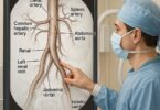
To truly appreciate the complexity of a distal radius fracture and the surgical skill required to fix it, one must first understand the anatomy involved. The wrist is not a single bone but a complex, synovial hinge joint that connects the forearm to the hand. Its stability and remarkable range of motion are the product of a delicate interplay between bones, ligaments, tendons, and nerves.
The Radius and Ulna: The forearm consists of two long bones: the radius and the ulna. The radius is the larger of the two on the thumb side (radial side) of the forearm. The term “distal” simply means “further away from the center of the body.” Therefore, the distal radius is the end of the radius bone that forms part of the wrist joint.
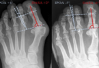
Articular Surface: The distal radius has a uniquely shaped articular surface that cradles the first row of carpal bones (the scaphoid and lunate). This surface is divided into two facets and is critical for the smooth, gliding motion of the wrist. When a fracture involves this joint surface (an intra-articular fracture), it is paramount that it be restored to near-perfect alignment; even a millimeter of step-off can lead to post-traumatic arthritis and permanent pain.
Ligaments and Tendons: A complex network of ligaments holds the carpal bones to the radius and to each other, providing stability. Numerous tendons—the cords that connect muscles to bones and allow for finger and wrist movement—cross the wrist joint. These include the tendons that flex the fingers and wrist on the palm side (volar) and those that extend them on the back-of-hand side (dorsal).
Nerves and Vessels: Three major nerves cross the wrist: the median, ulnar, and radial nerves. The median nerve, in particular, is of critical importance as it passes through the carpal tunnel on the volar side. A distal radius fracture can cause swelling that compresses this nerve, leading to carpal tunnel syndrome or acute median nerve neuropathy, characterized by pain, numbness, and tingling in the thumb, index, middle, and part of the ring finger. The radial artery, a major blood supplier to the hand, also runs near the distal radius.
A fracture in this anatomically dense area is not just a broken bone; it is a traumatic event that disrupts a finely tuned mechanical system. The goal of ORIF is not only to put the bone pieces back together but to restore the entire functional architecture of the wrist.
3. The Nature of the Break: Classifying Distal Radius Fractures
Not all distal radius fractures are created equal. Orthopedic surgeons use classification systems to describe the fracture pattern, which in turn guides treatment decisions—whether to cast or operate, and if operating, what surgical approach and hardware to use. The two most common classification systems are the Fernandez Classification (based on the mechanism of injury) and the AO/OTA Classification (a comprehensive alphanumeric system).
Common Fracture Types:
-
Colles’ Fracture: Perhaps the most well-known type. This is a dorsally displaced, extra-articular fracture, often with a characteristic “dinner fork” deformity when viewed from the side. It typically occurs from a fall on an outstretched hand (FOOSH injury).
-
Smith’s Fracture: Sometimes called a “reverse Colles’.” This is a volar displaced fracture, resulting from a fall onto a flexed wrist or a direct blow to the back of the wrist.
-
Barton’s Fracture: A fracture-dislocation of the wrist where the rim of the distal radius fractures, and the carpal bones dislocate along with the fragment. It can be volar or dorsal Barton’s.
-
Chauffeur’s Fracture (Hutchinson’s Fracture): A fracture of the radial styloid process. Historically, this injury occurred when a driver used a hand crank to start a car, and the crank kicked back, striking the styloid.
-
Die-Punch Fracture: An intra-articular fracture where a portion of the lunate facet of the articular surface is depressed or impacted into the metaphysis of the radius.
The key distinction for coding and treatment is whether the fracture is extra-articular (the fracture line does not enter the wrist joint) or intra-articular (the fracture line extends into and disrupts the joint surface). This distinction is what separates CPT code 25607 from its siblings, 25608 and 25609.
4. When Surgery is Necessary: Indications for ORIF
The decision to proceed with surgery is not taken lightly. Non-operative management with closed reduction (manually setting the bone) and casting is successful for many stable, non-displaced fractures. ORIF is indicated when the fracture is deemed unstable or has characteristics that predict poor healing with casting alone. The classic indications include:
-
Significant Displacement: Fracture fragments that are shifted too far apart to heal correctly. A general rule is more than 2-3 mm of shortening or more than 10 degrees of dorsal angulation.
-
Intra-articular Involvement: Any step-off or incongruity of the joint surface greater than 1-2 mm significantly increases the risk of post-traumatic arthritis and requires anatomic surgical reduction.
-
Severe Comminution: When the bone is broken into many small pieces, it is inherently unstable and unlikely to be held by a cast.
-
Open Fracture: When the bone breaks through the skin. This is a surgical emergency due to the high risk of infection.
-
Associated Neurovascular Injury: Fractures causing compression or injury to nerves (like the median nerve) or blood vessels often require surgical decompression and stabilization.
-
Polytrauma: Patients with multiple injuries may require surgical fixation of the wrist to allow for mobilization and use of crutches or walkers.
-
Failed Closed Reduction: When a fracture that was initially set and casted re-displaces during the healing process.
The ultimate goal of ORIF is to restore anatomy, stabilize the fracture to allow early motion, and maximize the patient’s long-term functional outcome.
5. The Surgical Marvel: A Step-by-Step Walkthrough of the ORIF Procedure
The term “ORIF” is a precise description of the procedure’s two main components: Open Reduction and Internal Fixation.
Preoperative Preparation: The patient receives regional or general anesthesia. The arm is prepped and draped sterilely. A tourniquet is almost always applied to the upper arm to create a bloodless surgical field, which is crucial for visualization.
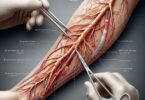
Step 1: Surgical Approach (The “Open” Part)
The most common approach for ORIF of a distal radius fracture today is the volar approach (Henry approach). This is because the volar surface is less complicated by tendons, and the development of volar locking plates has revolutionized fixation from this side.
-
Incision: A longitudinal incision is made over the flexor carpi radialis (FCR) tendon on the palm side of the wrist.
-
Dissection: The surgeon carefully dissects through the subcutaneous tissue, identifies and protects the median nerve and radial artery, and retracts the FCR tendon.
-
Access: The deep fascia is opened, and the flexor pollicis longus (FPL) muscle is retracted. The pronator quadratus muscle, which lies directly on the volar surface of the radius, is then elevated and peeled away to expose the fracture site.
In some cases, a dorsal approach may be necessary, particularly for certain intra-articular fragments or fracture patterns. This approach is more technically challenging due to the numerous extensor tendons that must be carefully navigated and protected.
Step 2: Reduction (The “Reduction” Part)
With the fracture site exposed, the surgeon directly visualizes the bone fragments. Using specialized instruments like elevators, dental picks, and traction, the surgeon meticulously maneuvers each fragment back into its correct anatomic position (reduction). This process is often guided by intraoperative fluoroscopy (a live X-ray) to confirm the alignment from multiple angles before fixation.
Step 3: Fixation (The “Internal Fixation” Part)
Once an anatomic reduction is achieved, it must be held in place. This is where the hardware comes in.
-
Plate and Screws: The workhorse of modern distal radius ORIF is the volar locking plate. This low-profile, anatomically contoured metal plate is placed on the volar surface of the radius. It has specially designed holes for screws. Locking screws thread into the plate, creating a fixed-angle construct that functions like an internal external fixator. This is exceptionally stable, even in osteoporotic bone, as it does not rely on friction between the plate and bone for stability.
-
Other Methods: While plates are most common, other forms of fixation can be used alone or in combination, including:
-
K-wires: Smooth pins used to hold fragments temporarily or for simple fractures.
-
Screws alone: For large, simple fragments (e.g., a radial styloid fracture).
-
External Fixation: A frame outside the body connected to pins in the bone, used for very severe, comminuted fractures or when the soft tissues are too damaged for immediate internal fixation.
-
Step 4: Closure
After final fluoroscopic images confirm perfect reduction and hardware placement, the tourniquet is deflated, and bleeding is controlled. The pronator quadratus muscle is often sutured back over the plate to provide a protective layer between the hardware and the flexor tendons. The subcutaneous tissue and skin are then closed with sutures or staples. A sterile dressing and a splint are applied to support the wrist initially.
6. Decoding CPT Code 25607: Open Treatment of Distal Radius Fracture, Internal Fixation
The American Medical Association’s (AMA) Current Procedural Terminology (CPT) code set is the universal language for describing medical, surgical, and diagnostic services. CPT code 25607 is defined specifically as: “Open treatment of distal radial extra-articular fracture or epiphyseal separation, with internal fixation.”
Let’s break down this definition word-by-word:
-
Open treatment: This mandates that the surgeon made an open incision to directly visualize the fracture site. This is distinct from percutaneous treatment, where fixation is placed through small stab incisions without opening the fracture, or closed treatment, where the bone is set without any incision.
-
Distal radial extra-articular fracture: This is the most critical part of the code’s description. Code 25607 is exclusively for fractures that do NOT involve the wrist joint. The fracture is contained to the metaphysis (the wider part of the bone near the end) of the distal radius.
-
With internal fixation: This confirms that hardware (plate, screws, etc.) was placed to hold the reduction.
Global Period and Modifiers:
CPT 25607 has a 90-day global surgical period. This means the reimbursement for this code is intended to cover all routine preoperative, intraoperative, and postoperative services related to the surgery for the following 90 days. This includes follow-up visits, dressing changes, and removal of sutures or staples. Separate reimbursement for these services within the 90-day window is generally not allowed.
Modifiers are two-digit codes appended to a CPT code to indicate that a service was altered in some way without changing the definition of the code itself. Common modifiers used with 25607 include:
-
-LT (Left side) / -RT (Right side): Essential for specifying which wrist was operated on.
-
-58 (Staged or Related Procedure): Used if a procedure (e.g., hardware removal) is performed during the postoperative period of a previous procedure and was planned or more extensive than the original.
-
-78 (Unplanned Return to the OR): Used if the patient must return to the operating room for a related procedure during the postoperative period (e.g., for an infection washout).
7. Coding Nuances: Differentiating 25607 from 25608 and 25609
CPT codes for distal radius fractures are organized by complexity. It is vital to choose the correct code to avoid billing inaccuracies, denials, or audits.
CPT Code 25607: As defined, for extra-articular fractures.
CPT Code 25608: “Open treatment of distal radial intra-articular fracture or epiphyseal separation; with internal fixation of 2 fragments.”
-
This code is used when the fracture extends into the joint (intra-articular) and the surgeon internally fixes exactly two main fragments.
CPT Code 25609: “Open treatment of distal radial intra-articular fracture or epiphyseal separation; with internal fixation of 3 or more fragments.”
-
This is the most complex code, used for comminuted intra-articular fractures where the surgeon fixes three or more fragments.
The reimbursement for these codes increases with complexity: 25607 < 25608 < 25609. The coder must carefully review the operative report to determine articular involvement and count the number of fragments that were fixed, not just the number of fracture lines.
Table: CPT Code Comparison for Distal Radius ORIF
| CPT Code | Fracture Type | Articular Involvement | Number of Fragments Fixed | Relative Complexity |
|---|---|---|---|---|
| 25607 | Extra-articular | No | N/A | Lower |
| 25608 | Intra-articular | Yes | 2 | Medium |
| 25609 | Intra-articular | Yes | 3 or more | Higher |
8. The Crucial Documentation: What Surgeons Must Note for Coders
The operative report is the foundation of accurate coding. For the coder to correctly assign 25607 (or 25608/25609), the surgeon’s documentation must be explicit. Key elements that must be included are:
-
Preoperative Diagnosis: Should clearly state “distal radius fracture.”
-
Postoperative Diagnosis: Must be consistent with the preoperative diagnosis.
-
Indication for Surgery: A brief note on why ORIF was chosen over non-operative management (e.g., “displaced and unstable fracture”).
-
Procedure Title: Should accurately reflect what was done (e.g., “ORIF left distal radius fracture”).
-
Description of Procedure (The Narrative):
-
Approach: Must state that an “open approach” was used (e.g., “a standard volar Henry approach was utilized”).
-
Fracture Description: This is the most critical part. The surgeon must document whether the fracture was extra-articular or intra-articular. Phrases like “the fracture was noted to be extra-articular” or “the fracture did not extend into the radiocarpal joint” are necessary for 25607. Conversely, “an intra-articular component was observed” is needed for 25608/25609.
-
Reduction: Should note that “open reduction” was performed.
-
Fixation: Must meticulously list all hardware used. “Internal fixation was achieved with a volar locking plate and multiple locking screws.” For 25608/25609, the report should describe the fragments (e.g., “the radial column, intermediate column, and ulnar corner fragments were reduced and fixed”).
-
-
Closure: Description of wound closure.
-
Imaging: A note that fluoroscopy was used and confirmed “anatomic alignment and satisfactory hardware position.”
Without clear documentation specifying the extra-articular nature of the fracture, a coder cannot legally assign 25607 and may be forced to assign a more complex (and incorrect) code, leading to compliance issues.
9. The Road to Recovery: Postoperative Care and Rehabilitation
The surgery is only the beginning of the healing journey. A well-structured postoperative protocol is essential for a successful outcome.
-
Immediate Post-Op (0-2 weeks): The wrist is immobilized in a splint. The focus is on controlling swelling (elevation, ice), managing pain, and encouraging gentle motion of the fingers, elbow, and shoulder to prevent stiffness.
-
Suture Removal (10-14 days): The splint and sutures/staples are removed. The patient is often placed into a removable wrist brace for comfort.
-
Early Motion (2-6 weeks): Under the guidance of a physical or occupational therapist, the patient begins active and passive range-of-motion exercises for the wrist. The brace is typically weaned off during this period.
-
Strengthening (6-12 weeks): As healing progresses confirmed by X-ray, therapy progresses to strengthening exercises with putty, light weights, and resistance bands.
-
Return to Activity (3-6 months): Most patients can return to light activities and desk work within 6-8 weeks. Full return to heavy labor, sports, and high-impact activities can take 4-6 months or longer.
The major advantage of stable ORIF with a locking plate is that it allows this early motion, which is crucial for preventing permanent wrist and hand stiffness.
10. Potential Complications: Risks and Mitigation Strategies
As with any major surgery, ORIF carries risks. Informed consent requires that patients understand these possibilities:
-
Infection: Risk is low (<2%) but can be superficial or deep.
-
Stiffness: The most common complication. Mitigated by stable fixation and early therapy.
-
Hardware Irritation: Tendons can be irritated by the plate or screws, causing pain or even rupture (especially of the extensor pollicis longus tendon with dorsal plates). Volar plates can irritate flexor tendons. This may require subsequent hardware removal after the fracture is fully healed.
-
Neurovascular Injury: Damage to nerves (median, radial, ulnar) or the radial artery can occur during surgery, though it is rare with experienced surgeons.
-
Complex Regional Pain Syndrome (CRPS): A poorly understood condition causing severe, chronic pain, swelling, and changes in skin color and temperature. Early mobilization is key to prevention.
-
Nonunion/Malunion: The fracture fails to heal (nonunion) or heals in a poor position (malunion). This is uncommon with modern techniques.
-
Post-traumatic Arthritis: Can occur if the joint surface was damaged and not perfectly restored.
11. Advancements in Technology: Volar Locking Plates and Beyond
The treatment of distal radius fractures has been revolutionized by the advent of volar locking plate systems in the early 2000s. Before this, fixation was often achieved with pins, external fixators, or non-locking plates that provided less stable fixation, especially in osteoporotic bone. Locking plate technology provides angular stability, allowing patients to start moving their wrists much earlier, leading to dramatically improved functional outcomes. Future advancements include:
-
Fragment-Specific Fixation: Smaller, low-profile plates designed to target specific fracture fragments.
-
Arthroscopic Assistance: Using a small camera inside the joint to aid in the visualization and reduction of intra-articular fractures without a large open approach.
-
Biologics: The use of bone grafts, bone graft substitutes, and growth factors to enhance healing in complex fractures with bone loss.
-
Patient-Specific Implants: 3D-printed plates and guides based on a patient’s CT scan, offering a perfect fit and potentially simplifying surgery.
12. Conclusion
CPT code 25607 precisely represents the open surgical treatment of an extra-articular distal radius fracture with internal fixation. Its accurate application hinges on a deep understanding of both the clinical procedure and the specific language required in surgical documentation. The evolution of locking plate technology has made ORIF a highly successful procedure, restoring function to one of the body’s most complex joints. Mastery of this code, from anatomy to reimbursement, is essential for the seamless operation of orthopedic care.
13. Frequently Asked Questions (FAQs)
Q1: How long does the surgery for a distal radius ORIF take?
A: The procedure itself typically takes between 60 and 90 minutes, though this can vary based on the fracture’s complexity. With preoperative preparation and anesthesia time, the patient may be in the operating room for 2-3 hours.
Q2: Will I have a visible scar?
A: Yes, there will be a scar from the incision. For a volar approach, it is usually about 3-4 inches long on the palm side of the wrist. Surgeons place incisions to minimize visibility, and scars will fade significantly over 6-12 months. Proper scar care can help its appearance.
Q3: When can I drive after ORIF surgery?
A: This is highly variable and depends on which wrist was operated on (dominant vs. non-dominant), your recovery speed, and pain medication use. Generally, patients cannot drive while in a splint or brace (2-6 weeks). You must have adequate strength, motion, and control to safely operate a vehicle. Always get clearance from your surgeon before resuming driving.
Q4: Is the hardware always removed?
A: No, hardware is typically not removed unless it causes problems. Many patients live with their plate and screws permanently without issue. Removal is only considered if the hardware causes pain, irritation, tendonitis, or if a patient is very young and active. This is a separate surgery, usually performed at least a year after the initial ORIF.
Q5: What is the long-term outlook after this surgery?
A: The vast majority of patients achieve excellent outcomes. Most can expect to regain nearly full function and return to all their previous activities. Some may experience minor, occasional stiffness or aches, particularly in cold weather. The outcome is best if the fracture heals in perfect alignment and the patient is dedicated to their postoperative therapy.
14. Additional Resources
-
American Academy of Orthopaedic Surgeons (AAOS): Patient education articles on distal radius fractures: https://orthoinfo.aaos.org
-
American Society for Surgery of the Hand (ASSH): Detailed resources on hand and wrist anatomy and conditions: https://www.assh.org
-
American Medical Association (AMA): CPT Code Set Information: https://www.ama-assn.org/amaone/cpt-current-procedural-terminology
-
PubMed.gov: A database of medical literature for in-depth clinical studies on ORIF outcomes and techniques.
15. Disclaimer
This article is for informational and educational purposes only. It is not intended to be a substitute for professional medical advice, diagnosis, or treatment. Always seek the advice of your surgeon, physician, or other qualified health provider with any questions you may have regarding a medical condition or procedure. The information on medical coding is provided as a guide and should be verified against the most current, official CPT codebook and payer-specific guidelines. The author and publisher disclaim any liability for any loss or damage incurred as a direct or indirect consequence of the use or application of any of the contents of this article.