In the intricate world of medical coding, a string of numbers like CPT 27822 might seem like a sterile, administrative artifact. To a healthcare provider, a biller, or a patient reviewing an explanation of benefits, it is simply the code for “Open treatment of trimalleolar ankle fracture, includes internal fixation, when performed, medial and/or lateral malleolus; without fixation of posterior lip.” Yet, behind this clinical description lies a profound human story—a story of traumatic injury, precise surgical artistry, grueling rehabilitation, and the hope of restored mobility. This code represents a pivotal event in a patient’s life, a complex procedure performed by skilled surgeons to reconstruct a shattered joint. This article aims to dissect every facet of CPT code 27822, transforming it from a five-digit number into a comprehensive narrative that encompasses anatomy, surgery, coding, recovery, and the very human experience at its core. We will journey from the moment of injury to the final step of rehabilitation, providing an exhaustive guide for medical professionals, students, and curious patients alike.

CPT Code 27822
2. Decoding the Ankle: A Primer on Anatomy and Trauma
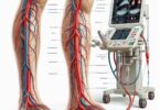
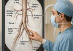
To understand the procedure coded as 27822, one must first understand the anatomy it seeks to repair. The ankle joint, or talocrural joint, is a sophisticated synovial hinge joint that bears the entire weight of the body. Its stability is paramount for ambulation and is maintained by a complex interplay of bones and ligaments.
The bony architecture consists of three parts:
-
The Tibia: The shin bone. Its distal end forms the medial malleolus (the inside bump of the ankle) and the articular surface (plafond) of the ankle joint.
-
The Fibula: The smaller, lateral bone of the lower leg. Its distal end forms the lateral malleolus (the outside bump of the ankle), which extends further posteriorly than the medial malleolus, providing critical stability against lateral motion.
-
The Talus: The critical “keystone” bone of the ankle that sits between the tibia and fibula above and the calcaneus (heel bone) below. It transmits forces from the leg to the foot.
These bones are held together by a robust network of ligaments, most notably the syndesmotic ligaments that bind the tibia and fibula together, and the deltoid ligament on the medial side.
An ankle fracture occurs when one or more of these bones break. The mechanism of injury is often a twisting or rolling motion, a fall, or a high-impact trauma like a car accident. The pattern of the fracture is directly related to the direction and magnitude of the force applied.
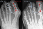
3. What is a Trimalleolar Ankle Fracture? The Anatomy of an Injury
The term “trimalleolar” is key to understanding CPT 27822. “Tri” means three, and “malleolus” refers to the bony prominences. A trimalleolar fracture is a severe injury involving breaks in all three malleoli that form the ankle joint:
-
The Lateral Malleolus: The end of the fibula.
-
The Medial Malleolus: The end of the tibia.
-
The Posterior Malleolus: The posterior (back) lip of the tibial plafond.
This injury is inherently unstable. The fracture of the posterior malleolus is particularly significant as it involves the weight-bearing surface of the tibia. Displacement of this fragment can lead to joint incongruity, where the talus no longer sits perfectly within the tibial socket. If left untreated, this misalignment almost guarantees the development of post-traumatic arthritis, chronic pain, and joint dysfunction. The goal of ORIF is to restore the native anatomy to within millimeter precision to avoid this devastating outcome.
4. The Indications for Surgery: When is ORIF Necessary?
Not all ankle fractures require surgery. Stable, non-displaced fractures can often be treated with casting and non-weight-bearing restrictions. However, for a trimalleolar fracture, surgical intervention is almost always indicated. The decision is based on several radiographic and clinical factors:
-
Displacement: Any significant displacement of the fracture fragments (typically more than 2-3 mm) disrupts the smooth articular surface of the ankle joint, necessitating surgical realignment.
-
Joint Instability: If the ankle joint is unstable—meaning the talus can shift abnormally within the mortise—surgery is required to restore stability. This is often assessed with stress-view X-rays.
-
Open Fracture: When the broken bone punctures the skin, it is an orthopedic emergency requiring immediate surgical debridement and fixation to prevent deep infection.
-
Neurovascular Compromise: If the fracture is compromising blood flow or nerve function to the foot, urgent surgery is needed.
-
Failure of Closed Reduction: If attempts to realign the fracture manually under sedation (closed reduction) are unsuccessful in achieving a stable, anatomical position.
5. The Surgical Team: Orthopedic Surgeons and Their Crucial Support
Performing an ORIF is not a solo endeavor. It is a symphony conducted by the orthopedic surgeon but played by an entire team:
-
Orthopedic Surgeon: The lead surgeon, specializing in the musculoskeletal system. Many surgeons have fellowship training in trauma or foot and ankle surgery.
-
Surgical First Assistant: Often another surgeon, a physician assistant (PA), or a registered nurse first assistant (RNFA) who aids in exposure, retraction, and closure.
-
Anesthesiologist/CRNA: Administers general or regional anesthesia (e.g., a spinal block) to ensure the patient is pain-free and immobile.
-
Circulating Nurse: A registered nurse who manages the sterile field, retrieves supplies, and documents the procedure.
-
Surgical Technologist: Prepares the sterile instruments, including the complex array of drills, screws, and plates, and passes them to the surgeon.
6. Preoperative Protocol: From ER to OR
Once the decision for surgery is made, a standardized protocol is followed:
-
Informed Consent: The surgeon discusses the procedure, its risks, benefits, and alternatives in detail with the patient or their proxy.
-
Medical Optimization: The patient’s overall health is assessed. This may involve cardiology or internal medicine consultations for patients with comorbidities like diabetes or heart disease to minimize surgical risk.
-
Imaging: Preoperative X-rays (AP, lateral, and mortise views) are standard. A CT scan is almost always obtained for a trimalleolar fracture to fully delineate the complexity of the fracture, especially the size and displacement of the posterior malleolar fragment.
-
NPO Status: The patient is instructed to have nothing by mouth (NPO) for typically 8 hours before surgery to reduce the risk of aspiration during anesthesia.
-
Prophylactic Antibiotics: Intravenous antibiotics are administered within 60 minutes of the incision to prevent surgical site infection.
7. Deep Dive into CPT Code 27822: The Procedure Step-by-Step
This is the core of the code itself. The description “Open treatment of trimalleolar ankle fracture…” is a succinct summary of a highly detailed operation.
7.1. Anesthesia and Positioning
The patient is placed supine (on their back) on the operating table. A bump may be placed under the hip to internally rotate the leg, bringing the ankle into a neutral position. For access to the posterior malleolus, the patient may be positioned prone (on their stomach). After adequate anesthesia, a high-thigh tourniquet is often applied to create a bloodless surgical field. The entire leg is prepped and draped in a sterile fashion.
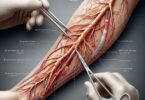
7.2. The Surgical Approach: Incisions and Exposure
Typically, two incisions are required to address all three malleoli while protecting critical structures.
-
Lateral Incision: A longitudinal incision is made over the lateral malleolus, carefully protecting the superficial peroneal nerve.
-
Medial Incision: A curved incision is made over the medial malleolus, carefully protecting the saphenous vein and nerve.
-
Posterior Approach: If the posterior malleolar fragment is large and requires direct fixation, a separate posterolateral incision may be made, retracting the peroneal tendons and protecting the sural nerve.
The fractures are exposed, and the hematoma (collection of blood) is evacuated. The fragments are carefully visualized, and any soft tissue interposition (e.g., tendon, ligament) blocking reduction is removed.
7.3. “Open Reduction”: The Art of Realigning the Fracture
Reduction is the process of anatomically realigning the fracture fragments. This is a meticulous and critical step. The surgeon uses specialized instruments like bone clamps, reduction forceps, and temporary K-wires (thin, smooth pins) to “reduce” the fragments into their original position. Fluoroscopy (a live X-ray machine) is used continuously throughout this process to confirm the accuracy of the reduction in multiple planes.
7.4. “Internal Fixation”: The Hardware of Stability
Once a perfect reduction is achieved, it must be held in place with implants. The choice of hardware depends on the fracture pattern and bone quality.
-
Lateral Malleolus: Typically fixed with a neutralization or locking plate and screws applied to the lateral surface of the fibula.
-
Medial Malleolus: Typically fixed with two cannulated, partially threaded lag screws that compress the fracture line. For smaller or comminuted fragments, a tension band wiring technique or a small plate may be used.
-
Posterior Malleolus: If the fragment involves more than 25-30% of the joint surface or is displaced, it requires fixation. This can be done with anteroposterior (AP) lag screws placed from the front of the tibia into the posterior fragment or, more directly, with posterior-to-anterior screws or a plate through a posterior approach.
-
Syndesmotic Injury: A key part of the procedure is testing the stability of the syndesmosis (the tibia-fibula ligamentous connection). If unstable, it must be stabilized with one or two syndesmotic screws or a suture-button device, which hold the bones together while the ligaments heal.
Table 1: Common Implants Used in Ankle ORIF (27822)
| Implant Type | Function | Common Location of Use |
|---|---|---|
| Cannulated Lag Screw | Compresses fracture fragments across a fracture line. | Medial malleolus, posterior malleolus. |
| Neutralization Plate | Protects a fracture from bending or rotational forces after lag screw fixation. | Lateral malleolus (fibula). |
| Locking Plate | Creates a fixed-angle construct, ideal for osteoporotic bone. | Lateral malleolus in elderly patients. |
| K-wire (Kirschner wire) | Provides temporary fixation before permanent hardware is placed. | All malleoli. |
| Syndesmotic Screw | Stabilizes the distal tibiofibular joint (syndesmosis). | Through fibula into tibia. |
| Suture-Button Device | Dynamic stabilization of the syndesmosis. | Alternative to syndesmotic screw. |
7.5. Irrigation, Closure, and Dressing
After all hardware is placed and final fluoroscopic images confirm a perfect reduction and implant position, the surgical site is copiously irrigated with saline to remove any debris. The incisions are closed in layers: deep fascia, subcutaneous tissue, and finally the skin with staples or sutures. A sterile dressing and a well-padded splint are applied to immobilize the ankle in a neutral 90-degree position.
8. Coding and Billing Nuances: Beyond 27822
Accurately coding 27822 is essential for appropriate reimbursement and legal compliance.
8.1. Modifiers and Their Critical Importance
-
Modifier -LT (Left side) or -RT (Right side): This is mandatory. CPT 27822 is a unilateral code. Failing to append LT or RT will result in a claim denial.
-
Modifier -58 (Staged or Related Procedure): If the patient returns to the operating room during the postoperative period for a planned procedure (e.g., hardware removal after fracture healing), this modifier is appended to the second procedure’s code.
-
Modifier -78 (Unplanned Return to OR): If the patient requires an unplanned return to the OR for a related complication (e.g., irrigation and debridement for an infection) during the global period, this modifier is used.
8.2. Bundled Services and Global Periods
CPT 27822 has a 90-day global surgical period. This means that all routine postoperative care related to the surgery—including office visits, dressing changes, and suture removal—within those 90 days is bundled into the payment for the procedure itself and cannot be billed separately.
8.3. Common Coding Pitfalls and How to Avoid Them
-
Coding for Isolated Malleoli: Do not report 27822 if only one or two malleoli are fractured. Use:
-
CPT 27766 for isolated lateral malleolus fracture.
-
CPT 27762 for isolated medial malleolus fracture.
-
CPT 27814 for bimalleolar fractures (medial and lateral).
-
-
Including Syndesmotic Fixation: The fixation of the syndesmosis is included in 27822 if performed. It should not be reported with a separate code (e.g., 27829).
-
Documentation is Key: The operative report must clearly document the treatment of all three malleoli to justify the use of 27822. Precise documentation of the approach, reduction, and fixation is critical for supporting the code billed.
9. The Immediate Postoperative Journey: Hospital Stay and Recovery
Postoperatively, the patient is monitored in the recovery room. Pain is managed with a multimodal approach (nerve blocks, IV and oral medications). The leg is elevated to control swelling. Patients typically stay in the hospital for 1-2 nights. Physical therapists will instruct the patient on non-weight-bearing ambulation using crutches or a walker before discharge. The importance of strict non-weight-bearing is emphasized to protect the fixation.
10. Long-Term Rehabilitation: The Road to Functional Recovery
Rehabilitation is a long and phased process, often spanning 4-6 months or more.
-
Phase 1 (0-6 weeks): Strict non-weight-bearing. Splint/cast immobilization. Focus is on controlling edema, maintaining range of motion of the hip and knee, and isometric exercises.
-
Phase 2 (6-12 weeks): Transition to a removable walking boot. Weight-bearing is gradually advanced as tolerated, guided by X-ray evidence of healing. Formal physical therapy begins, focusing on restoring ankle range of motion, strength, and proprioception.
-
Phase 3 (3+ months): Full weight-bearing. Therapy intensifies to include balance training, gait training, and functional exercises to prepare for a return to normal activities and work.
11. Potential Complications: Risks and Mitigation Strategies
As with any major surgery, risks exist:
-
Infection: Mitigated by prophylactic antibiotics and sterile technique.
-
Nonunion/Malunion: Failure of the bone to heal or healing in a poor position. Mitigated by sound surgical technique and patient compliance with weight-bearing restrictions.
-
Hardware Irritation: Plates and screws can be felt under the skin and sometimes cause pain, requiring subsequent removal after full healing.
-
Post-traumatic Arthritis: The primary long-term risk, directly related to the initial cartilage damage at the time of injury and the precision of the surgical reduction.
-
Blood Clots (DVT/PE): Mitigated with early mobilization, mechanical compression devices, and sometimes anticoagulant medication.
-
Nerve Injury: Risk to superficial nerves around the incision sites, which can cause numbness or pain.
12. The Patient’s Perspective: Life After an ORIF
Beyond the clinical details, an ORIF is a significant life event. Patients contend with pain, frustration from loss of independence, financial stress from medical bills and time off work, and anxiety about their recovery. A successful outcome depends not only on surgical skill but also on a strong support system, clear communication from the healthcare team, and the patient’s resilience and dedication to their rehabilitation program.
13. Advancements in Technology: Navigating the Future of Ankle Trauma Care
The field is continually evolving. Locking plate technology has improved outcomes in osteoporotic bone. Intraoperative 3D fluoroscopy provides CT-like images in the OR, allowing surgeons to confirm reduction and hardware placement before closing. Pre-contoured, low-profile implants minimize soft tissue irritation. Biological adjuncts like platelet-rich plasma (PRP) and bone morphogenetic proteins (BMP) are being studied to enhance healing. The future may hold more personalized approaches with patient-specific instruments and guides.
14. Conclusion: Synthesizing the Art and Science of Ankle ORIF
CPT code 27822 represents a highly sophisticated orthopedic procedure that restores function from severe trauma. It demands a surgeon’s anatomical expertise, technical precision, and a comprehensive perioperative team. Accurate coding is vital, reflecting the procedure’s complexity through careful documentation and modifier use. Ultimately, successful patient outcomes hinge on this seamless integration of surgical art, clinical science, and dedicated rehabilitation.
15. Frequently Asked Questions (FAQs)
Q1: How long will the hardware from my ankle ORIF stay in? Does it need to be removed?
A: Hardware is typically meant to be permanent. It is only removed if it becomes a source of pain, irritation, or infection, which usually occurs no sooner than 12-18 months after the initial surgery, once the bone is fully healed. Removal is a separate surgical procedure.
Q2: When can I drive after ankle ORIF surgery?
A: This is highly variable. If you have surgery on your left ankle and drive an automatic transmission, you may be able to drive once you are off strong pain medication and can comfortably bear weight in a boot (often 6-8 weeks). For right ankle surgery, it is typically longer, around 10-12 weeks or until you have regained sufficient reaction time and control to safely operate the pedals.
Q3: Will I develop arthritis in my ankle now that I’ve had a fracture?
A: There is an increased risk of post-traumatic arthritis following any intra-articular fracture (where the break line enters the joint surface). The goal of ORIF is to minimize this risk by restoring the joint surface as perfectly as possible. Your long-term risk depends on the severity of the initial injury, the accuracy of the reduction, and factors like your weight and activity level.
Q4: What is the difference between CPT 27822 and 27823?
A: CPT 27822 is for a trimalleolar fracture without fixation of the posterior lip (posterior malleolus). CPT 27823 is for a trimalleolar fracture with fixation of the posterior lip. The choice of code is determined by whether the surgeon directly addressed the posterior fragment with screws or a plate.
16. Additional Resources
-
American Academy of Orthopaedic Surgeons (AAOS): OrthoInfo website provides excellent patient-friendly information on ankle fractures and recovery.
-
American Medical Association (AMA): The definitive source for CPT code definitions, guidelines, and updates.
-
National Institutes of Health (NIH) – PubMed: A database of medical literature for those seeking advanced clinical studies on ankle fracture outcomes.
17. Disclaimer
This article is for informational and educational purposes only. It is not intended to be a substitute for professional medical advice, diagnosis, or treatment. Always seek the advice of your orthopedic surgeon or other qualified health provider with any questions you may have regarding a medical condition or treatment. The information on medical coding is provided as a guide and should be verified with the current year’s CPT codebook and payer-specific policies for accurate billing and reimbursement. The author assumes no liability for any errors or omissions in the content.