Imagine a diagnostic tool so powerful it can peer into the most complex structure in the known universe—the human brain—without making a single incision, without using ionizing radiation, and with breathtaking anatomical detail. This is not science fiction; this is the reality of magnetic resonance imaging (MRI). For decades, physicians relied on crude shadows from X-rays or the clinical intuition of a neurological exam to understand the brain’s mysteries. Today, the non-contrast MRI of the brain, precisely defined by the Current Procedural Terminology (CPT) code 70551, stands as a cornerstone of modern neurology, neurosurgery, and psychiatry. It is a testament to human ingenuity, a fusion of quantum physics, advanced computing, and medical science that has revolutionized our ability to diagnose, monitor, and understand a vast array of neurological conditions.
This article serves as the ultimate guide to CPT code 70551. We will embark on a deep dive that goes far beyond a simple definition. We will explore the intricate physics that make the scan possible, walk through the patient’s journey from scheduling to results, decipher the complex language of radiology reports, and demystify the critical processes of medical coding and billing that ensure this technology remains accessible. Whether you are a healthcare provider seeking a deeper understanding, a medical coder aiming for precision, or a patient preparing for an upcoming scan, this comprehensive resource is designed to provide clarity, insight, and appreciation for one of medicine’s most indispensable diagnostic tools.
2. Decoding the CPT Code: What is 70551?
At its core, a CPT code is a universal medical language used to describe specific procedures and services provided by healthcare professionals. Maintained by the American Medical Association (AMA), this standardized system is essential for communication between providers, patients, and payers (insurance companies). It is the foundation upon which billing and reimbursement are built.
CPT Code 70551 is explicitly defined as: “Magnetic resonance (eg, proton) imaging, brain (including brain stem); without contrast material.”
Let’s deconstruct this definition:
-
Magnetic resonance imaging: This specifies the modality. It is not a CT scan (which uses X-rays), an ultrasound, or a PET scan.
-
Brain (including brain stem): This defines the anatomical region of interest. The code encompasses the entire cerebrum, cerebellum, and brainstem. It is distinct from codes for the orbits (eyes), internal auditory canals (ears), or pituitary gland, which are considered separate structures, though often imaged concurrently.
-
Without contrast material: This is the most critical differentiator. “Contrast material” refers to a gadolinium-based dye injected intravenously to enhance the visibility of certain pathologies like tumors, inflammation, or infection. Code 70551 is used when this injection is not administered.
It is crucial to distinguish 70551 from its counterpart, CPT Code 70553: “…with contrast material.” Furthermore, if a study is performed both without and with contrast, a third code, 70552, is used. The correct application of these codes is not a matter of choice but of strict adherence to the service actually rendered. Miscoding can lead to claim denials, audits, and compliance issues.
When is 70551 Used?
This code is reported for the technical and professional components of performing and interpreting a non-contrast MRI of the brain. The technical component covers the overhead costs of operating the MRI scanner, including the use of the machine, technologist’s time, and expensive maintenance. The professional component covers the radiologist’s expertise in interpreting the images and generating a diagnostic report.
3. The Clinical Powerhouse: Indications for a Non-Contrast MRI Brain
The non-contrast MRI brain is a first-line investigative tool for a staggering variety of neurological symptoms and conditions. Its ability to exquisitely detail soft tissue structures makes it superior to CT for most non-traumatic, non-emergent brain pathologies. The following table outlines the primary clinical indications, categorized by symptom and suspected pathology.
Table 1: Clinical Indications for a Non-Contrast MRI Brain (70551)
| Category | Common Symptoms | Suspected Pathology/Indication | Why Non-Contrast is Often Sufficient |
|---|---|---|---|
| Cerebrovascular | Sudden weakness, slurred speech, vision loss, dizziness | Acute ischemic stroke, chronic small vessel ischemic disease, cerebral aneurysms (using MRA), vascular malformations | Excellent for detecting acute ischemia (diffusion-weighted imaging), chronic infarcts, and leukoaraiosis (white matter disease). MRA visualizes blood vessels without contrast. |
| Headache | New, severe, or worsening headache; “thunderclap” headache; headache with neurological deficits | Rule out mass, idiopathic intracranial hypertension, sinus disease, Arnold-Chiari malformation | Highly sensitive for detecting tumors, structural causes of increased ICP, and congenital anomalies. Often the first step in a headache workup. |
| Seizure/Epilepsy | Unexplained seizures, spells of altered consciousness | Structural epilepsy focus (e.g., cortical dysplasia, mesial temporal sclerosis, tumor), evaluation for surgery | The gold standard for identifying subtle cortical malformations and hippocampal sclerosis that are often the source of seizures. |
| Trauma | Head injury with persistent neurological symptoms (when CT is negative or inconclusive) | Diffuse axonal injury (DAI), small contusions, microhemorrhages (using SWI/GRE sequences) | Far more sensitive than CT for detecting non-hemorrhagic shearing injuries (DAI) and tiny hemorrhages. |
| Neurodegenerative | Memory loss, cognitive decline, personality changes, movement disorders (e.g., tremor, rigidity) | Alzheimer’s disease, frontotemporal dementia, vascular dementia, normal pressure hydrocephalus (NPH) | Used to assess patterns of atrophy, rule out other causes, and support a clinical diagnosis. |
| Demyelinating | Episodes of neurological dysfunction (e.g., optic neuritis, numbness, weakness) | Multiple Sclerosis (MS), ADEM | Can detect classic demyelinating plaques (ovoid, periventricular, callososeptal) and monitor disease burden over time. |
| Infectious/Inflammatory | Headache, fever, confusion (when meningitis/encephalitis is suspected, but often followed by contrast) | Encephalitis, abscess (late stage), meningitis complications | Can show patterns of inflammation and complications like hydrocephalus or empyema, though contrast is usually added for full evaluation. |
| Oncologic | New-onset seizures, focal neurological deficits, signs of increased ICP | Primary brain tumor (e.g., glioma, meningioma), metastasis | Excellent for initial detection and characterization of many tumors. However, contrast-enhanced MRI (70553/70552) is almost always needed for staging, surgical planning, and monitoring treatment. |
| Congenital/Developmental | Developmental delay in children, known or suspected congenital syndrome | Evaluation of brain maturation, neuronal migration disorders (e.g., lissencephaly, heterotopia), corpus callosum anomalies | The definitive test for visualizing brain anatomy and diagnosing a wide range of congenital malformations. |
As the table illustrates, 70551 is an incredibly versatile tool. It is often the initial study of choice because it provides a vast amount of diagnostic information on its own. In many cases, such as evaluating headaches, dementia, or uncomplicated epilepsy, a non-contrast study is all that is required. In other scenarios, it serves as the essential baseline upon which a contrast-enhanced study is later added if findings are suspicious.
4. A Journey Through the Magnet: The Patient Experience from Start to Finish
For a patient, an MRI can be an intimidating prospect. Understanding the process can significantly alleviate anxiety.
Step 1: Preparation and Scheduling
The journey begins with an order from a physician. The scheduling team will confirm the order for a “Brain MRI without Contrast” and collect necessary insurance information. They will provide detailed instructions, which typically include:
-
Screening Questionnaire: A thorough safety screening is paramount. Patients must disclose any implanted medical devices (pacemakers, aneurysm clips, cochlear implants), metal fragments (especially in the eye), pregnancy, or history of claustrophobia.
-
What to Wear: Patients are instructed to wear loose, comfortable clothing without metal zippers, snaps, or underwires. Often, they are asked to change into a hospital gown.
-
Food and Medications: Typically, there are no restrictions on food or medications for a non-contrast brain MRI. This is a key difference from studies requiring contrast.
Step 2: Arrival and Check-In
Upon arrival, the patient will complete registration and provide a copy of their insurance card. The MRI technologist will review the safety questionnaire again in person—a critical double-check for patient safety.
Step 3: In the Scanner Room
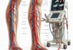
The patient will remove all metallic objects (jewelry, watches, hairpins, hearing aids, etc.) and empty their pockets. Even a small piece of metal can become a dangerous projectile or distort the images. The technologist will then help the patient onto the scanner bed. For a brain MRI, a specialized head coil—a plastic cage-like device—is placed around the head. This is not restrictive; it is a radiofrequency antenna designed to improve image quality. Earplugs or headphones are provided to protect hearing from the loud knocking noises the scanner makes and to allow for communication with the technologist.
Step 4: The Scan Itself
The bed moves into the magnet bore, which is a long, narrow tube. The most important instruction for the patient is to remain perfectly still. Even the slightest movement can blur the images, rendering them nondiagnostic. The scan is painless. The patient will hear a series of loud, rhythmic knocking and humming sounds as different pulse sequences are run. Each sequence can take from 2 to 8 minutes. A typical non-contrast brain MRI protocol may consist of 5-7 sequences and last between 30 to 45 minutes. The technologist can see and hear the patient at all times and will provide updates and check in through a two-way intercom system.
Step 5: After the Scan
Once the scan is complete, the bed slides out, and the patient is free to leave. There is no recovery period or lingering effects. They can resume their normal activities, including driving, immediately. The images are sent to a picture archiving and communication system (PACS) for a radiologist to interpret.
5. The Technology Behind the Image: Physics and Pulse Sequences Demystified
The magic of MRI lies in its exploitation of the quantum properties of hydrogen atoms in water and fat, which are abundant in the human body. The process involves three main steps:
-
Alignment: The powerful superconducting magnet (typically 1.5 Tesla or 3.0 Tesla, tens of thousands of times stronger than Earth’s magnetic field) causes the protons in hydrogen atoms to align with its magnetic field.
-
Excitation: A precise radiofrequency (RF) pulse is broadcast into the body, knocking these aligned protons out of alignment.
-
Relaxation and Signal Detection: When the RF pulse is turned off, the protons relax back to their aligned state, releasing energy in the form of a radio signal. This signal is detected by the receiver coils (the head coil) and sent to a powerful computer.
The computer analyzes the timing and strength of these signals from millions of points (voxels) to construct a detailed cross-sectional image. The timing of the RF pulses (the “pulse sequence”) is what creates different types of image weightings, each highlighting specific tissues and pathologies.
Key Pulse Sequences in a Non-Contrast Brain MRI (70551):
-
T1-Weighted Imaging: Excellent for viewing normal anatomy. Cerebrospinal fluid (CSF) is dark, while white matter is brighter than gray matter. It provides a clear anatomical roadmap.
-
T2-Weighted Imaging: Fluid (CSF, edema, cysts) is very bright. This is excellent for detecting pathologies that cause swelling or contain fluid, such as tumors, strokes, MS plaques, and inflammation.
-
FLAIR (Fluid-Attenuated Inversion Recovery): A special type of T2 weighting where the signal from free water (CSF) is suppressed, making it dark. This allows lesions next to fluid-filled spaces (like MS plaques around the ventricles) to “light up” brilliantly, making them impossible to miss.
-
DWI (Diffusion-Weighted Imaging) & ADC (Apparent Diffusion Coefficient): This is the most critical sequence for diagnosing an acute ischemic stroke, often within minutes of its onset. It detects the restriction of water molecule movement within dying brain cells (cytotoxic edema). Areas of acute infarction appear bright on DWI and dark on ADC maps.
-
SWI (Susceptibility Weighted Imaging) or GRE (Gradient Echo): Exquisitely sensitive to blood products (hemorrhage), calcium, and iron. It is used to detect microbleeds from trauma (DAI), cerebral amyloid angiopathy, cavernous malformations, and other vascular abnormalities.
-
MRA (Magnetic Resonance Angiography): Can be performed without contrast (Time-of-Flight technique) to visualize the major arteries of the circle of Willis. It is used to screen for aneurysms, stenosis, and other vascular abnormalities.
A radiologist synthesizes the information from all these sequences to form a complete diagnostic picture. A lesion might be dark on T1, bright on T2 and FLAIR, and show restricted diffusion—a signature pattern for an acute stroke.
6. The Art of Interpretation: What Radiologists See
The raw images are just the beginning. The value is unlocked by the trained eye of a radiologist. Their systematic approach involves assessing:
-
Symmetry: Is the brain symmetrical? Asymmetry can indicate atrophy or mass effect from a tumor.
-
Gray-White Matter Differentiation: Is the clear boundary between the brain’s gray and white matter preserved? Loss of this differentiation is an early sign of stroke.
-
Ventricular Size and Shape: Are the fluid-filled ventricles normal in size, or are they enlarged (hydrocephalus) or compressed?
-
Sulcal and Gyral Pattern: Are the folds of the brain (sulci and gyri) normal, or are they thickened (as in some infections) or atrophied?
-
Midline Structures: Is the midline shift present, indicating pressure from one side of the brain?
-
Signal Abnormalities: The radiologist looks for areas that are too bright or too dark on the various sequences, correlating these findings with the clinical history to narrow the differential diagnosis.
They then compile their findings into a structured report, which includes:
-
Technique: A description of the sequences performed.
-
Comparison: To any prior studies, if available.
-
Findings: A detailed, objective description of what is seen in each anatomical area.
-
Impression/Conclusion: A concise summary of the most significant findings and their likely clinical significance, often with recommendations for further imaging or follow-up.
7. Navigating the Healthcare System: Coding, Billing, and Reimbursement
The accurate use of CPT code 70551 is vital for the financial sustainability of a radiology practice. The process is complex and involves several key players and concepts:
-
Medical Coder: A professional who translates the radiology report into the correct CPT and ICD-10 codes.
-
ICD-10-CM Codes: These are diagnosis codes that represent the medical necessity for the procedure. For 70551 to be paid, it must be linked to a covered, appropriate ICD-10 code (e.g., R51.9 Headache, unspecified; G45.9 Transient cerebral ischemic attack, unspecified; R41.82 Altered mental status). Mismatched codes are a primary reason for claim denials.
-
Claim Submission: The practice’s billing department submits the claim (CPT 70551 + ICD-10 code(s)) to the patient’s insurance payer.
-
Reimbursement: The payer evaluates the claim based on the patient’s plan benefits and a fee schedule. Reimbursement rates for 70551 are set by Medicare (via the Physician Fee Schedule) and negotiated with private insurers. They are intended to cover both the technical and professional components.
-
Audits and Compliance: Incorrect coding, whether accidental (upcoding) or deliberate (fraud), can trigger audits from payers or government agencies like the OIG, resulting in hefty fines and penalties. Therefore, coding accuracy is both a financial and a legal imperative.
8. Safety First: Absolute and Relative Contraindications
MRI is exceptionally safe for the vast majority of patients as it does not use ionizing radiation. However, the powerful magnetic field presents unique dangers.
Absolute Contraindications (Cannot have an MRI):
-
Certain implanted electronic devices: Pacemakers, implantable cardioverter-defibrillators (ICDs) – though some newer models are “MRI-conditional.”
-
Metallic foreign bodies in the eye (if there is any history and it cannot be ruled out by an X-ray).
-
Cochlear implants (most are not safe).
-
Certain aneurysm clips (particularly older types).
Relative Contraindications (Risks and benefits must be weighed):
-
Claustrophobia: Can often be managed with sedation or a “open” MRI scanner (though these have lower image quality).
-
First trimester of pregnancy: While no adverse effects have been proven, MRI is generally avoided unless the clinical information is critical and cannot be obtained by ultrasound.
-
Severe obesity: Patients may not fit into the scanner bore.
-
MRI-conditional implants: Devices like joint replacements, stents, or some cardiac devices may be safe under specific conditions (e.g., a certain magnet strength). The radiology team must verify the exact model and its conditions.
For a non-contrast MRI, the significant safety concerns related to gadolinium-based contrast agents (allergic-like reactions, nephrogenic systemic fibrosis) are completely avoided, making 70551 an even lower-risk procedure.
9. The Future is Now: Advancements in MRI Technology
The field of MRI is not static. Continuous innovation is pushing the boundaries of what we can see and measure.
-
Higher Field Strengths: 7 Tesla (7T) scanners are moving from research to clinical use, offering unprecedented resolution to visualize tiny structures and lesions.
-
Artificial Intelligence (AI): AI algorithms are being developed to accelerate scan times (reducing motion artifact), enhance image quality, and even assist radiologists by automatically highlighting potential abnormalities, measuring tumors, or predicting tissue characteristics.
-
Functional MRI (fMRI): While not part of a standard 70551, it maps brain activity by detecting changes in blood flow, used pre-surgically to map eloquent cortex (e.g., motor or speech areas).
-
Diffusion Tensor Imaging (DTI): A advanced form of DWI that maps the white matter tracts of the brain, crucial for neurosurgical planning to avoid damaging critical pathways.
-
Quantitative MRI: Moving beyond qualitative pictures to extracting precise numerical measurements of tissue properties, which could lead to more objective diagnosis and monitoring of diseases like MS.
10. Conclusion: The Indispensable Role of the Non-Contrast MRI Brain
CPT code 70551 represents far more than a billing tool; it signifies a safe, powerful, and versatile diagnostic pillar of modern neurology. From detecting life-threatening strokes to unraveling the mysteries of neurodegenerative disease, its value is immeasurable. Its continued evolution through AI and higher-field technology promises to further solidify its role as an indispensable window into the human brain, improving patient outcomes for generations to come.
11. Frequently Asked Questions (FAQs)
Q1: How is an MRI without contrast different from one with contrast?
A: A non-contrast MRI (70551) relies on the body’s natural tissue properties to create images. A contrast-enhanced MRI (70553) involves injecting a gadolinium-based dye that highlights areas with increased blood flow or a broken blood-brain barrier, such as active tumors, infections, or inflammation. The non-contrast scan is often used first; if something suspicious is found, contrast may be recommended for further characterization.
Q2: I’m claustrophobic. What are my options?
A: Inform your doctor and the imaging center beforehand. Options include: 1) Taking a prescribed anti-anxiety medication before the scan (you will need someone to drive you), 2) Using an “open” MRI scanner, which has larger, more open sides (though image resolution may be lower), or 3) Some centers offer premium “wide-bore” 3T scanners that are shorter and wider, reducing the claustrophobic feeling.
Q3: Why did my doctor order a non-contrast MRI for my headaches?
A: A non-contrast MRI is an excellent first test to rule out serious structural causes of headaches, such as a brain tumor, hydrocephalus, or a vascular malformation. In the vast majority of cases, these scans come back normal, which is reassuring for both you and your doctor and allows them to focus on other causes like migraines or tension headaches.
Q4: How long does it take to get results?
A: The images are available immediately, but they must be interpreted by a radiologist. A finalized report is typically sent to your referring physician within 24-48 hours. Your doctor will then contact you to discuss the results in the context of your overall health.
Q5: Is there any radiation involved in an MRI?
A: No. Unlike CT scans and X-rays, which use ionizing radiation, MRI uses a powerful magnetic field and radio waves. There is no known harmful radiation exposure associated with an MRI scan.
12. Additional Resources
-
American College of Radiology (ACR): https://www.acr.org – Provides patient-friendly information on MRI safety and appropriateness criteria.
-
RadiologyInfo.org: https://www.radiologyinfo.org – A joint resource by the ACR and RSNA with detailed guides on MRI procedures for the public.
-
American Society of Neuroradiology (ASNR): https://www.asnr.org – Focuses on advancements in brain and spine imaging.
-
National Institute of Biomedical Imaging and Bioengineering (NIBIB) – MRI: https://www.nibib.nih.gov/science-education/science-topics/magnetic-resonance-imaging-mri – Explains the science behind MRI in an accessible way.