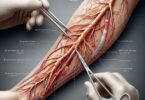
Imagine a non-invasive window into the human chest, capable of revealing the intricate architecture of the heart, the delicate branching of the airways, and the hidden structures nestled behind the breastbone. This is not the realm of science fiction but the everyday reality of modern medicine, made possible by Computed Tomography (CT). At the heart of this diagnostic power for thoracic conditions lies a specific procedural code: CPT Code 71271. This code is far more than a mere billing identifier; it is a precise language that communicates a complex, contrast-enhanced journey into the thorax, capturing dynamic processes that static images cannot.
This article serves as the definitive guide to CPT code 71271. We will embark on a detailed exploration that moves beyond the basic definition, delving into the clinical scenarios that demand its use, the exact technical protocols it entails, the art and science of interpreting its results, and the critical compliance landscape that governs its application. Whether you are a healthcare provider seeking to understand the optimal use of this tool, a medical coder navigating its nuances, or a patient curious about what to expect, this comprehensive resource is designed to provide clarity, depth, and expert insight into one of radiology’s most vital diagnostic procedures.
CPT Code 71271
2. Decoding the Jargon: What is CPT Code 71271?
CPT Code 71271 is defined by the American Medical Association (AMA) as: “Computed tomography, thorax, diagnostic; with contrast material(s)”.
This succinct description contains several layers of critical information that must be unpacked to fully understand the procedure’s scope.
-
Computed Tomography (CT): This is the imaging modality. A CT scanner uses a rotating X-ray tube and a ring of digital detectors to capture a series of cross-sectional images (slices) of the body. A powerful computer then processes these slices to generate two-dimensional and three-dimensional reconstructions, providing exceptional detail free from the superimposition of structures that plagues conventional X-rays.
-
Thorax: This specifies the anatomical region of interest. The thorax, or chest, encompasses a vast and vital territory, including:
-
The lungs and tracheobronchial tree
-
The heart and great vessels (aorta, pulmonary arteries, superior vena cava)
-
The mediastinum (the central compartment containing the heart, esophagus, thymus, lymph nodes, and nerves)
-
The chest wall, including the ribs, muscles, and breasts
-
The upper abdomen (e.g., the adrenal glands, upper liver), which is often included in the field of view.
-
-
Diagnostic: This key term differentiates the procedure from a screening exam. A “diagnostic” CT is performed to investigate specific signs, symptoms, or known abnormalities. It implies a medically necessary investigation based on a clinical question, as opposed to a broad, population-based screening test for asymptomatic individuals (like a lung cancer screening CT, which has its own specific codes).
-
With Contrast Material(s): This is the most crucial differentiator for 71271. The administration of contrast media is not an optional add-on; it is intrinsic to the code’s definition. Contrast material, typically an iodine-based solution, is injected intravenously. It temporarily alters how X-rays interact with blood vessels and tissues, causing them to appear brighter (enhance) on the scan. This enhancement is paramount for:
-
Vascular Assessment: Visualizing blood vessels to detect aneurysms, dissections, blockages (pulmonary emboli), or malformations.
-
Characterizing Masses: Distinguishing between highly vascular tumors, cysts, abscesses, and normal tissue. Malignancies often create new, leaky blood vessels (angiogenesis), leading to a specific pattern of contrast uptake.
-
Evaluating Inflammation: Identifying areas of infection or inflammation, which also receive increased blood flow.
-
Delineating Anatomy: Clearly separating lymph nodes, vessels, and other mediastinal structures from one another.
-
It is essential to distinguish 71271 from other related thoracic CT codes:
-
CPT 71250: CT thorax, diagnostic; without contrast.
-
CPT 71260: CT thorax, diagnostic; without contrast, followed by with contrast.
-
CPT 71275: CT thorax, for lung cancer screening, without contrast.
Using 71271 appropriately requires that the study is both diagnostic and performed with contrast material. If no contrast is used, 71250 is the correct code. If both non-contrast and contrast-enhanced phases are acquired, 71260 is used.
3. The Clinical Powerhouse: Indications and Medical Necessity
The decision to order a CT thorax with contrast is never taken lightly. It is driven by a need to answer a specific clinical question that cannot be adequately addressed by simpler, less expensive, or less irradiating tests like a chest X-ray or ultrasound. Medical necessity is the cornerstone of appropriate imaging, and for 71271, the indications are vast and critical.
A. Pulmonary Embolism (PE) and Thromboembolic Disease:
This is one of the most common and urgent indications. A CT Pulmonary Angiogram (CTPA), performed using the 71271 protocol, is the gold standard for diagnosing PE. The contrast is timed to achieve peak opacification of the pulmonary arteries, allowing radiologists to clearly identify filling defects (clots) that appear as dark areas within the bright contrast-filled vessels. Rapid and accurate diagnosis is life-saving, guiding immediate anticoagulation therapy or intervention.
B. Oncology: The Three C’s (Characterization, Staging, and Restaging)
-
Characterization: When a chest X-ray or CT without contrast reveals a suspicious lung nodule or mass, a contrast-enhanced CT is crucial. The pattern and degree of contrast uptake (enhancement) help differentiate benign from malignant lesions. For instance, a benign hamartoma may show fat density, while a malignant tumor like adenocarcinoma often shows strong, heterogeneous enhancement.
-
Staging: For confirmed lung cancer, 71271 is indispensable for determining the TNM stage.
-
T (Tumor): Assesses the size of the primary tumor and its invasion into nearby structures like the chest wall or mediastinum.
-
N (Nodes): Evaluates mediastinal and hilar lymph nodes for enlargement, a sign of metastatic spread.
-
M (Metastasis): Identifies distant metastases to the adrenal glands, liver, bones, or other parts of the lungs.
-
-
Restaging/Treatment Response: After chemotherapy, radiation, or surgery, contrast-enhanced CT is used to monitor tumor shrinkage, progression, or recurrence.
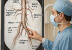
C. Aortic Pathologies:
The high-flow dynamics of contrast are perfect for evaluating the aorta.
-
Aortic Dissection: Contrast will highlight the true lumen and false lumen, with the intimal flap often visible as a thin, dark line separating the two contrast-filled channels.
-
Aortic Aneurysm: Precisely measures the diameter of a dilated aorta and assesses for risk of rupture, such as the presence of a penetrating atherosclerotic ulcer or intramural hematoma.
-
Traumatic Aortic Injury: In major trauma, a CT with contrast can quickly identify life-threatening tears or transections of the aorta.
D. Mediastinal Mass Evaluation:
The mediastinum is a complex space where subtle abnormalities can have significant consequences. Contrast helps to:
-
Distinguish solid vascular masses (e.g., thymoma, lymphoma, thyroid goiter) from cystic lesions (e.g., bronchogenic cyst).
-
Define the relationship of a mass to major vessels, the airway, and the heart, which is critical for surgical planning.
E. Infectious and Inflammatory Processes:
-
Pneumonia: While often diagnosed on X-ray, contrast-enhanced CT is superior for identifying complications like lung abscesses (which show ring enhancement) or empyema (pus in the pleural space).
-
Inflammatory Diseases: For conditions like sarcoidosis or granulomatosis with polyangiitis, CT can reveal characteristic patterns of lymph node involvement and lung inflammation that are best seen with contrast.
F. Pre-Surgical and Pre-Procedural Planning:
Before lung resection surgery or a biopsy, a detailed map of the vascular anatomy is essential to avoid catastrophic bleeding. A contrast-enhanced CT provides a 3D road map for surgeons and interventional radiologists.
Table 1: Common Clinical Indications for CPT 71271
| Clinical Scenario | Primary Diagnostic Question | Role of Contrast Enhancement |
|---|---|---|
| Suspected Pulmonary Embolism | Is there a clot blocking the pulmonary arteries? | Opacifies pulmonary arteries to directly visualize filling defects. |
| Lung Nodule/Mass | Is the nodule benign or malignant? | Assesses vascularity; malignancies often enhance significantly. |
| Lung Cancer Staging | What is the size, location, and spread of the tumor? | Defines tumor boundaries, evaluates lymph node involvement, and detects distant metastases. |
| Chest Pain (Suspected Aortic Dissection) | Is there a tear in the aortic wall? | Delineates the true and false lumens, highlighting the intimal flap. |
| Unexplained Hemoptysis (Coughing Blood) | Where is the bleeding coming from? | May identify abnormal vascularity (e.g., AVM) or a bleeding tumor. |
| Mediastinal Mass on X-ray | What is the nature and extent of the mass? | Characterizes tissue (solid vs. cystic) and defines relationship to great vessels. |
| Fever of Unknown Origin & Immunocompromise | Is there a hidden abscess or infection? | Enhances walls of abscesses and areas of inflammation. |
4. A Journey Through the Scanner: The Patient Experience
For a patient, undergoing a CT scan can be an anxiety-provoking experience. Understanding the process can significantly alleviate fear and ensure a successful exam.
A. Pre-Procedure Preparation:
-
Fasting: Patients are typically instructed to fast for 2-4 hours before the exam. This minimizes the risk of nausea and vomiting if contrast is administered and reduces potential artifacts from food in the esophagus.
-
Lab Work: A recent serum creatinine level is often required, especially for patients with known kidney disease, diabetes, hypertension, or who are over a certain age (e.g., 60). This assesses kidney function to ensure it is safe to administer iodinated contrast, which is cleared by the kidneys.
-
Medication Review: The patient will be screened for medications like metformin (a common diabetes drug). While often safe, its use may need to be paused temporarily after contrast administration in patients with impaired renal function due to a rare risk of lactic acidosis.
-
Allergy Screening: A history of prior allergic-like reactions to iodinated contrast media is crucial. While severe reactions are rare, premedication with corticosteroids and antihistamines may be prescribed for those with a history of reaction.
-
Pregnancy Screening: For women of childbearing age, a pregnancy test may be performed due to the potential radiation risk to a fetus.
B. The Day of the Exam:
-
Check-In and Consent: The patient arrives, completes paperwork, and changes into a gown free of metal snaps or zippers. The procedure, including the benefits and risks of contrast, is explained, and informed consent is obtained.
-
IV Placement: A nurse or technologist places an intravenous (IV) catheter, typically in a vein in the arm. A larger bore IV (e.g., 18-20 gauge) is often used to accommodate the high flow rate of the contrast injection.
-
Positioning: The patient lies supine (on their back) on the motorized CT table. Arms are raised above the head to avoid artifacts in the image. Straps and pillows may be used to help the patient maintain the correct position and remain still.
-
Breathing Instructions: The technologist provides clear breathing instructions via an intercom. “Take a breath in, hold your breath!” is a common command. Holding breath is critical to prevent motion blur, especially at the lung bases. The command “Breathe!” will signal when the patient can resume normal breathing. The patient must practice these instructions carefully.
-
The Injection and Scan: The technologist initiates the scan from the control room. The patient may hear a whirring sound as the scanner rotates. A power injector connected to the IV will automatically deliver the contrast material. The patient will likely feel:
-
A metallic taste in the mouth.
-
A warm, flushing sensation throughout the body, particularly in the pelvic region. This is normal and lasts for about a minute.
-
A feeling like they have urinated on themselves (they have not).
The actual scan itself is very quick, often completed in a single breath-hold of 5 to 15 seconds.
-
-
Post-Procedure: The IV is removed, and pressure is applied to the site. The patient is encouraged to drink plenty of fluids to help flush the contrast material from their system. If not premedicated, they can typically resume normal activities and diet immediately.
5. Behind the Glass: The Technologist’s Protocol
The radiology technologist is the engineer of the exam, translating the radiologist’s diagnostic needs into a set of technical parameters. The protocol for a 71271 is highly customized based on the clinical indication.
A. Contrast Administration:
-
Type of Contrast: Non-ionic, low-osmolar iodinated contrast is the modern standard due to its superior safety profile.
-
Dose: The volume is weight-based, typically ranging from 75-120 mL for an average adult.
-
Flow Rate: The rate of injection is critical and varies by indication. A CTPA for PE requires a very fast rate (4-5 mL/sec) to achieve a sharp “bolus” of contrast in the pulmonary arteries. A routine chest CT for cancer staging may use a slower rate (2-3 mL/sec).
-
Saline Chaser: A bolus of saline (sterile salt water) is almost always injected immediately after the contrast. This “pushes” the entire contrast column into the venous system, ensuring efficient use and a sharper bolus.
B. Timing is Everything: Bolus Tracking
Initiating the scan at the precise moment the contrast arrives in the target vessel is paramount. This is achieved through bolus tracking. A low-dose monitoring scan is performed at a specific level (e.g., the main pulmonary artery for a CTPA, the aortic arch for a dissection protocol). A region of interest (ROI) is placed over the target vessel. The software monitors the Hounsfield unit (HU) density in real-time. Once the density reaches a pre-set threshold (e.g., 100 HU), signifying the arrival of the contrast bolus, the scanner automatically triggers the diagnostic acquisition after a short, pre-programmed delay.
C. Image Acquisition and Reconstruction:
-
Helical (Spiral) Scanning: The modern standard where the table moves continuously through the rotating gantry, creating a volumetric data set.
-
Slice Thickness: The thorax is typically scanned with thin slices (0.625mm to 1.25mm). This allows for the creation of high-resolution multi-planar reconstructions (MPRs) and 3D models.
-
Radiation Dose Modulation: Automatic exposure control (AEC) techniques are used to automatically adjust the radiation dose based on the patient’s size and the density of the tissue being imaged (e.g., lower dose through the lungs, higher dose through the shoulders), minimizing unnecessary exposure without sacrificing image quality.
D. Post-Processing:
The raw data is reconstructed into images for interpretation. This includes:
-
Axial Images: The primary source images, viewed in the transverse plane.
-
Multi-Planar Reformat (MPR): Reconstructing the data into coronal (front-to-back) and sagittal (side-to-side) planes. This is invaluable for assessing structures like the aorta, pulmonary arteries, and spine.
-
Maximum Intensity Projection (MIP): A technique that projects only the brightest voxels (3D pixels) into a 2D image. It is exceptionally useful for evaluating vascular structures and tracking the course of vessels, making tiny nodules more conspicuous.
-
3D Volume Rendering: Creates detailed, rotatable 3D models of the vasculature (CT angiography), bones, and airways, providing an unparalleled surgical roadmap.
6. The Radiologist’s Canvas: Interpretation and Findings
The contrast-enhanced CT study provides the radiologist with a rich dataset to analyze. The pattern, degree, and timing of contrast enhancement provide vital diagnostic clues.
A. Systematic Approach to Interpretation:
A seasoned radiologist follows a disciplined search pattern to ensure no abnormality is missed:
-
Mediastinum and Great Vessels: Assess the aorta, pulmonary arteries, SVC, and heart for size, contour, and the presence of dissection or clot. Evaluate mediastinal lymph nodes using size and morphological criteria (e.g., short-axis diameter >1cm is often considered enlarged).
-
Lungs: Review the lung parenchyma using both lung window settings (to see fine details of the airways and lung tissue) and soft tissue windows (to evaluate nodules and masses). Look for nodules, masses, consolidation, emphysema, fibrosis, and interstitial disease.
-
Pleura and Chest Wall: Check the pleural spaces for fluid (effusion) or air (pneumothorax). Examine the ribs, spine, and soft tissues for lesions or fractures.
-
Upper Abdomen: Since the lower slices often include the upper abdomen, the liver, adrenal glands, and upper kidneys are scrutinized for incidental findings, which are common and can be significant (e.g., adrenal metastasis).
B. Key Findings and Their Appearance with Contrast:
-
Malignant Lung Nodule: Often shows irregular, spiculated margins and heterogeneous enhancement >15-20 Hounsfield Units (HU) after contrast administration.
-
Pulmonary Embolus: Appears as a filling defect within a contrast-opacified pulmonary artery—a dark clot surrounded by bright contrast.
-
Aortic Dissection: Visualized as an intimal flap separating the true and false lumens. The false lumen may have slower flow or thrombus.
-
Lung Abscess: Typically a thick-walled cavity with irregular inner margins that shows “rim enhancement” and may contain an air-fluid level.
-
Lymphoma: Manifests as a large, homogeneously enhancing anterior mediastinal mass or widespread lymphadenopathy.
-
Thymoma: A well-defined, enhancing mass in the anterior mediastinum.
The radiologist synthesizes all these findings into a structured report, providing a diagnosis or a differential diagnosis that directly answers the referring physician’s clinical question, guiding the next steps in patient management.
7. Navigating the Rules: Coding, Billing, and Compliance
Correctly applying CPT 71271 is a complex process that hinges on strict adherence to coding rules and payer policies to avoid denials, audits, and potential legal issues.
A. The Golden Rule: Medical Necessity and Documentation
The entire claim rests on the foundation of medical necessity. The indication for the scan must be clearly documented in the patient’s medical record by the ordering physician. The radiology report must then corroborate this necessity. A coder cannot change the code based on the findings; they must code based on what was ordered and performed. If a study is performed with contrast, it must be coded as such, even if the findings are normal.
B. Bundling and Separately Identifiable Services:
The global fee for 71271 includes the technical component (equipment, technologist time, contrast material) and the professional component (radiologist’s interpretation). It also includes the routine administration of IV contrast and local sedation. However, certain services may be billed separately if they are significant and separately identifiable:
-
IV Placement: Typically not billed separately as it is considered part of the procedure.
-
Contrast Material: The cost of the contrast itself is usually bundled into the technical component. Some facilities use a separate HCPCS Level II code (e.g., Q9958-Q9960) for the contrast supply, but payer policies on this vary widely.
-
Power Injection: The use of a power injector is included in the procedure code.
C. The Role of Modifiers:
Modifiers provide additional information about the circumstances of the service.
-
Modifier -26 (Professional Component): Used by the radiologist when billing only for the interpretation of the scan, not the technical costs (e.g., if the scan was performed at a hospital-owned facility).
-
Modifier -TC (Technical Component): Used by the facility when billing only for the technical costs of performing the scan, not the interpretation.
-
Modifier -59 (Distinct Procedural Service): Used to indicate that a procedure or service was distinct or independent from other services performed on the same day. This is rarely used with 71271 unless it is performed in a completely separate session from another CT scan.
D. Compliance Pitfalls and Audit Risks:
-
Incorrect Code Selection: Using 71271 when no contrast was administered (should use 71250) or when both non-contrast and contrast phases were performed (should use 71260) is a major error.
-
Lack of Medical Necessity: Performing a contrast-enhanced CT without a clear clinical indication documented in the order. Payers have specific coverage determinations (e.g., Local Coverage Determinations from Medicare Administrative Contractors) that list approved indications.
-
Incomplete Documentation: The radiology report must explicitly state that intravenous contrast was administered, including the type and amount. Failure to document this can lead to a downcode or denial.
Table 2: Coding Scenarios for Thoracic CT
| Scenario | Correct CPT Code | Rationale |
|---|---|---|
| A patient with chest pain and suspected PE receives a CT scan with IV contrast timed for the pulmonary arteries. | 71271 | The study is diagnostic and performed with contrast material. |
| A patient with a known lung nodule has a CT scan first without contrast, then immediately after with contrast. | 71260 | The code specifically describes a study performed both without and with contrast. |
| A patient undergoes a low-dose CT scan for lung cancer screening as a former smoker. No contrast is used. | 71275 | This is the specific code for lung cancer screening without contrast. |
| A patient with hemoptysis has a CT scan. No IV access is obtained, so no contrast is given. | 71250 | The diagnostic study was performed without contrast material. |
8. Weighing the Scales: Risks, Benefits, and Radiation Safety
Like all medical interventions, a CT thorax with contrast involves a careful risk-benefit analysis.
A. Benefits:
The benefits are profound: accurate and rapid diagnosis of life-threatening conditions (PE, dissection), precise cancer staging that directly guides treatment, and minimally invasive evaluation that often eliminates the need for diagnostic surgery. The information gained is invaluable and often unequivocally outweighs the risks.
B. Risks:
-
Ionizing Radiation: CT scans involve a higher dose of radiation than conventional X-rays. The effective dose of a chest CT is approximately 7 mSv, which is comparable to the background radiation a person receives from the environment over about two years. This exposure carries a small, cumulative increased lifetime risk of developing cancer. This risk is higher for younger patients and is a primary driver of the “As Low As Reasonably Achievable” (ALARA) principle in radiology, which emphasizes dose reduction techniques without compromising diagnostic quality.
-
Contast-Induced Nephropathy (CIN): A rare but serious condition where iodinated contrast causes a acute drop in kidney function, typically in patients with pre-existing renal impairment. Careful screening of renal function (creatinine levels) and hydration protocols are used to mitigate this risk.
-
Allergic-Like Reactions: Reactions to modern non-ionic contrast are uncommon (1-3%) and usually mild (itching, hives). Severe, anaphylactoid reactions are very rare (<0.04%). Premedication protocols are highly effective for patients with a known history of reaction.
-
Extravasation: The accidental leakage of contrast material from the vein into the surrounding soft tissue. This can cause pain, swelling, and, in very rare cases with large volumes, tissue damage. Using correct IV placement techniques and monitoring during injection minimizes this risk.
9. The Future is Now: Technological Advancements in Thoracic CT
The field of thoracic CT is not static; it is evolving rapidly, enhancing both safety and diagnostic power.
-
Low-Dose CT (LDCT): Advanced iterative reconstruction algorithms allow for the creation of diagnostic-quality images from data acquired with significantly less radiation—often reducing dose by 50-75% compared to traditional techniques. This is the backbone of lung cancer screening programs.
-
Dual-Energy CT (DECT): This revolutionary technology uses two different X-ray energy spectra simultaneously. It allows radiologists to “see beyond the standard image” by:
-
Iodine Mapping: Creating maps that show the precise distribution of iodine, improving the detection of subtle lung perfusion defects from PE and better characterizing masses.
-
Virtual Non-Contrast (VNC) Images: Algorithmically subtracting iodine from contrast-enhanced images, potentially eliminating the need for a true non-contrast acquisition phase and thus reducing radiation exposure. This calls into question the future need for combination codes like 71260.
-
-
Artificial Intelligence (AI) and Machine Learning: AI is transforming radiology in several ways:
-
Automated Nodule Detection: AI algorithms can analyze a CT scan in seconds, identifying and measuring pulmonary nodules with superhuman sensitivity, acting as a powerful second reader for radiologists to reduce perceptual errors.
-
Quantitative Imaging: AI can provide precise, reproducible measurements of tumor volume, density, and texture, offering more sensitive markers of treatment response than simple linear measurements.
-
Workflow Optimization: AI can automate protocoling, dose monitoring, and preliminary triage of critical findings (e.g., flagging a study as positive for PE or pneumothorax), speeding up care.
-
These advancements are making CPT 71271 an even safer, more precise, and more informative tool, solidifying its role as a cornerstone of thoracic diagnosis.
10. Conclusion
CPT Code 71271 represents far more than a billing tool; it encapsulates a sophisticated, essential diagnostic journey into the human chest. Its power lies in the dynamic visualization provided by contrast enhancement, enabling the detection and characterization of life-threatening vascular, oncologic, and inflammatory diseases. From the precise technical protocols and expert interpretation to the strict adherence to coding compliance, every aspect of this procedure is designed to provide critical answers that directly guide patient care. As technology advances with low-dose techniques, dual-energy imaging, and artificial intelligence, the future of thoracic CT promises even greater diagnostic confidence with ever-increasing safety, ensuring its indispensable role in modern medicine for years to come.
11. Frequently Asked Questions (FAQs)
Q1: I have a shellfish allergy. Can I still have a CT scan with contrast?
A: Yes, in the vast majority of cases. The belief that shellfish allergy predicts an iodine contrast reaction is a common misconception. Shellfish allergies are triggered by a protein (tropomyosin), not iodine. However, it is crucial that you inform your doctor and the radiology team of any and all allergies so they can assess your overall risk and premedicate you if necessary.
Q2: How long does the contrast stay in my body?
A: The iodine-based contrast is cleared from your bloodstream by your kidneys and excreted in your urine. For a person with normal kidney function, most of it is eliminated within 24 hours. Drinking plenty of water after your scan helps flush it out more quickly.
Q3: What’s the difference between a CT with contrast and an MRI with contrast?
A: They are fundamentally different technologies. CT uses X-rays and iodine-based contrast to visualize anatomy, blood flow, and tissue density. MRI uses powerful magnets and radio waves, often with a gadolinium-based contrast agent, to evaluate soft tissue structure, inflammation, and water content with exceptional detail without using ionizing radiation. The choice between them depends on the specific clinical question.
Q4: My doctor ordered a CT with contrast, but the report says “no acute findings.” Does that mean the scan was useless?
A: Absolutely not. A “negative” or “normal” result is a highly valuable finding. It rules out serious and life-threatening conditions, allowing your doctor to confidently pursue other causes for your symptoms and providing you with significant peace of mind.
Q5: Can I have a CT scan if I am pregnant?
A: CT is generally avoided during pregnancy, especially in the first trimester, due to the potential radiation risk to the developing fetus. If a CT scan is absolutely necessary for a life-threatening maternal condition (e.g., suspected pulmonary embolism), it will be performed with precautions to minimize fetal dose. The benefits of making a critical diagnosis in the mother are considered to outweigh the potential risks to the fetus. Ultrasound and MRI (without contrast) are preferred imaging methods during pregnancy when possible.
12. Additional Resources
-
American College of Radiology (ACR) Appropriateness Criteria: An evidence-based guidelines to help providers choose the most appropriate imaging exam for hundreds of clinical conditions. (https://www.acr.org/Clinical-Resources/ACR-Appropriateness-Criteria)
-
RadiologyInfo.org: A public resource co-sponsored by the ACR and the Radiological Society of North America (RSNA) that provides easy-to-understand information on all radiology procedures for patients. (https://www.radiologyinfo.org/)
-
American Medical Association (AMA) CPT® Network: The official source for information on CPT codes (requires subscription). (https://www.ama-assn.org/practice-management/cpt)
-
Society of Thoracic Radiology (STR): A professional organization dedicated to thoracic imaging, offering resources for both clinicians and patients. (https://www.thoracicrad.org/)