A sudden twist on a staircase, the relentless ache after a long run, the concerning swelling in a diabetic patient—these common clinical presentations often lead a physician to the same fundamental diagnostic starting point: the X-ray. In the realm of podiatry, orthopedics, emergency medicine, and primary care, the radiographic foot series is an indispensable tool. But to the uninitiated, the process of ordering, performing, coding, and billing for this service can seem like a cryptic language of its own. At the heart of this process for a comprehensive initial evaluation is a specific, five-digit code: CPT Code 73630.
This article is designed to be the definitive guide to CPT code 73630—Radiologic examination, foot; complete, minimum of 3 views. We will move beyond a simple definition and embark on a detailed exploration that spans clinical medicine, radiological technique, administrative coding, and healthcare economics. Whether you are a medical student, a radiology technologist perfecting your craft, a coder navigating complex payer policies, a physician ensuring documentation compliance, or simply a curious patient, this deep dive will illuminate every facet of this common but crucial procedure. Our journey will reveal that this “simple X-ray” is, in fact, a sophisticated interplay of anatomy, physics, medicine, and information technology, all converging to answer a critical clinical question and guide patient care.

2. The Clinical Imperative: Why Image the Foot?
The Foot’s Complex Architecture
To understand the necessity of a multi-view approach, one must first appreciate the anatomical complexity of the human foot. It is a mechanical marvel comprising 26 bones, 33 joints, and over a hundred muscles, tendons, and ligaments. These structures are organized into three key regions:
- The Hindfoot: Contains the talus and calcaneus (heel bone), forming the foundation of the foot and absorbing the initial impact of walking.
- The Midfoot: A pyramid-like collection of bones (navicular, cuboid, and three cuneiforms) that acts as a shock absorber and stabilizer.
- The Forefoot: Includes the metatarsals and phalanges (toes), responsible for propulsion and balance.
This intricate arrangement allows for weight-bearing, mobility, and adaptation to uneven surfaces. However, this complexity also means that injury or disease in one area can have profound biomechanical consequences. A single two-dimensional image is wholly insufficient to evaluate this three-dimensional structure, as bones will inevitably overlap, obscuring vital details and potential pathology.
Common Indications for a 3-View Foot Series
A physician orders a complete foot series based on a patient’s history and physical exam findings. Common indications include:
- Trauma: The most frequent reason. Following acute injury from falls, twists, or direct impact to assess for fractures (e.g., metatarsal, navicular, calcaneal) or dislocations.
- Pain: Unexplained, chronic, or worsening pain in the foot, with or without a remembered injury.
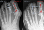
- Deformity: Evaluation of congenital or acquired deformities such as pes planus (flat foot), pes cavus (high arch), bunions (hallux valgus), or hammertoes.
- Pre- and Post-Operative Assessment: Planning for surgical intervention (e.g., bunionectomy, fusion) and evaluating the results post-surgery (alignment, hardware position, healing).
- Infection: In diabetic patients or those with open wounds, to detect osteomyelitis (bone infection), which presents as bone destruction and demineralization.
- Arthritis: To diagnose and monitor degenerative (osteoarthritis) or inflammatory (rheumatoid arthritis, gout) joint diseases, evidenced by joint space narrowing, erosions, and osteophyte formation.
- Foreign Body: Locating radiopaque (visible on X-ray) objects like glass or metal embedded in the soft tissues.
3. A Deep Dive into CPT Code 73630: The Technical Specifics
Code Definition and Official Descriptor
CPT (Current Procedural Terminology) code 73630 is defined by the American Medical Association as: “Radiologic examination, foot; complete, minimum of 3 views.”
This descriptor is intentionally specific. The word “complete” signifies that this is a standalone study designed to provide a comprehensive diagnostic evaluation of the entire foot. The phrase “minimum of 3 views” is a technical requirement, establishing the baseline for what constitutes a “complete” exam. It is distinct from a limited study, which is coded as 73620 and is typically reserved for a single specific view, such as a follow-up image to check healing of a known fracture.
Unbundling the “3 Views”: Standard Projections Explained
The power of the 3-view series lies in the orthogonal (right-angle) relationship of the images, which allows the radiologist to mentally reconstruct a 3D model of the foot. The standard projections are:
- Anteroposterior (AP) View: The X-ray beam enters the front (anterior) of the foot and exits the back (posterior), striking the detector. This view provides an excellent overview of the forefoot and midfoot, clearly showing the metatarsals and phalanges, and the joints between the metatarsals and the midfoot bones. It is essential for evaluating alignment, joint spaces, and fractures in these areas.
- How it’s done: The patient is typically seated or supine on the X-ray table with the knee flexed and the sole of the foot flat on the detector. The beam is directed vertically.
- Oblique View: This is the crucial view that “unlocks” the midfoot. By rotating the foot internally (medial oblique) or externally (lateral oblique), the bones that are superimposed on each other in the AP view are splayed apart.
- How it’s done: For the standard medial oblique view, the foot is rotated inward approximately 30-45 degrees. This view excellently visualizes the cuboid, the tarsometatarsal joints (Lisfranc joint complex), and the sinus tarsi, making it indispensable for diagnosing Lisfranc injuries, which are often missed on AP and lateral views alone.
- Lateral View: The X-ray beam passes from one side of the foot to the other. This is the primary view for assessing the calcaneus, the talus, the ankle joint, and the longitudinal arch of the foot. It is critical for evaluating fractures of the calcaneus, assessing plantar fascia integrity, and viewing any soft tissue swelling.
- How it’s done: The patient typically lies on their side with the medial aspect of the foot against the detector. The beam is directed horizontally.
* Standard Projections in a 3-View Foot Series (CPT 73630)*
| Projection | Primary Anatomical Areas Visualized | Key Clinical Utility | Patient Positioning |
|---|---|---|---|
| Anteroposterior (AP) | Metatarsals, Phalanges, Tarsometatarsal joints | Fractures, joint alignment, arthritis | Foot flat on detector, beam vertical |
| Oblique (Medial) | Cuboid, Cuneiforms, Lisfranc joint complex | Lisfranc injuries, midfoot fractures | Foot rotated inward 30-45°, beam vertical |
| Lateral (Mediolateral) | Calcaneus, Talus, Ankle joint, Longitudinal arch | Calcaneal fractures, plantar fasciitis, soft tissue swelling | Patient on side, medial foot on detector, beam horizontal |
Modifiers: When and How to Use Them
Modifiers are two-digit codes appended to a CPT code to provide additional information about the circumstances of the service. Common modifiers used with 73630 include:
- -LT (Left side) and -RT (Right side): These are essential laterality modifiers. They must always be used to specify which foot was imaged. Billing without a laterality modifier is a common cause of claim denial.
- -TC (Technical Component): Used by the facility (e.g., hospital, imaging center) that owns the equipment and employs the technologist who performed the scan. It covers the cost of the equipment, film, technologist’s salary, etc.
- -26 (Professional Component): Used by the radiologist who interprets the images, creates a written report, and bills for their expert diagnostic service.
- -59 (Distinct Procedural Service): Used to indicate that a procedure or service was distinct or independent from other services performed on the same day. This might be necessary if, for example, both feet are imaged on the same day. However, payer policies on -59 are strict, and it should only be used if the services are performed at separate sessions or on separate anatomical sites.
4. Coding in Practice: Documentation, Medical Necessity, and Compliance
The Pillar of Medical Necessity
The cornerstone of medical coding and reimbursement is medical necessity. An insurer will only pay for a service if it is deemed “reasonable and necessary” for the diagnosis or treatment of an illness or injury. Ordering a 3-view foot series for a routine screening without symptoms would not be considered medically necessary. The ordering physician’s documentation in the patient’s medical record must clearly support the need for the study.
Documentation Requirements for Clean Claims
For a claim for 73630 to be processed smoothly (“clean claim”), the medical record must contain specific elements:
- Order: A signed order from the treating physician that includes the specific exam (“X-ray foot 3 views” or “Radiologic exam foot complete”).
- Reason for Exam: The clinical indication, such as “foot pain after fall,” “evaluate for bunion,” or “rule out Lisfranc injury.”
- History & Physical Findings: Supporting documentation like point of tenderness, swelling, ecchymosis, decreased range of motion, or difficulty bearing weight.
- Radiology Report: The final interpreted report by the radiologist must describe the views obtained (e.g., “AP, oblique, and lateral views of the right foot were obtained”) and provide a diagnosis or impression.
Navigating Common Denials and How to Avoid Them
- Denial: “Lack of Medical Necessity.”
- Prevention: Ensure the referring provider’s note is detailed and links the patient’s symptoms directly to the need for the imaging.
- Denial: “Missing/Invalid Modifier.”
- Prevention: Always append the correct laterality modifier (-LT or -RT). If billing for a component, ensure -26 or -TC is used appropriately.
- Denial: “Bundled Service.”
- Prevention: If imaging both feet, use modifier -59 on the second code (e.g., 73630-RT and 73630-LT-59) if the payer’s policy requires it. Be prepared to provide documentation.
5. The Radiologic Technologist’s Perspective: Capturing the Perfect Image
The radiologic technologist (RT) is the healthcare professional who brings the physician’s order to life. Their expertise is critical in obtaining diagnostic-quality images while minimizing patient discomfort and radiation exposure.
Patient Positioning and Comfort: The RT must expertly guide the patient, who may be in pain, into the precise positions required for each view. This requires clear communication, patience, and a deep understanding of anatomy. For a lateral view of a painful trauma patient, for instance, the RT might use a positioning sponge to support the leg without moving the injured foot.
Technical Factors: kVp and mAs: The RT must manually select or choose from pre-set protocols the technical exposure factors:
- Kilovoltage peak (kVp): Controls the energy (penetrating power) of the X-ray beam. A higher kVp is used for larger, denser body parts.
- Milliampere-seconds (mAs): Controls the quantity (number) of X-ray photons. It directly influences the image darkness and patient dose.
Choosing the correct combination is an art form that balances image contrast and clarity with the ALARA principle (As Low As Reasonably Achievable) for radiation dose.
6. The Radiologist’s Read: Interpreting the 3-View Series
Once the images are captured, they are sent digitally to a Picture Archiving and Communication System (PACS). The radiologist then performs a systematic search pattern to avoid missing subtle findings.
A Systematic Approach to Interpretation:
- Adequacy and Alignment: Are all three views present? Is the positioning adequate to see the necessary anatomy?
- Bones: Check each bone individually for continuity of cortex, looking for lucent (dark) fracture lines or sclerotic (white) areas. Assess bone density.
- Joints: Evaluate each joint space for symmetry and width. Narrowing suggests arthritis; widening suggests ligamentous injury or effusion.
- Soft Tissues: Look for swelling, which appears as increased opacity (whiteness) or loss of normal fat planes. Look for foreign bodies or gas within the soft tissues, which can indicate infection.
7. Beyond the Basic 3-View: Advanced Imaging and Complementary Codes
Sometimes, the 3-view series is only the beginning. If the standard views are inconclusive or reveal a complex injury, further imaging may be required.
- Additional Views: A “weight-bearing” series (AP and lateral views taken while the patient is standing) is crucial for evaluating dynamic deformities and ligamentous instability, particularly in the Lisfranc joint.
- Computed Tomography (CT): CT provides exquisitely detailed cross-sectional images and 3D reconstructions. It is the gold standard for characterizing complex fractures (e.g., calcaneal, intra-articular), assessing fracture healing, and evaluating bone tumors. Relevant CPT codes are in the 77000 series (e.g., 71250 for CT foot without contrast).
- Magnetic Resonance Imaging (MRI): MRI is unparalleled in visualizing soft tissues—ligaments, tendons, cartilage, and bone marrow. It is the study of choice for diagnosing tendon tears, ligament injuries (like a full Lisfranc ligament tear), osteochondral defects, and osteomyelitis. Relevant CPT codes are in the 77000 series (e.g., 73721 for MRI foot without contrast).
8. The Financial Landscape: Reimbursement and Payer Policies
The reimbursement for 73630 is not a fixed number. It is calculated using the Resource-Based Relative Value Scale (RBRVS). This system assigns a value to a service based on:
- Physician Work (PW): The time, skill, and intensity required by the radiologist to interpret the study.
- Practice Expense (PE): The overhead costs for the facility (equipment, supplies, technologist salaries).
- Malpractice (MP): The cost of professional liability insurance.
These values are combined into Total Relative Value Units (RVUs), which are then multiplied by a yearly conversion factor (CF) set by the Centers for Medicare & Medicaid Services (CMS) to determine the payment amount. This amount varies by geographic region and payer (Medicare, Medicaid, private insurers).
9. The Future of Foot Radiography: AI, Technology, and Trends
The field is rapidly evolving. Artificial Intelligence (AI) is being developed to act as a “second reader,” using algorithms to flag potential fractures or abnormalities, potentially reducing missed diagnoses. Digital Radiography has largely replaced film, offering instant images, lower radiation doses, and powerful post-processing tools. Point-of-Care Ultrasound (POCUS) is also being used by emergency physicians and podiatrists for dynamic, real-time assessment of tendons and ligaments without any radiation.
10. Conclusion: The Enduring Value of a Foundational Study
CPT code 73630 represents far more than a simple transaction; it encapsulates a complete diagnostic process vital for patient care. From the initial clinical question to the technologist’s skilled positioning, the radiologist’s expert analysis, and the coder’s precise translation into data, the 3-view foot series remains a fundamental, powerful, and cost-effective tool in modern medicine. Its continued relevance is a testament to the enduring need for clear, multi-perspective visualization of the human body’s intricate structures.
11. Frequently Asked Questions (FAQs)
Q1: What is the difference between CPT 73620 and 73630?
A: CPT 73620 is a “limited” exam, typically one or two views, often used for follow-up on a known condition (e.g., “check healing of a known 5th metatarsal fracture”). CPT 73630 is a “complete” exam, requiring a minimum of three distinct views, and is used for a comprehensive initial diagnostic evaluation.
Q2: How long does it take to get results from a foot X-ray?
A: The images themselves are available instantly to the technologist. However, the formal interpreted report from a radiologist can take anywhere from a few hours to 24-48 hours, depending on the facility’s workflow and urgency. In an emergency room setting, preliminary results are often communicated much faster.
Q3: Is the radiation from a foot X-ray dangerous?
A: The radiation dose from a foot X-ray is extremely low—equivalent to the natural background radiation everyone receives from the environment in a few hours to a day. The benefits of an accurate diagnosis far outweigh this minimal risk. Technologists adhere to the ALARA principle to keep doses “As Low As Reasonably Achievable.”
Q4: Can I get an X-ray if I might be pregnant?
A: You must always inform your doctor and the technologist if there is any possibility of pregnancy. While the dose to the foot is negligible and directed far from the abdomen, shielding with a lead apron will be used as an extra precaution, or the physician may consider postponing the exam if it is not urgent.
Q5: Why did I get a bill from the hospital and a separate bill from a doctor for the same X-ray?
A: This is due to the component billing explained earlier. The hospital bill is for the Technical Component (TC) – the use of the room, equipment, and technologist. The separate doctor’s bill is for the Professional Component (26) – the radiologist’s interpretation and report.
12. Additional Resources
- American Medical Association (AMA): For the complete and official CPT code set and guidelines. https://www.ama-assn.org/
- American College of Radiology (ACR): Provides practice parameters and appropriateness criteria for medical imaging. https://www.acr.org/
- RadiologyInfo.org: A public resource developed by the ACR and RSNA that explains imaging procedures in patient-friendly language. https://www.radiologyinfo.org/
- Centers for Medicare & Medicaid Services (CMS): For official Medicare coverage policies and reimbursement information. https://www.cms.gov/
- American Society of Podiatric Medical Assistants (ASPMA): Offers resources on podiatric-specific coding and assisting.
Date: September 9, 2025
Author: The MediCoders Team
Disclaimer: The information contained in this article is for educational and informational purposes only and is not intended as medical advice, diagnosis, or treatment. Always consult with a qualified healthcare provider for any health concerns or before making any decisions related to your health or treatment. CPT® is a registered trademark of the American Medical Association. This article is not affiliated with or endorsed by the AMA.