CPT Code 78801 refers to “Radiopharmaceutical localization of tumor, inflammatory process, or infection (including vascular graft infection); planar, single area (e.g., head, neck, chest, pelvis), single-day imaging.” This code is used in nuclear medicine to describe imaging procedures that help detect tumors, infections, or inflammatory conditions using radiopharmaceuticals.
This imaging technique is essential in oncology, infectious disease management, and inflammatory disorder diagnostics. Unlike traditional imaging methods, radiopharmaceutical localization provides functional insights, allowing physicians to assess metabolic activity rather than just anatomical structures.
This article explores the clinical significance, procedural details, billing considerations, and future advancements related to CPT Code 78801.

CPT Code 78801
2. Understanding Radiopharmaceutical Localization
What Are Radiopharmaceuticals?
Radiopharmaceuticals are radioactive compounds used in diagnostic imaging and therapeutic procedures. They consist of:
-
A radioisotope (e.g., Technetium-99m, Gallium-67)
-
A pharmaceutical component that targets specific tissues
When injected into the body, these compounds accumulate in areas of abnormal metabolic activity, such as tumors or infections, emitting gamma rays that are captured by imaging devices.
Types of Radiopharmaceutical Imaging
-
Planar Imaging (2D) – Used in CPT 78801, providing a single-view image.
-
SPECT (3D Imaging) – Offers cross-sectional views (coded separately under 78802-78803).
-
PET Scans – Used for metabolic activity assessment (different CPT codes apply).
3. Clinical Applications of CPT Code 78801
CPT 78801 is primarily used for:
A. Tumor Localization
-
Detecting primary and metastatic cancers (e.g., lymphoma, neuroendocrine tumors).
-
Monitoring treatment response in oncology patients.
B. Infection and Inflammation Detection
-
Diagnosing osteomyelitis (bone infections).
-
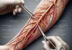
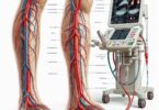
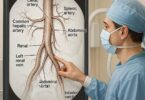
Identifying vascular graft infections.
-
Assessing inflammatory bowel disease (IBD) and rheumatoid arthritis.
C. Post-Surgical Evaluations
-
Detecting post-operative abscesses.
-
Evaluating prosthetic joint infections.
4. Procedure Breakdown: How 78801 is Performed
| Step | Description |
|---|---|
| 1. Patient Preparation | Fasting may be required; hydration is encouraged. |
| 2. Radiopharmaceutical Injection | Administered intravenously; uptake time varies (1-4 hours). |
| 3. Imaging Acquisition | Gamma camera captures planar images of the targeted area. |
| 4. Image Interpretation | A nuclear medicine specialist analyzes the results. |
| 5. Report Generation | Findings are documented for the referring physician. |
5. Comparison with Related CPT Codes
| CPT Code | Description | Key Differences |
|---|---|---|
| 78801 | Planar, single area, single-day imaging | Basic 2D imaging |
| 78802 | SPECT, single area | 3D cross-sectional imaging |
| 78803 | SPECT with CT fusion | Combines functional & anatomical data |
| 78830-78832 | PET imaging | Higher resolution, different isotopes |
6. Billing and Reimbursement Guidelines
Key Considerations:
-
Medical Necessity Documentation is crucial for claim approval.
-
Correct Modifiers (e.g., -26 for professional component).
-
Bundling Rules: Ensure no duplicate billing with related services.
Reimbursement Rates (2024 Estimates)
-
Medicare: ~$250-$400 per study.
-
Private Insurance: Varies by provider (may require prior authorization).
7. Common Challenges and Compliance Issues
-
Denials Due to Lack of Medical Necessity – Ensure proper ICD-10 coding (e.g., C79.9 for metastatic cancer).
-
Incorrect Code Selection – Confusing 78801 with SPECT or PET codes.
-
Radiation Safety Compliance – Adhering to ALARA (As Low As Reasonably Achievable) principles.
8. Case Studies and Real-World Examples
Case Study 1: Diagnosing Osteomyelitis
A 55-year-old diabetic patient with a non-healing foot ulcer underwent a 78801 scan with Tc-99m HDP, confirming osteomyelitis and guiding antibiotic therapy.
Case Study 2: Lymphoma Staging
A Gallium-67 scan (78801) helped identify metastatic lymphoma lesions not visible on CT, altering the treatment plan.
9. Future Trends in Radiopharmaceutical Imaging
-
AI-Assisted Image Analysis – Improving diagnostic accuracy.
-
New Radiotracers – Targeting specific cancer biomarkers.
-
Theranostics – Combining diagnosis and therapy (e.g., Lu-177 PSMA).
10. Conclusion
CPT Code 78801 is a vital tool in nuclear medicine for detecting tumors, infections, and inflammatory diseases. Proper documentation, accurate coding, and adherence to compliance guidelines ensure successful reimbursement. Advances in radiopharmaceuticals and AI will further enhance its diagnostic capabilities.
11. Frequently Asked Questions (FAQs)
Q1: What is the difference between 78801 and 78803?
-
78801 is planar (2D) imaging, while 78803 includes SPECT with CT fusion (3D).
Q2: Does Medicare cover CPT 78801?
-
Yes, if medically necessary (e.g., infection or tumor evaluation).
Q3: How long does a 78801 procedure take?
-
Typically 2-4 hours, including radiopharmaceutical uptake time.
12. Additional Resources
-
Society of Nuclear Medicine & Molecular Imaging (SNMMI) – www.snmmi.org
-
American College of Radiology (ACR) Guidelines – www.acr.org
-
CMS Medicare Coverage Database – www.cms.gov