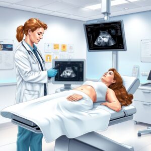
For a patient, a pelvic ultrasound is a deeply personal experience. It can be a procedure filled with hope during a pregnancy, anxiety during a diagnostic search for pain, or necessity in monitoring a known condition. The focus is rightly on the sonographer’s warm gel, the black-and-white images on the screen, and the reassuring words of the radiologist or physician.
But after the appointment ends, a different language takes over—a language of numbers and acronyms that ultimately determines the financial aspect of your healthcare journey. At the heart of this administrative process is a five-digit code: the Current Procedural Terminology (CPT) code.
This article aims to bridge the gap between the clinical and the administrative. We will demystify the CPT codes for pelvic ultrasound, primarily 76856 and 76857. This is not just a dry explanation for medical coders and billers. It is an empowering guide for patients who want to understand their medical bills, for healthcare providers who strive for accurate documentation, and for anyone interested in the intricate ecosystem of modern healthcare. By understanding the “why” behind the code, we can all become more informed participants in the medical process, ensuring that the focus remains where it should be: on effective, compassionate, and fairly compensated patient care.
Pelvic Ultrasound CPT Code
2. Understanding the Fundamentals: What is a Pelvic Ultrasound?
Before we can understand the code, we must fully understand the procedure it represents.
A pelvic ultrasound is a non-invasive, painless diagnostic imaging test that uses high-frequency sound waves to create real-time pictures of the organs and structures within the female pelvis. Unlike X-rays or CT scans, it does not use ionizing radiation, making it a very safe option for all patients, including pregnant women.
The Science of Sound: How Ultrasound Imaging Works
The principle behind ultrasound is called echolocation, similar to what bats and submarines use. The ultrasound transducer (or probe) is placed on the skin or inside the vagina. This transducer both emits sound waves and records them as they bounce back.
-
Sound Wave Emission: The transducer generates high-frequency sound waves that travel into the body.
-
Echo Reception: These sound waves hit boundaries between tissues (e.g., between fluid and soft tissue, soft tissue and bone) and echo back to the transducer.
-
Image Processing: A computer measures the strength of the returning echoes and the time it took for them to return. Using this data, it generates a two-dimensional grayscale image on a monitor in real-time.
The density of the tissue determines how many sound waves are reflected back. Dense tissues (like bone) appear white (hyperechoic), as they reflect most sound waves. Fluid-filled structures (like a bladder or a cyst) appear black (hypoechoic or anechoic), as they allow most sound waves to pass through. Soft tissues show up in various shades of gray.
Indications for a Pelvic Ultrasound: Why is it Ordered?
A physician may order a pelvic ultrasound for a multitude of reasons, which can be broadly categorized into obstetric and gynecologic.
Obstetric Reasons:
-
Confirming pregnancy and determining the baby’s gestational age.
-
Checking the baby’s heartbeat, growth, and anatomy.
-
Identifying multiple pregnancies (twins, triplets).
-
Screening for birth defects or chromosomal abnormalities (e.g., nuchal translucency scan).
-
Locating an intrauterine device (IUD).
-
Investigating vaginal bleeding or pelvic pain during pregnancy.
-
Checking the placenta’s location and health (e.g., for placenta previa).
-
Guiding procedures like amniocentesis.
Gynecologic Reasons:
-
Investigating pelvic pain, abnormal bleeding, or menstrual irregularities.
-
Evaluating infertility by monitoring follicle development in the ovaries or examining the uterus.
-
Diagnosing conditions like uterine fibroids, polyps, or adenomyosis.
-
Identifying ovarian cysts or tumors.
-
Diagnosing pelvic inflammatory disease (PID) or endometriosis.
-
Evaluing congenital anomalies of the uterus.
-
Guiding procedures like egg retrieval for IVF or endometrial biopsy.
3. The Core of Medical Billing: Introduction to CPT Codes
What is the CPT Code System?
The Current Procedural Terminology (CPT) code set is a uniform coding system created and maintained by the American Medical Association (AMA). It is used to describe medical, surgical, and diagnostic services provided by healthcare professionals. Think of it as a universal language that allows physicians, coders, patients, insurance companies, and government payers (like Medicare and Medicaid) to communicate accurately about what service was performed.
Each CPT code is a five-digit number that corresponds to a specific service or procedure. For example, 99213 is an office visit, 81000 is a urinalysis, and 76856 is a complete pelvic ultrasound.
Why Accurate Coding is Crucial: Compliance, Reimbursement, and Patient Care
The accuracy of CPT coding is not merely an administrative detail; it is the linchpin of the entire healthcare revenue cycle and a matter of legal compliance.
-
Precise Reimbursement: Insurance companies use CPT codes to determine how much to pay a provider for a given service. Each code is linked to a payment rate based on the complexity, time, and resources required. An incorrect code can lead to underpayment (hurting the provider) or overpayment (which must be paid back, often with penalties).
-
Regulatory Compliance: Incorrect coding, whether unintentional or deliberate, can be construed as fraud or abuse under federal laws like the False Claims Act. Consistently “upcoding” (using a code for a more complex service than was performed) or “unbundling” (billing separately for services that are meant to be billed as one) can result in massive fines, exclusion from government programs, and even criminal charges.
-
Data Tracking and Public Health: Aggregated CPT code data is used for public health monitoring, research, and policy planning. It helps track the prevalence of certain procedures, disease outbreaks, and the overall utilization of healthcare services.
-
Patient Understanding: When patients can match the codes on their bill to the services they received, it fosters transparency and allows them to spot potential billing errors.
4. A Deep Dive into Pelvic Ultrasound CPT Codes: 76856, 76857, and Beyond
The CPT codes for non-obstetric pelvic ultrasound are found in the Radiology section, under “Ultrasound, Pelvis.” The two primary codes are defined by the extent of the examination.
CPT 76856: Pelvic Ultrasound, Complete
-
CPT Code Definition: “Ultrasound, pelvic (non-obstetric), real-time with image documentation; complete.”
-
What it entails: This code is used for a comprehensive evaluation of the entire female pelvis. The required elements, as defined by the American College of Radiology (ACR) and standard practice, must include:
-
Uterus: Longitude and transverse views, including measurements of the length, height, and width. Evaluation of the endometrial stripe (its thickness and appearance).
-
Ovaries: Visualization and measurement of both the right and left ovaries (if present). If an ovary is not visualized, the sonographer must document the reason (e.g., “left ovary not visualized despite patient repositioning, likely obscured by bowel gas”).
-
Bladder: Assessment of the urinary bladder.
-
Cul-de-sac: Evaluation of the pouch of Douglas (the area behind the uterus) for the presence of free fluid or other abnormalities.
-
Adnexa: Examination of the areas adjacent to the uterus (the adnexa), which include the fallopian tubes and supporting structures.
-
-
Documentation is Key: To bill for 76856, the medical record (the sonographer’s worksheet and the radiologist’s report) must clearly document the evaluation of all these structures. Images of each required element must be saved and stored in the patient’s record.
CPT 76857: Pelvic Ultrasound, Limited or Follow-Up
-
CPT Code Definition: “Ultrasound, pelvic (non-obstetric), real-time with image documentation; limited or follow-up.”
-
What it entails: This code is for a focused, less comprehensive exam. It is used when the provider’s question is specific and does not require a full evaluation of all pelvic structures. Common scenarios include:
-
Follow-up: “Follow-up on a known simple ovarian cyst seen on a previous exam to ensure it has resolved.”
-
Focused Evaluation: “Measure the thickness of the endometrial stripe in a postmenopausal patient with bleeding.” The exam may focus only on the uterus and not attempt to fully visualize the ovaries if they are not relevant.
-
Guidance: “Guide the placement of a needle for follicle aspiration during IVF.” The exam is only used for real-time needle guidance, not for diagnostic evaluation of all organs.
-
Incomplete Exam: If a structure cannot be visualized despite best efforts (e.g., ovaries obscured by bowel gas and not the primary focus), the exam may be billed as limited.
-
-
Documentation is Key (Again): The report must clearly state the reason for the limited exam (e.g., “Limited ultrasound performed for follow-up of right ovarian cyst. The remainder of the pelvis was not evaluated.”).
The Transducer Debate: Transabdominal vs. Transvaginal and How to Code
A common point of confusion is how the type of ultrasound affects the CPT code.
-
CPT Codes are Technique-Agnostic: The codes 76856 and 76857 do not specify how the exam is performed. They are based on the extent of the exam (complete vs. limited), not the method.
-
Combined Exams are Common: It is extremely common to begin a “complete” pelvic ultrasound with a transabdominal (TA) approach. If the TA view does not provide sufficient diagnostic information (e.g., the uterus is not well seen due to patient body habitus), a transvaginal (TV) approach is then performed.
-
How to Code a Combined TA/TV Exam: When both approaches are used to complete a full evaluation of the pelvis, you bill only one unit of 76856. The TV approach is considered an integral part of the complete exam, not a separate procedure. You cannot bill 76856 for the TA and another code for the TV. The complete exam is a single service, regardless of how many transducers are used to acquire the necessary diagnostic information.
Real-World Clinical Scenarios: Choosing the Right Code
-
Scenario A: A 32-year-old patient presents with chronic pelvic pain. The physician orders a pelvic ultrasound to evaluate for fibroids, cysts, or other abnormalities. The sonographer performs a transabdominal scan, then a transvaginal scan to get clearer images. The radiologist’s report describes the size and appearance of the uterus, endometrium, both ovaries, the bladder, and the cul-de-sac.
-
Correct Code: 76856 (A complete evaluation was performed and documented).
-
-
Scenario B: A 45-year-old patient had a pelvic ultrasound 6 weeks ago that showed a 2cm simple cyst on the right ovary. Her physician orders a follow-up ultrasound to see if it has resolved. The sonographer focuses the exam on the right ovary, confirms the cyst is gone, takes images of the ovary, and ends the exam.
-
Correct Code: 76857 (A limited, follow-up exam was performed for a specific purpose).
-
-
Scenario C: A 28-year-old patient is undergoing infertility treatment. Today, she is in the clinic for monitoring of follicular development. The sonographer uses the transvaginal probe to count and measure the follicles on each ovary. The report states: “Follicle monitoring exam performed. Three dominant follicles in the right ovary measuring 18mm, 16mm, and 15mm. The uterus and cul-de-sac were not formally evaluated.”
-
Correct Code: 76857 (This is a limited exam focused on a specific task).
-
5. The Technical vs. Professional Component: A Critical Distinction
This is one of the most important concepts in radiology billing. A single CPT code can be broken down into two parts, often referred to as “TC” and “PC.”
-
Technical Component (TC): This covers the equipment, the facility overhead, the supplies (e.g., gel), and the technologist’s time and expertise to perform the scan. The TC is billed by the entity that owns the equipment and employs the technologist (e.g., a hospital or an outpatient imaging center).
-
Professional Component (PC): This covers the physician’s service—specifically, the interpretation of the images, the analysis of the findings, and the creation of a formal written report. The PC is billed by the physician (e.g., a radiologist) who performs this interpretation.
Technical vs. Professional Component
| Component | What It Includes | Who Typically Bills for It | Modifier |
|---|---|---|---|
| Technical (TC) | Ultrasound machine, maintenance, transducer, gel, exam room, electricity, salaried technologist who performs the scan. | Hospital, Outpatient Imaging Center, Clinic | -TC |
| Professional (PC) | Physician’s cognitive work: reviewing images, comparing to prior studies, dictating a detailed diagnostic report. | Radiologist, Referring Physician (if they read their own scans) | -26 |
| Global Service | Both the technical and professional components. This is the standard, full service. | A practice that owns the machine and has its own physician read the scan. | (No Modifier) |
Example: A patient gets an ultrasound at a hospital. The hospital owns the machine and employs the sonographer, so it bills for the Technical Component (76856-TC). A radiologist who is not a hospital employee reads the scan from their remote office and bills for the Professional Component (76856-26). If the patient had the scan at a private radiology group’s office and their radiologist read it, the group would bill the global service (76856) with no modifier.
6. The Patient’s Journey: From Scheduling to Payment
Understanding this process can demystify the often-frustrating experience of medical bills.
-
Order & Scheduling: Your physician determines a pelvic ultrasound is medically necessary and gives you an order. You schedule the appointment at a hospital or imaging center.
-
Registration: You provide your insurance information. The facility verifies your benefits and obtains any necessary pre-authorization from your insurance company—a critical step to ensure coverage.
-
The Procedure: The sonographer performs the exam, documenting findings and saving images.
-
Interpretation & Report: A radiologist analyzes the images and dictates a report sent to your referring doctor.
-
Coding & Billing: A medical coder at the facility reviews the radiologist’s report and assigns the appropriate CPT code (76856 or 76857). The billing department submits the claim to your insurance company.
-
Adjudication: The insurance company processes the claim. They check if the code is billed correctly, if it was medically necessary, and if you have met your deductible. They then apply your co-insurance and determine the allowed amount they will pay.
-
Explanation of Benefits (EOB): You receive an EOB from your insurer. This is not a bill. It is a statement showing what was billed, what the insurer allowed, what they paid, and what you may owe.
-
The Bill: You receive a bill from the healthcare provider for your patient responsibility (e.g., co-pay, co-insurance, deductible amount).
7. Navigating Insurance and Reimbursement
How Insurance Companies Use CPT Codes
Insurers use CPT codes to cross-reference their own “fee schedules.” They have a pre-determined amount they are willing to pay for each CPT code in a given geographic region. This is called the “allowed amount.” If the provider charges $500 for 76856 but the insurer’s allowed amount is $300, the insurer will only consider $300 for payment. The provider must write off the remaining $200 if they are “in-network” with that insurer.
Common Reasons for Claim Denials and How to Appeal
-
Lack of Medical Necessity: The insurer doesn’t believe the reason for the scan met their criteria. This is why the referring physician’s documentation is crucial.
-
Coding Errors: Incorrect or mismatched codes.
-
Missing Authorization: The provider did not get pre-approval.
-
Patient Eligibility: Insurance had lapsed at the time of service.
If your claim is denied, you have the right to an appeal. Start by calling your insurer and the provider’s billing office to understand the exact reason. Gather all supporting documents (your doctor’s order, medical records justifying the need) and submit a formal appeal in writing.
The Role of Modifiers
Modifiers are two-digit codes added to a CPT code to provide more information. We already discussed -26 and -TC.
-
Modifier -59: Used to indicate that a procedure was distinct or independent from other services performed on the same day. This is rarely needed for a standalone pelvic ultrasound but might be used if, for example, a pelvic ultrasound and a bladder ultrasound were performed for two completely separate reasons on the same day.
8. Coding Compliance: Avoiding Fraud, Waste, and Abuse
Consistently billing 76856 when the documentation only supports 76857 is a serious compliance issue. Healthcare organizations have compliance officers and perform regular internal audits to ensure their coding practices are accurate and defensible. “When in doubt, document” is the mantra. The medical record must always support the level of service billed.
9. The Future of Ultrasound and Medical Coding
AI in Ultrasound Imaging: Artificial intelligence is beginning to assist sonographers by helping to identify standard anatomy planes, take measurements automatically, and even flag potential abnormalities for the radiologist’s review. This could improve consistency and efficiency, but the CPT coding structure will remain based on the physician’s interpreted service.
Evolving CPT Codes and Telemedicine: The AMA updates the CPT code set annually. As ultrasound technology evolves (e.g., with 3D/4D imaging becoming more standard), codes may be updated or added. Furthermore, the rise of teleradiology (where a radiologist reads scans from a remote location) is already commonplace and is perfectly handled by the Professional Component modifier (-26).
10. Conclusion: Empowering Through Understanding
The CPT code for a pelvic ultrasound is far more than a billing code. It is a precise language that encapsulates a complex medical service, ensuring accurate communication across the healthcare system. Understanding the difference between 76856 and 76857 hinges on the scope of the exam, not the technology used. Recognizing the split between technical and professional components brings clarity to medical bills. For patients, this knowledge is power—power to ask informed questions and confidently navigate the financial aspects of their care.
11. Frequently Asked Questions (FAQs)
Q1: I had both a transabdominal and a transvaginal ultrasound. Why did I only get one charge?
A: This is correct coding. The CPT code is based on the completeness of the exam, not the number of transducers used. Both approaches were likely necessary to complete the single, comprehensive exam (76856). You should not be billed separately for the transvaginal portion.
Q2: My bill has two lines for the same ultrasound, one with a “-26” and one with a “-TC”. What does this mean?
A: This means the technical and professional components were billed separately. One entity (e.g., the hospital) billed for performing the scan (-TC), and a different entity (e.g., a radiology group) billed for reading and interpreting the scan (-26). This is a standard practice.
Q3: My insurance denied my ultrasound claim as “not medically necessary.” What can I do?
A: First, contact your insurer to get the specific reason. Then, work with your referring physician’s office. They can often provide additional clinical information (symptoms, exam findings) to your insurer to justify the medical necessity through an appeal process.
Q4: Is a pelvic ultrasound always covered by insurance?
A: Most are when ordered by a physician for a diagnosable medical reason. However, coverage depends entirely on your specific insurance plan. Elective or screening ultrasounds without symptoms may not be covered. Always check with your insurer about coverage and pre-authorization requirements before your appointment.
Q5: What’s the difference between a pelvic ultrasound and an abdominal ultrasound?
A: They examine different areas. A pelvic ultrasound specifically looks at the reproductive organs (uterus, ovaries) and bladder within the pelvis. An abdominal ultrasound examines organs in the upper abdomen, such as the liver, gallbladder, spleen, kidneys, and pancreas. They have completely different CPT codes.
12. Additional Resources
-
American College of Radiology (ACR): Provides practice parameters and guidelines for performing and interpreting ultrasound exams. https://www.acr.org
-
American Medical Association (AMA): The owner and publisher of the CPT code set. They offer resources on proper coding practices. https://www.ama-assn.org
-
Centers for Medicare & Medicaid Services (CMS): Provides Medicare-specific fee schedules and coverage determinations. https://www.cms.gov
-
Your Insurance Company’s Website: Often has a tool to look up coverage for specific procedures and CPT codes.
13. Disclaimer
This article is for informational and educational purposes only. It is not intended as medical advice, legal advice, or coding advice. The information contained herein is based on current CPT guidelines and is subject to change. The ultimate responsibility for correct coding lies with the healthcare provider based on the specific clinical circumstances and complete documentation in the medical record. For specific medical concerns, always consult with a qualified healthcare professional. For specific coding and billing questions, healthcare providers should consult the current, official CPT codebook and/or a certified professional coder.


