In the intricate world of medical coding, a series of five digits can tell a profound story. It can describe a complex diagnostic procedure, represent the expertise of a skilled radiologist, and signify a critical step in a patient’s healthcare journey. For medical coders, billers, radiologists, and urology specialists, understanding the CPT codes for renal ultrasound is far more than a clerical task—it is an essential bridge between clinical medicine and healthcare administration. Accurate coding ensures that providers are justly compensated for their services, that valuable data is collected for public health, and that the entire financial ecosystem of a medical practice remains healthy and compliant.
This comprehensive guide is designed to be the definitive resource on CPT codes for renal ultrasound. We will move beyond simple code definitions and delve into the rich clinical context that gives these codes meaning. We will explore the technical nuances that differentiate one code from another, master the modifiers that clarify who performed which service, and decode the documentation requirements that make or claim successful. Whether you are a seasoned coder looking for a refresher or a new healthcare administrator seeking clarity, this article will provide the depth, detail, and professional insight you need to navigate this vital area of diagnostic imaging with confidence.
CPT Codes for Renal Ultrasound
2. Understanding the Fundamentals: What is a Renal Ultrasound?
Before we can accurately code a procedure, we must first understand what it entails from a clinical perspective. A renal ultrasound, also known as a kidney ultrasound or renal sonogram, is a non-invasive, painless diagnostic imaging test that uses high-frequency sound waves to produce detailed pictures of the kidneys, bladder, and, in complete studies, other surrounding structures.
The Physics of Sound: How Ultrasound Creates an Image
A handheld device called a transducer is placed on the patient’s skin, typically over the flank or abdomen. This transducer both emits sound waves and records the echoes as they bounce back from internal organs and structures. Dense tissues, like a renal stone or the renal capsule, return stronger echoes (appearing white or hyperechoic on the image), while fluid-filled spaces, like a simple cyst or the renal pelvis, return few echoes (appearing black or anechoic). A computer translates these echoing sound waves in real-time into a dynamic, grayscale image on a monitor. The lack of ionizing radiation makes it an exceptionally safe modality, ideal for vulnerable populations, including pregnant women and children.
Key Anatomical Structures Visualized
A renal ultrasound examination is designed to evaluate:
-
Kidneys: Size, shape, and parenchymal echotexture.
-
Renal Cortex and Medulla: The outer and inner parts of the kidney.
-
Renal Sinus: The central compartment containing the collecting system, renal pelvis, and adipose tissue.
-
Renal Pelvis and Ureters (proximal): The funnel-like structure where urine collection begins and the tubes leading to the bladder (often only the proximal portions are visible if not dilated).
-
Urinary Bladder: Wall thickness, volume, and for the presence of masses or stones.
-
Perirenal Space: The area immediately surrounding the kidneys.
Common Indications for the Procedure
A physician may order a renal ultrasound for a wide variety of reasons, including but not limited to:
-
Flank or abdominal pain, potentially indicating kidney stones.
-
Hematuria (blood in the urine).
-
Recurrent urinary tract infections (UTIs).
-
Impaired renal function or abnormal lab values (e.g., elevated creatinine).
-
Evaluation of a suspected mass or cyst found on another exam.
-
Monitoring known conditions like hydronephrosis (swelling of a kidney due to a backup of urine), polycystic kidney disease, or renal cysts.
-
Follow-up of a renal transplant.
-
Guidance for a procedure such as a biopsy or nephrostomy tube placement.
-
Post-void residual measurement to assess bladder emptying.
3. The CPT Code System: A Primer for Medical Professionals
The Current Procedural Terminology (CPT®) code set is a medical code system developed and maintained by the American Medical Association (AMA). It is the uniform language for describing medical, surgical, and diagnostic services and is used by physicians, coders, insurance companies, and accreditation organizations across the United States.
The Role of the American Medical Association (AMA)
The AMA’s CPT Editorial Panel is responsible for maintaining the CPT code set. This panel, composed of physicians, meets multiple times a year to review requests for new codes, revisions to existing codes, and deletions of obsolete codes. This process ensures that the code set evolves alongside medical technology and practice. It is crucial to note that CPT codes are intellectual property of the AMA. Healthcare organizations and individuals using CPT codes for billing are legally required to purchase a license from the AMA to use the most current, updated codes. Using outdated or unlicensed codes can lead to significant billing errors and compliance issues.
Understanding Code Modifiers and Units
CPT codes can be enhanced with two-digit modifiers that provide additional information about the service performed.
-
Modifiers clarify circumstances such as which provider performed the service (e.g., Modifier 26 for the professional interpretation by the radiologist, Modifier TC for the technical performance of the scan by the facility), whether a procedure was bilateral (Modifier 50), or if it was a repeat procedure by the same or different physician. We will explore renal-specific modifiers in depth later.
-
Units refer to the number of times a service was performed. For most renal ultrasounds, the unit is “1,” as the procedure is typically performed once per session. However, understanding units is critical for other services.
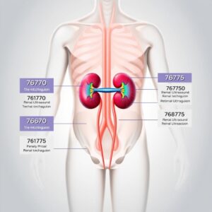
4. Deconstructing the Primary Renal Ultrasound CPT Codes: 76770, 76775, and 76776
This is the core of renal ultrasound coding. Correctly distinguishing between these three codes is paramount. The differentiation hinges entirely on the anatomic scope of the examination as documented in the radiology report.
CPT 76770: The Complete Renal Ultrasound
-
CPT Description: “Ultrasound, retroperitoneal (e.g., renal, aorta, nodes), real-time with image documentation; complete.”
-
What it entails: This is the most comprehensive code. A complete renal ultrasound must include imaging of both kidneys and the urinary bladder. Furthermore, the report must document an evaluation of the abdominal aorta in the region of the kidneys. The examination is a complete survey of the retroperitoneal structures relevant to urological and nephrological concerns.
-
Documentation Must Show: The final report must explicitly mention the evaluation of the kidneys (right and left), the urinary bladder, and the abdominal aorta. Phrases like “the aorta was visualized and appears normal in caliber without aneurysm” or “the abdominal aorta was examined and is unremarkable” are necessary to justify billing 76770. If the aorta is not mentioned, the examination defaults to a limited study.
CPT 76775: The Limited Renal Ultrasound
-
CPT Description: “Ultrasound, retroperitoneal (e.g., renal, aorta, nodes), real-time with image documentation; limited.”
-
What it entails: This code is used for a focused examination of one or more specific organs. It is not simply a “single kidney” code. A limited study might involve:
-
One or both kidneys, but not the urinary bladder.
-
The urinary bladder alone (e.g., for a post-void residual check).
-
One kidney and the bladder, but not the aorta.
-
A follow-up study for a known abnormality (e.g., “limited US to measure known renal cyst”).
-
An examination that is truncated for any reason (e.g., patient body habitus limits visualization, patient cannot tolerate the full exam).
-
-
Documentation Must Show: The report should clearly indicate the limited nature of the study. It may state “limited ultrasound performed to assess the right kidney only” or “focused evaluation of the bladder for wall thickening.” The key is that it does not meet the criteria for a “complete” exam.
CPT 76776: The Retroperitoneal Ultrasound
-
CPT Description: “Ultrasound, transplanted kidney, real-time with image documentation.”
-
What it entails: This is a highly specific code used only for evaluating a transplanted kidney, which is typically located in the iliac fossa (lower abdomen), not the retroperitoneal space. This code encompasses a complete ultrasound evaluation of the transplanted kidney itself, including its parenchyma, collecting system, and vascular anastomosis site. It often includes a limited view of the bladder but is distinct from codes 76770 and 76775.
-
Documentation Must Show: The report must specify that the examination is of a “transplanted kidney” or “renal transplant.” It is incorrect to use 76770 or 76775 for a transplanted kidney.
Comparative Table: 76770 vs. 76775 vs. 76776
| Feature | CPT 76770 (Complete) | CPT 76775 (Limited) | CPT 76776 (Transplant) |
|---|---|---|---|
| Anatomic Scope | Must include: Both Kidneys, Urinary Bladder, Abdominal Aorta | Focused on: One or more organs, but not the full set required for 76770. (e.g., 1 kidney, or bladder only, or kidneys without aorta). | Transplanted kidney (in iliac fossa), includes anastomosis site and often bladder. |
| Billing | Billed once per session, regardless of findings. | Billed once per session for the limited exam. | Billed once per session for the transplant exam. |
| Common Use Case | Comprehensive initial workup for hematuria, renal failure, pain. | Follow-up of a known cyst, post-void residual, focused pain evaluation. | Routine monitoring of a renal transplant for rejection, hydronephrosis, or fluid collections. |
| Key Documentation | Report must mention all three structures: kidneys, bladder, aorta. | Report specifies a limited goal (e.g., “right kidney only,” “bladder volume”). | Report explicitly states “transplanted kidney.” |
Table 1: Key Differences Between Primary Renal Ultrasound CPT Codes
5. The Technical and Professional Components: A Deep Dive into Modifiers
In medical billing, a single procedure can be broken down into who provided the service and what that service entailed. This is the concept of component coding, governed by modifiers.
Modifier 26: Professional Component
The professional component refers to the interpretation and report of the ultrasound examination. This is the work done by the radiologist or other qualified physician. It involves:
-
Reviewing the images captured by the sonographer.
-
Analyzing the findings.
-
Dictating or generating a formal written report with a diagnosis or impression.
-
Who bills with -26? A radiologist in a private practice who interprets an ultrasound performed at a hospital would bill CPT 76770-26. The physician is only billing for their intellectual effort of interpretation, not for the use of the equipment or the sonographer’s time.
Modifier TC: Technical Component
The technical component encompasses all the physical and operational aspects of performing the test. This includes:
-
The cost of the ultrasound machine and its maintenance.
-
The salary of the sonographer who performs the scan.
-
The cost of supplies (e.g., gel, towels).
-
The overhead of the room where the procedure is done.
-
Who bills with -TC? A hospital or an outpatient imaging center that owns the equipment and employs the sonographer would bill CPT 76770-TC. They are billing for the “technical” execution of the procedure but not for the physician’s interpretation.
Global Service: No Modifier
When the same entity provides both the technical and professional components, it is billed as a global service. This means they submit the code without any modifier (e.g., 76770).
-
Example: A freestanding imaging center owned by a radiology group performs the ultrasound on their machine with their sonographer, and one of their radiologists interprets it. They bill the global fee (76770) as they provided the entire service.
Case Scenarios and Billing Examples
-
Scenario 1: A patient goes to City Hospital for a complete renal ultrasound. A hospital employee sonographer performs the scan. The images are sent to Radiology Group, Inc., an independent practice, where Dr. Smith interprets them and sends a report back to the hospital.
-
City Hospital Bills: 76770-TC (Technical Component)
-
Radiology Group, Inc. Bills: 76770-26 (Professional Component)
-
-
Scenario 2: A patient goes to the outpatient office of Radiology Group, Inc. The group owns the building, the ultrasound machine, and employs the sonographer and radiologist.
-
Radiology Group, Inc. Bills: 76770 (Global Service)
-
-
Scenario 3: A urologist in their office has a small ultrasound machine and performs a limited ultrasound on a patient to check for bladder retention after voiding. The urologist performs the scan and interprets it immediately.
-
Urologist Bills: 76775 (Global Service – assuming they own the equipment)
-
6. Coding for Complexity: Doppler and Duplex Scanning (93975 & 93976)
Sometimes, a standard grayscale ultrasound is insufficient. To assess blood flow—critical for evaluating renal artery stenosis, renal vein thrombosis, or the viability of a transplant—Doppler ultrasound is employed.
The Role of Doppler in Renal Diagnostics
Doppler technology uses the Doppler effect to measure the velocity and direction of blood flow within vessels. It provides hemodynamic information that anatomy alone cannot. It is an essential tool for determining if a kidney is receiving adequate blood supply.
CPT 93975: Duplex Scan of Arterial Inflow and Venous Outflow
-
CPT Description: “Duplex scan of arterial inflow and venous outflow of abdominal, pelvic, scrotal contents and/or retroperitoneal organs; complete study.”
-
What it entails: This code represents a complete Doppler evaluation of the native kidneys’ vasculature. It involves a spectral Doppler analysis of the main renal arteries and veins from their origins (at the aorta for arteries, IVC for veins) to the hilum of the kidney. It is a complex, time-consuming study requiring significant operator skill.
-
Coding Rules: This code is separately reportable when performed in conjunction with a complete (76770) or limited (76775) renal ultrasound. You would bill both 76770 (for the anatomy) and 93975 (for the vascular flow) if both were performed. Append modifier 59 (Distinct Procedural Service) to 93975 if necessary to indicate it was a separate and distinct service from the grayscale exam, though many payers’ policies automatically recognize this.
CPT 93976: Complete Duplex Evaluation of a Transplanted Kidney
-
CPT Description: “Duplex scan of arterial inflow and venous outflow of abdominal, pelvic, scrotal contents and/or retroperitoneal organs; limited study.”
-
What it entails: This code is used for a complete duplex evaluation of a transplanted kidney. Despite the word “limited” in its code description, it is the primary code for a full Doppler exam of a transplant, including the anastomosis sites, intrarenal arteries (e.g., segmental, interlobar), and venous outflow.
-
Coding Rules: This code is typically billed alongside the anatomic code for a transplant kidney, 76776. Therefore, for a full transplant ultrasound with Doppler, you would bill:
-
76776 – Ultrasound, transplanted kidney (anatomic)
-
93976 – Duplex scan, transplanted kidney (vascular)
-
Do not use 93975 for a transplanted kidney.
-
7. Documentation is King: What Must Be in the Report for Accurate Coding
The radiology report is the legal document that describes the procedure and its findings. It is also the sole source of information for the medical coder. Without adequate documentation, a code cannot be justified, and a claim will be denied.
Documentation Requirements for 76770
The report must contain explicit statements confirming the evaluation of:
-
Kidneys: Description of both the right and left kidney (size, shape, echotexture, cortical thickness, presence of masses, stones, or hydronephrosis).
-
Urinary Bladder: Description of the bladder (wall thickness, contour, presence of masses or stones).
-
Abdominal Aorta: A statement that the abdominal aorta was visualized and assessed (e.g., “The abdominal aorta was visualized in the region of the kidneys and appears normal in caliber without dilation or aneurysm.”).
Documentation Requirements for 76775
The report should state the limited nature of the exam. Examples:
-
“Focused ultrasound of the right kidney was performed to evaluate for hydronephrosis.”
-
“Limited retroperitoneal ultrasound was performed; the aorta was not fully assessed due to patient body habitus.”
-
“Ultrasound of the bladder was performed for pre- and post-void volumes.”
Documentation Requirements for Doppler Studies
The report for a Doppler exam (93975 or 93976) must go beyond saying “Doppler was performed.” It must include:
-
Indication for the Doppler (e.g., “evaluate for renal artery stenosis,” “assess transplant perfusion”).
-
Description of vessels interrogated (e.g., main renal arteries, intrarenal arcuate arteries, renal veins).
-
Spectral Doppler waveforms and measurements (e.g., Peak Systolic Velocity, Resistive Index, Acceleration Time).
-
Color Doppler findings.
-
An interpretation of the hemodynamic findings.
The Sonographer’s Worksheet and the Radiologist’s Report
The sonographer’s worksheet, which contains detailed measurements and images, supports the radiologist’s final report. However, the radiologist’s signed report is the definitive document for coding purposes. The coder must code based on what the radiologist has documented as being performed and interpreted.
8. Navigating Common Coding Challenges and Denials
Even with a firm grasp of the codes, real-world scenarios present challenges.
Bilateral vs. Unilateral Studies
The codes 76770 and 76775 are inherently “complete” or “limited” studies of the retroperitoneal area; they are not coded per kidney. You bill 76770 once whether you find one kidney or two. You bill 76775 once for a limited exam, even if it involves both kidneys (but not the bladder or aorta). Modifier 50 (Bilateral Procedure) is not used with these codes.
“Rule Out” Diagnoses and Medical Necessity
Payers cover services they deem “medically necessary.” A diagnosis code like “R10.9 – Unspecified abdominal pain” may lead to a denial. The referring physician’s order and the radiologist’s report should align with covered indications. The best practice is to use the most specific diagnosis code available. If the patient has hematuria, code R31.9 (Hematuria, unspecified) is better than unspecified pain. If a stone is suspected, code N20.0 (Calculus of kidney) is even better.
Dealing with Incomplete Studies
If a patient cannot tolerate a full exam or body habitus prevents adequate visualization of the aorta, the study must be coded as limited (76775). The report should document the reason for the limitation (e.g., “The abdominal aorta could not be adequately visualized due to overlying bowel gas and body habitus, therefore this is a limited retroperitoneal ultrasound.”).
Appealing Denials Effectively
If a claim is denied, a strong appeal includes:
-
A copy of the radiology report with the key elements highlighted.
-
A cover letter citing the specific CPT code guidelines and how the documentation meets them.
-
Copies of relevant AMA CPT guidelines or payer-specific policies.
-
The patient’s medical records supporting medical necessity.
9. The Clinical Perspective: How Findings Influence Patient Management
Coding is not done in a vacuum. Understanding the clinical impact of the procedure adds a layer of meaning to the codes we assign.
From Image to Diagnosis: Hydronephrosis, Stones, Masses, and Cysts
-
Hydronephrosis: The ultrasound finding of a dilated collecting system immediately changes patient management. It can lead to further investigations like a CT urogram or procedures to relieve the obstruction.
-
Nephrolithiasis: The detection of an echogenic focus with posterior acoustic shadowing confirms a renal stone. Its size and location determine whether the patient is managed with conservative therapy, lithotripsy, or surgery.
-
Renal Masses: The ultrasound can characterize a simple cyst (benign, requiring no follow-up) versus a complex solid mass (suspicious for malignancy, requiring CT/MRI and biopsy).
-
Renal Cysts: Monitoring the size and complexity of cysts in patients with polycystic kidney disease is crucial for predicting and managing renal decline.
The Critical Role in Renal Transplant Monitoring
Ultrasound is the first-line imaging modality for transplant patients. It can diagnose:
-
Rejection: Elevated resistive indices on Doppler.
-
Anastomotic stenosis: Narrowing of the surgical connection point of the artery or vein.
-
Urinary obstruction: Hydronephrosis of the transplant.
-
Fluid collections: Lymphoceles, hematomas, or urinomas that may require drainage.
Coding 76776 and 93976 correctly ensures this vital monitoring service is sustainably funded.
Guiding Interventional Procedures
Ultrasound provides real-time, radiation-free guidance for procedures like:
-
Renal biopsy: Precise needle placement into the renal cortex.
-
Nephrostomy tube placement: Draining an obstructed kidney.
-
Abscess drainage. These procedures have their own set of CPT codes (e.g., 50200 for renal biopsy), but the renal ultrasound is the foundational tool that makes them possible.
10. The Future of Renal Ultrasound and Coding
The field of medical imaging is constantly advancing, and with it, the CPT code set must evolve.
Advancements in Elastography and Contrast-Enhanced US
-
Elastography: This technology measures tissue stiffness, which can help characterize masses and assess the fibrotic changes of chronic kidney disease non-invasively. As it becomes standard of care, new CPT codes may be created.
-
Contrast-Enhanced Ultrasound (CEUS): Using microbubble contrast agents, CEUS provides detailed vascular mapping of lesions, often with similar accuracy to CT or MRI but without radiation. Specific codes for CEUS of abdominal organs already exist and may see expanded renal applications.
The Impact of Artificial Intelligence (AI) on Imaging and Coding
AI algorithms are being developed to assist with:
-
Image acquisition: Helping sonographers capture standardized, high-quality images.
-
Image analysis: Automatically measuring kidney size, volume, and cystic disease burden, or flagging suspicious areas for radiologist review.
-
Automated reporting: Drafting reports based on findings. The coding implications are profound. Will an AI-assisted exam be coded differently? How will the professional component be defined if AI provides the initial interpretation? The coding world will need to adapt.
Evolving Payer Policies and Regulations
Medicare, Medicaid, and private insurers constantly update their coverage policies and reimbursement rates. Staying current with resources like the Medicare National Correct Coding Initiative (NCCI) edits and Local Coverage Determinations (LCDs) is not optional; it is a required part of compliant coding practice.
11. Conclusion
Mastering CPT codes for renal ultrasound requires a synthesis of technical coding knowledge, a deep understanding of clinical medicine, and meticulous attention to documentation detail. The codes 76770, 76775, and 76776, along with their Doppler counterparts, form a precise language that describes vital patient care. By moving beyond mere memorization and embracing the clinical story behind each code, medical professionals can ensure accuracy, optimize reimbursement, and ultimately contribute to the sustainable delivery of high-quality diagnostic services. In the ever-evolving landscape of healthcare, this expertise is more valuable than ever.
12. Frequently Asked Questions (FAQs)
Q1: Can I bill both 76770 and 76775 during the same session for the same patient?
A: No. These codes are mutually exclusive. An examination is either complete (76770) or limited (76775). You must choose the single code that best describes the service that was actually performed and documented.
Q2: A patient has a native right kidney and a transplanted kidney in the left iliac fossa. We performed a full ultrasound on both. What codes do I use?
A: This is a complex scenario. You would need to bill for each distinct anatomic area:
-
76775 for the limited exam of the native right kidney (as the bladder and aorta were likely not the focus).
-
76776 for the complete exam of the transplanted kidney.
Append modifier 59 to the second code to indicate a distinct procedural service. Always check payer-specific guidelines for billing multiple ultrasound codes.
Q3: The sonographer’s worksheet notes that the aorta was visualized and measured, but the radiologist’s final report does not mention the aorta. Can I code 76770?
A: No. Coding must be based solely on the radiologist’s signed final report. If the aorta is not mentioned in the impression or the body of the final report, you must default to coding a limited retroperitoneal ultrasound (76775). The radiologist’s report is the legal document of record.
Q4: Is a bladder volume measurement (pre- and post-void) included in 76770?
A: Yes, a standard evaluation of the bladder is included in a complete renal ultrasound (76770). However, if a bladder volume measurement is the only reason for the study, it would be billed as a limited retroperitoneal ultrasound (76775). If a post-void residual is performed as an add-on to a complete renal exam, it is not separately billable.
Q5: How often are these CPT codes updated?
A: The AMA releases a new CPT code book annually. While the core renal ultrasound codes (76770, 76775, 76776) have been stable for many years, their official definitions and parenthetical notes can change. It is imperative to use the current year’s CPT manual for accurate coding.
13. Additional Resources
-
The American Medical Association (AMA): The primary source for the CPT code set. Purchase the current CPT manual and subscribe to their newsletters for updates. https://www.ama-assn.org
-
The American College of Radiology (ACR): Provides practice parameters, technical standards, and extensive resources on the appropriate use of ultrasound and other radiological procedures. https://www.acr.org
-
The Society of Diagnostic Medical Sonography (SDMS): An excellent resource for sonographers and coders, offering educational materials on proper scanning technique and protocol, which directly informs coding. https://www.sdms.org
-
The Centers for Medicare & Medicaid Services (CMS): Provides access to the National Correct Coding Initiative (NCCI) edits, Local Coverage Determinations (LCDs), and other vital Medicare billing policies. https://www.cms.gov
-
The American Academy of Professional Coders (AAPC): A premier organization for medical coders, offering certifications, training, networking, and up-to-date coding news. https://www.aapc.com
Date: August 28, 2025
Author: The Medical Coding Specialist Team
Disclaimer: This article is intended for informational and educational purposes only. It is not a substitute for professional medical advice, diagnosis, or treatment. Always seek the advice of your physician or other qualified health provider with any questions you may have regarding a medical condition or procedure. The CPT codes and descriptions discussed are based on the proprietary work of the American Medical Association (AMA) and are subject to change. Always refer to the most current, official AMA CPT code books for accurate billing and coding guidance.


