Imagine the unsettling sensation of tightness, the constant, low-grade itch that nags at your consciousness, the visual of flaky, lackluster skin that no amount of superficial cream seems to fix. This is the daily reality for millions of individuals living with xeroderma, the medical term for abnormally dry skin. Often dismissed as a mere cosmetic nuisance, xeroderma is, in fact, a complex dermatologic condition with significant implications for an individual’s physical comfort, skin integrity, and overall quality of life. It is not a single disease but a symptom, a final common pathway for a multitude of internal and external insults that disrupt the skin’s delicate barrier function.
The journey into understanding xeroderma is a journey into the very essence of our body’s largest organ. It forces us to look beyond the surface and appreciate the sophisticated biological machinery that works tirelessly to keep us hydrated, protected, and resilient. When this machinery falters, the consequences are far-reaching. What begins as simple dryness can escalate into inflammation, fissuring, secondary infections, and a relentless cycle of itching and scratching that can lead to lichenification and even psychological distress.
This article aims to be the definitive guide on xeroderma. We will dissect it from every angle: from the precise medical classification using the ICD-10 coding system that allows healthcare providers to speak a common language, to the microscopic breakdown of the skin’s barrier. We will explore the environmental villains and internal culprits, chart the clinical presentation from mild to severe, and build a comprehensive therapeutic ladder for effective management. Our goal is to empower patients, clinicians, and students with a deep, nuanced understanding of xeroderma, transforming it from a frustrating mystery into a manageable condition.
icd 10 code xeroderma
2. Decoding the Diagnosis: The ICD-10 Framework for Xeroderma
In the world of modern medicine, precision is paramount. The International Classification of Diseases, 10th Revision (ICD-10) is the global standard for diagnostic health information. It provides a unified language that enables physicians, researchers, and insurers to accurately record, track, and manage diseases.
The Purpose and Power of ICD-10
The ICD-10 system transforms clinical descriptions of diseases and health problems into an alphanumeric code. This standardization is crucial for:
-
Epidemiology: Tracking the incidence and prevalence of conditions like xeroderma across populations.
-
Billing and Reimbursement: Ensuring healthcare providers are compensated correctly for their services.
-
Clinical Research: Facilitating the study of patient outcomes and treatment efficacy.
-
Quality Assurance: Monitoring the standard of care provided for specific conditions.
For a condition as common as xeroderma, accurate coding is essential for understanding its burden on the healthcare system and for allocating resources effectively.
Navigating the L85 Category: Xerosis in Detail
Xeroderma and its more severe or specific forms are classified under Chapter XII of the ICD-10 manual, which covers diseases of the skin and subcutaneous tissue. The primary code is found within the block L80-L99, “Other disorders of the skin and subcutaneous tissue.”
The most relevant code for generalized, uncomplicated dry skin is:
-
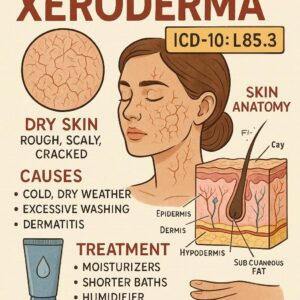
L85.3 – Xerosis cutis: This is the default and most frequently used code for diagnosing xeroderma. It signifies “dry skin” without specifying a particular underlying cause or complication beyond the inherent dryness.
However, the ICD-10 system allows for greater specificity. Depending on the clinical presentation, a physician might use one of the following codes:
-
L85.0 – Acquired ichthyosis: This code is used when the dry skin presents with fish-like scales, resembling the genetic disorder ichthyosis, but is acquired later in life due to conditions like malignancy (e.g., lymphoma), sarcoidosis, or certain medications.
-
L85.1 – Acquired keratosis [keratoderma] palmaris et plantaris: This refers to specific, localized dry, thickened skin on the palms and soles.
-
L85.9 – Epidermal thickening, unspecified: A less common code that might be used if the dryness has led to significant thickening of the epidermis, but the presentation doesn’t fit the other categories more precisely.
It is critical to distinguish xerosis from other common dermatologic conditions that may present with dryness but have different pathophysiologies and codes, such as:
-
Atopic Dermatitis (L20.-): A chronic, inflammatory condition where dry skin is a key feature, but it is accompanied by significant erythema (redness), pruritus (itching), and eczematous lesions.
-
Contact Dermatitis (L23-L25): Dryness can be a symptom, but it is directly caused by an allergen or irritant.
-
Psoriasis (L40.-): While scaling is a hallmark, the plaques are typically well-demarcated, erythematous, and have a different underlying inflammatory pathway.
Clinical Documentation: The Key to Accurate Coding
The accuracy of the assigned ICD-10 code hinges entirely on the clinician’s documentation. Vague notes like “patient has dry skin” are insufficient. A robust clinical note should include:
-
Location: Is it generalized or localized (e.g., lower legs, arms)?
-
Severity: Mild, moderate, or severe?
-
Morphology: Is the skin simply dry (xerosis), scaly (ichthyosis-like), or cracked (fissured)?
-
Associated Symptoms: Is there pruritus? Pain? Bleeding?
-
Etiology, if known: Is it due to aging (senile xerosis), environmental factors, or an underlying systemic disease?
This level of detail ensures the correct code (e.g., L85.3) is selected, painting an accurate picture for all stakeholders involved in the patient’s care.
3. The Skin We Live In: A Primer on Structure and Function
To comprehend why xeroderma occurs, one must first understand the elegant biology of healthy skin. The skin is a dynamic, multi-layered organ, and its outermost layer, the epidermis, is the primary battlefield in the fight against dryness.
The Stratum Corneum: The Barrier Guardian
The very top layer of the epidermis is the stratum corneum, often described as a “brick and mortar” structure.
-
The Bricks (Corneocytes): These are anucleate, flattened, dead keratinocytes filled with keratin filaments. They are tough and provide structural integrity.
-
The Mortar (Lipid Matrix): The spaces between the corneocytes are filled with a meticulously organized lipid matrix composed of ceramides, cholesterol, and free fatty acids. This lipid mortar is the primary barrier that prevents Transepidermal Water Loss (TEWL)—the passive evaporation of water from the deeper skin layers to the external environment.
When the stratum corneum is intact and healthy, it maintains optimal hydration, keeping the skin soft, supple, and resilient.
Natural Moisturizing Factor (NMF) and Lipids: The Hydration Engine
Within the corneocytes resides another critical component: the Natural Moisturizing Factor (NMF). NMF is a collection of water-soluble compounds, including amino acids, pyrrolidone carboxylic acid, lactate, urea, and salts. These compounds are hygroscopic, meaning they attract and bind water from the atmosphere and the dermis below, acting like microscopic sponges within the skin cells.
The health of the skin barrier is, therefore, a function of both the intercellular lipids (preventing water loss) and the intracellular NMF (holding onto water). Xeroderma fundamentally occurs when there is a deficiency in either of these components, or when the rate of TEWL exceeds the skin’s ability to replenish its water content. This leads to a state of subclinical inflammation, impaired desquamation (shedding of dead skin cells), and the characteristic rough, flaky, and tight appearance of dry skin.
4. The Multifaceted Etiology of Xeroderma: Unraveling the Causes
Xeroderma is rarely the result of a single factor. It is typically the consequence of a complex interplay between exogenous (external) triggers and endogenous (internal) predispositions.
Exogenous Factors: The Assault from Outside
-
Low Humidity and Cold Weather: Winter months are notorious for exacerbating dry skin. Cold air holds less moisture, and indoor heating further depletes ambient humidity, creating an environment that actively pulls water from the skin.
-
Hot, Dry Winds: Can have a similar desiccating effect.
-
Ultraviolet (UV) Radiation: Chronic sun exposure damages keratinocytes and degrades collagen and elastin, impairing the skin’s structural integrity and barrier function over time.
-
Chemical Irritants:
-
Harsh Soaps and Detergents: These are often alkaline and designed to dissolve oils. They strip the skin of its natural sebum and can damage the lipid bilayer.
-
Alcohol-based Sanitizers and Astringents: These rapidly degrease the skin.
-
Occupational Exposures: Constant exposure to solvents, chemicals, or cement can be profoundly damaging.
-
-
Frequent Hot Bathing or Showering: Prolonged contact with hot water, especially, washes away natural lipids and NMF components.
-
Mechanical Friction: From abrasive clothing or repetitive physical tasks.
Endogenous Factors: The Internal Imbalance
-
Advanced Age (Asteatotic Eczema): As we age, skin undergoes “senile xerosis.” There is a natural decline in the production of sebum, a slowdown in epidermal cell turnover, and a reduction in the synthesis of ceramides and NMF components. The skin becomes thinner and more fragile.
-
Genetic Predisposition: Individuals with a personal or family history of atopic dermatitis, ichthyosis, or keratosis pilaris have a inherently weaker skin barrier.
-
Hormonal Changes: Fluctuations in estrogen, such as during menopause or pregnancy, can affect skin hydration and sebum production.
-
Nutritional Deficiencies: Deficiencies in essential fatty acids, vitamins (especially A, C, D, and E), and zinc can compromise skin barrier repair and function.
Xeroderma as a Manifestation of Systemic Disease
Sometimes, xeroderma is a cutaneous clue to an internal medical condition. This makes a thorough medical history vital.
-
Hypothyroidism: A slowed metabolic rate leads to reduced sweating and sebum production, resulting in dry, coarse, and cool skin.
-
Chronic Kidney Disease (CKD): Uremia and electrolyte imbalances can directly affect the skin, causing xerosis and severe pruritus.
-
Diabetes Mellitus: Diabetic neuropathy can lead to anhydrosis (lack of sweating), and microvascular complications can impair skin nutrition.
-
Malignancies: Particularly lymphomas (e.g., Hodgkin’s disease) can present with acquired ichthyosis or severe pruritus.
-
Autoimmune Diseases: Sjögren’s syndrome, which attacks moisture-producing glands, often causes severe xerosis.
-
HIV/AIDS: Dermatologic manifestations are common, including severe xerosis and seborrheic dermatitis.
-
Anorexia Nervosa: Malnutrition and dehydration directly impact skin health.
5. Clinical Presentation: Recognizing the Signs and Symptoms
The presentation of xeroderma exists on a spectrum, from a mild, asymptomatic state to a severe, debilitating condition.
From Roughness to Rhagades: The Spectrum of Dryness
-
Mild: The skin may feel rough or gritty to the touch. There may be slight scaling, often described as “ashy” skin, particularly in darker skin tones. The primary sensation is one of tightness, especially after bathing.
-
Moderate: Scaling becomes more pronounced and visible. The skin may appear dull and lackluster. Fine lines may be more noticeable. Pruritus (itching) often begins at this stage.
-
Severe: Large, flaky scales adhere to the skin surface. The skin loses its elasticity and can develop rhagades—painful, deep fissures or cracks that can bleed and pose a risk for secondary bacterial infection. Erythema (redness) indicates underlying inflammation, a state sometimes referred to as eczematous xerosis or asteatotic eczema.
Itch-Scratch Cycle: The Vicious Circle of Pruritus
Pruritus is one of the most distressing symptoms of xeroderma. Dry skin stimulates nerve endings in the epidermis. Scratching, while providing momentary relief, causes further damage to the stratum corneum. This damage:
-
Releases inflammatory mediators that worsen the itch.
-
Further compromises the barrier, increasing TEWL.
This creates a self-perpetuating itch-scratch cycle that can be incredibly difficult to break and can lead to lichenification—a thickening of the skin with accentuated skin markings—as a result of chronic scratching and rubbing.
Beyond the Surface: The Psychosocial Impact
The impact of chronic xeroderma extends beyond physical discomfort. Persistent itching can lead to:
-
Sleep deprivation and chronic fatigue.
-
Difficulty concentrating.
-
Irritability and anxiety.
-
Social embarrassment and reduced self-esteem due to the visible appearance of the skin.
Acknowledging this burden is a critical part of holistic patient care.
6. A Systematic Approach to Diagnosis and Differential Diagnosis
Diagnosing xeroderma is primarily clinical, based on a thorough history and physical examination.
The Patient History: A Detective’s Tool
A clinician will explore:
-
Timeline: Onset, duration, and frequency.
-
Location: Where did it start? Is it widespread?
-
Aggravating/Alleviating Factors: Does it worsen with bathing, in winter, or with certain soaps? Does it improve with moisturizers?
-
Associated Symptoms: Itch, pain, bleeding?
-
Past Medical History: Any history of atopy, thyroid, kidney, or other systemic diseases?
-
Medication Review: Drugs like diuretics, retinoids, or some cholesterol-lowering agents can cause dry skin.
-
Social/Occupational History: Hobbies, work environment, skincare habits.
Physical Examination: A Head-to-Toe Assessment
A full skin exam is performed under good lighting. The clinician assesses for:
-
Distribution: Is it symmetrical? Is it on the extensor surfaces (common in simple xerosis) or flexural areas (more suggestive of atopic dermatitis)?
-
Morphology: The type of scaling (fine vs. large), presence of fissures, erythema, excoriations (scratch marks), or lichenification.
When to Investigate Further
For typical, uncomplicated xeroderma, no tests are needed. However, if an underlying cause is suspected, the following may be ordered:
-
Blood Tests:
-
Thyroid-Stimulating Hormone (TSH) for hypothyroidism.
-
Basic Metabolic Panel (BMP) for renal function.
-
Complete Blood Count (CBC) for signs of lymphoma.
-
HbA1c for diabetes.
-
-
Skin Biopsy: Rarely needed, but may be performed to rule out other conditions like psoriasis or cutaneous T-cell lymphoma if the presentation is atypical.
7. The Therapeutic Ladder: A Comprehensive Management Strategy
Managing xeroderma is a proactive and continuous process. The approach can be visualized as a therapeutic ladder, starting with foundational measures and escalating as needed.
Foundation Level: Lifestyle and Environmental Modifications
-
Humidification: Using a humidifier, especially in the bedroom during winter, can significantly reduce TEWL.
-
Bathing Modifications:
-
Use lukewarm, not hot, water.
-
Limit shower/bath time to 5-10 minutes.
-
Use a mild, syndet (synthetic detergent) or soap-free cleanser with a pH close to that of skin (pH 4.5-5.5).
-
Pat skin dry; do not rub vigorously.
-
-
Avoid Irritants: Wear gloves for wet work and when handling chemicals. Choose clothing made of soft, breathable fabrics like cotton.
First-Line Therapy: Emollients, Humectants, and Occlusives
This is the cornerstone of treatment. Moisturizers are not all created equal; they work through different mechanisms, and understanding the difference is key.
The Triad of Effective Moisturization
| Component Type | Mechanism of Action | Key Ingredients | Ideal Use Case |
|---|---|---|---|
| Humectants | Attract water from the dermis and environment into the stratum corneum. | Glycerin, Hyaluronic Acid, Urea, Alpha-Hydroxy Acids (AHAs like Lactic Acid), Sorbitol | All types of dry skin, but best used under an occlusive to prevent water from evaporating. Excellent for “plumping” the skin. |
| Emollients | Smooth and soften the skin by filling the spaces between desquamating corneocytes with lipids. | Squalene, Jojoba Oil, Caprylic/Capric Triglyceride, Diisopropyl Dimer Dilinoleate | For rough, flaky skin to improve texture and flexibility. They feel “cosmetically elegant.” |
| Occlusives | Form a hydrophobic, inert film on the skin surface to physically block TEWL. | Petrolatum, Lanolin, Mineral Oil, Dimethicone, Shea Butter, Beeswax | For moderate to severe xeroderma, very dry climates, or for use overnight. Petrolatum is the gold standard, reducing TEWL by over 98%. |
The most effective moisturizers, often called “barrier repair creams,” contain a balanced combination of all three components. The “soak and seal” method is highly effective: applying a moisturizer to damp skin immediately after bathing to trap the absorbed water.
Second-Line Therapy: Topical Pharmacologic Agents
When moisturizers alone are insufficient, targeted topical prescriptions may be used.
-
Topical Corticosteroids: Low-potency steroids (e.g., 1% hydrocortisone) are used for short courses to reduce inflammation and break the itch-scratch cycle in eczematous xerosis.
-
Topical Calcineurin Inhibitors: Such as tacrolimus and pimecrolimus, can be used in sensitive areas (face, groin) as steroid-sparing agents.
-
Prescription Urea (e.g., 40%): Used for severe scaling and hyperkeratosis, it is a potent keratolytic (breaks down scales) and humectant.
-
Topical Retinoids: May be used with caution for conditions like acquired ichthyosis to normalize keratinization.
Third-Line Therapy: Systemic and Advanced Treatments
For the most severe, refractory cases, or when xeroderma is secondary to a systemic disease:
-
Systemic Antipruritics: Antihistamines (e.g., hydroxyzine, cetirizine) can help control itch, particularly sedating ones at night to improve sleep.
-
Phototherapy: Narrowband UVB therapy can have an anti-inflammatory and immunomodulatory effect, reducing pruritus and improving the skin barrier in chronic cases.
-
Treating the Underlying Cause: This is the most critical step when xeroderma is a symptom. Initiating thyroid hormone replacement, better glycemic control in diabetes, or treating the underlying malignancy is paramount.
8. Special Populations and Considerations
Xeroderma in the Elderly: Geriatric Xerosis
This is almost a universal finding. Management emphasizes gentle cleansing, frequent and generous application of thick, occlusive-rich emollients (like petrolatum-based ointments), and environmental modifications. The fragility of aged skin makes it more prone to fissuring and infection, so prevention is key.
Pediatric Dry Skin
Children, especially infants, have a skin barrier that is not fully matured. Harsh products and overwashing should be avoided. Simple, fragrance-free emollients are safest. Persistent dry skin in a child, especially with itch, should be evaluated for atopic dermatitis.
Xeroderma in Compromised Skin: The Atopic and Diabetic Patient
In atopic dermatitis, the barrier defect is fundamental. A rigorous, daily moisturizing regimen with barrier repair creams is not just symptomatic treatment but a primary strategy to prevent flares. In diabetic patients, meticulous foot care is essential, as xerosis can lead to fissures that serve as a portal for infection, potentially leading to serious complications.
9. The Future of Dry Skin Management: Innovations and Research
The field of dermatology is rapidly evolving, and xeroderma management is benefiting from these advances.
-
Biologic and Targeted Therapies: While used for severe eczema (e.g., Dupilumab), research into how these drugs repair the barrier may lead to new insights for treating complex xerosis.
-
Microbiome Modulation: The skin’s microbiome plays a role in barrier health. Pre- and post-biotic topical formulations designed to support beneficial bacteria are an emerging area.
-
Advanced Delivery Systems: Technologies like liposomal and nanoparticle encapsulation are being explored to deliver key moisturizing ingredients like ceramides deeper and more effectively into the stratum corneum.
10. Conclusion
Xeroderma, coded in the ICD-10 system as L85.3, is a pervasive dermatological condition far beyond a simple cosmetic concern. Its development is a complex interplay of external environmental aggressors and internal physiological disruptions that compromise the skin’s vital barrier function. Effective management demands a holistic, multi-faceted strategy, beginning with foundational lifestyle adjustments and escalating to sophisticated topical and systemic therapies tailored to the individual’s needs. Ultimately, a proactive and consistent regimen centered on understanding and replenishing the skin’s barrier is the cornerstone of transforming dry, uncomfortable skin into a healthy, resilient state.
11. Frequently Asked Questions (FAQs)
Q1: What is the single most important thing I can do for my dry skin?
A: The most impactful habit is to apply a thick, fragrance-free moisturizer containing both humectants and occlusives (like a cream or ointment) to slightly damp skin within 3 minutes of bathing. This “soak and seal” method is unparalleled in its effectiveness at trapping hydration.
Q2: Is there a difference between “xerosis” and “xeroderma”?
A: In clinical practice, the terms are used interchangeably. Both refer to abnormal dryness of the skin. “Xerosis cutis” is the full Latin term, with “cutis” meaning skin. ICD-10 uses “Xerosis cutis” (L85.3) as the official diagnosis.
Q3: Can drinking more water cure my dry skin?
A: While systemic hydration is important for overall health, it is not a direct cure for xeroderma. If you are adequately hydrated, increasing water intake will not significantly moisturize a compromised skin barrier. Topical therapy to repair the barrier and prevent water loss is far more critical.
Q4: Why is my dry skin so much worse in the winter?
A: This is primarily due to low humidity levels both outdoors (cold air holds less moisture) and indoors (due to heating systems). This dry air creates a gradient that pulls water from your skin into the atmosphere (increased TEWL). Hot showers and baths, which are more common in winter, exacerbate the problem.
Q5: When should I see a doctor about my dry skin?
A: You should seek medical advice if:
-
The dryness and itching are severe and interfere with sleep or daily activities.
-
Your skin is cracked, bleeding, or shows signs of infection (increased redness, warmth, pus, pain).
-
Over-the-counter products provide no relief after several weeks of consistent use.
-
You suspect it may be related to an underlying medical condition or medication.
12. Additional Resources
-
American Academy of Dermatology (AAD): www.aad.org – An excellent source for patient-friendly information on dry skin and eczema, with a “Find a Dermatologist” tool.
-
National Eczema Association (NEA): nationaleczema.org – While focused on eczema, their resources on skin barrier function and moisturizer selection are invaluable for anyone with chronic dry skin.
-
World Health Organization (WHO): ICD-10 Online Browser – To understand the official structure and coding of diseases.
Disclaimer: This article is for informational purposes only and is not a substitute for professional medical advice, diagnosis, or treatment. Always seek the advice of your physician or other qualified health provider with any questions you may have regarding a medical condition. The ICD-10 codes provided are for educational use; always consult with a certified medical coder for official billing and diagnostic purposes.
Date: November 04, 2025