Imagine a skin condition so common that it affects nearly half of the world’s adolescent population and a significant portion of adults, yet remains one of the most misunderstood and often dismissed dermatological concerns. This is the reality for millions living with Keratosis Pilaris (KP). Often trivialized as mere “chicken skin,” KP is a chronic, genetic keratinization disorder that manifests as rough, bumpy patches on the skin, primarily on the upper arms, thighs, cheeks, and buttocks. While medically benign, its impact is far from trivial. For many, these persistent bumps are a source of self-consciousness, frustration, and a relentless cycle of trial-and-error with over-the-counter remedies. In the intricate world of modern healthcare, where precision is paramount, this common condition is distilled into a specific, universal language: the International Classification of Diseases, Tenth Revision (ICD-10) code. This article delves deep into the world of ICD-10 Codes for Keratosis Pilaris, moving beyond the surface-level understanding to explore its pathology, its human impact, and the critical administrative backbone of its care—the ICD-10 code L11.0. We will unravel why this simple alphanumeric sequence is so much more than a billing tool; it is a key that unlocks appropriate care, facilitates research, and validates the experiences of those living with this ubiquitous skin condition.
ICD-10 Codes for Keratosis Pilaris
Understanding the Foe: What is Keratosis Pilaris?
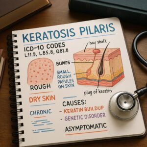
To effectively manage and code for Keratosis Pilaris, one must first understand its fundamental nature. KP is a disorder of keratinization, a process where the skin produces keratin, a tough, protective protein. In healthy skin, keratin helps form a resilient barrier. However, in KP, this process goes awry. The root of the problem lies in the hair follicles. Excess keratin, combined with sometimes abnormal sebum, forms a plug that blocks the opening of the hair follicle. This plug is known as a hyperkeratotic plug. Instead of a hair growing out smoothly, the follicle becomes occluded, leading to the formation of a small, raised bump.
Beneath the surface, a microscopic examination would reveal a dilated hair follicle filled with a keratotic plug, often accompanied by a coiled or trapped hair. The surrounding skin may show mild inflammation, which accounts for the redness (erythema) often seen around the bumps, a variant known as Keratosis Pilaris Rubra. It is crucial to understand that KP is not an infection, it is not contagious, and it is not a result of poor hygiene. It is fundamentally a genetic condition. The predisposition to produce excess keratin in the follicular openings is inherited in an autosomal dominant pattern, meaning a child has a 50% chance of inheriting the condition if one parent has it. The expression of the gene can vary widely, which is why one sibling may have severe KP while another has none.
The prevalence of KP peaks during adolescence, a time of hormonal fluctuations that can stimulate sebum production and exacerbate keratinization. It often improves in adulthood but can persist indefinitely. Its course is also highly influenced by environmental factors; it typically worsens in the winter months when humidity is low and indoor heating dries the skin, and often improves during the humid summer months. Understanding this pathophysiology—the faulty keratin production and follicular plugging—is the first step in demystifying the condition and appreciating the logic behind both its treatment and its classification in medical coding systems.
The Clinical Picture: Signs, Symptoms, and Subtypes
Keratosis Pilaris presents with a characteristic set of signs and symptoms that, while variable in severity, form a recognizable clinical pattern. The primary lesion is a fine, pinpoint, skin-colored or erythematous (red) papule. These papules feel like sandpaper to the touch, creating a rough, bumpy texture. They are typically 1-2 mm in diameter and are often described as “goosebumps” or “chicken skin” that do not resolve.
Common Locations:
-
Upper Outer Arms: This is the most classic and common site for KP.
-
Anterior Thighs and Buttocks: Another very frequent location.
-
Cheeks: Particularly common in children, often giving the cheeks a perpetually flushed, rough appearance.
-
Back and Trunk: Less common, but can occur.
While KP is primarily asymptomatic, meaning it doesn’t usually cause physical discomfort, it is not entirely without symptoms. Some individuals experience:
-
Mild Pruritus (Itching): Especially when the skin is dry.
-
Skin Dryness: The affected areas are often concomitantly dry.
-
Cosmetic Concern: This is the most significant “symptom” for the vast majority of patients, leading to psychological distress.
Beyond the classic presentation, several clinical subtypes of KP are recognized, which are important for clinical documentation:
-
Keratosis Pilaris Rubra: This variant is characterized by prominent erythema (redness) surrounding the bumps, which may be more widespread. The papules are often more inflammatory and can be mistaken for acne. It is frequently seen on the cheeks in younger individuals.
-
Keratosis Pilaris Alba: This is the non-inflammatory form. The bumps are skin-colored with little to no redness, and the primary complaint is the rough, dry texture.
-
Keratosis Pilaris Atrophicans: This is a more severe and rarer spectrum of disorders that includes Keratosis Pilaris atrophicans faciei (affecting the face and eyebrows) and Atrophoderma vermiculata (honeycomb-like scarring on the cheeks). These conditions can lead to follicular scarring and atrophy, resulting in permanent pitting or loss of eyebrows.
-
Erythromelanosis Follicularis Faciei et Colli: A variant presenting with reddish-brown pigmentation and follicular papules on the face and neck.
Recognizing these subtypes is part of a thorough clinical assessment, even though they all fall under the same umbrella ICD-10 code.
The Diagnostic Pathway: How Keratosis Pilaris is Identified
The diagnosis of Keratosis Pilaris is overwhelmingly a clinical one. This means it is made based on the characteristic history and physical examination findings, without the need for invasive or expensive laboratory tests. A healthcare provider, typically a dermatologist or a primary care physician, will arrive at a diagnosis through a straightforward process.
1. Patient History: The clinician will ask about the onset of the bumps (often in childhood or adolescence), their location, and any associated symptoms like itching. A family history is a key question, as a positive history strongly supports the diagnosis. They will also inquire about what makes it better or worse (e.g., seasonal changes, specific skincare products).
2. Physical Examination: This is the cornerstone of diagnosis. The provider will visually inspect the skin, noting the distribution (bilateral arms, thighs, etc.) and the morphology of the lesions (fine, keratotic, follicular papules). The “sandpaper” feel is a classic tactile finding. The presence of little to no inflammation beyond the immediate follicle and the absence of comedones (blackheads and whiteheads) help distinguish it from acne.
3. Dermoscopy: If there is any uncertainty, a dermatoscope—a handheld skin microscope—can be used. Dermoscopy of KP typically reveals whitish, coiled hairs within the keratotic plugs and perifollicular erythema, providing a magnified view of the pathology.
Biopsy is rarely performed for KP. It is only considered in atypical, severe, or diagnostically challenging cases where other conditions like follicular eczema, lichen spinulosus, or milia are strong possibilities. A histopathological examination of a biopsy specimen would confirm the diagnosis by revealing the tell-tale signs: a dilated follicular infundibulum plugged with keratin, and possibly a twisted hair shaft.
The simplicity of the diagnostic process underscores a key point: the clinical picture is paramount. This clinical certainty is what allows for the confident assignment of the specific ICD-10 code, streamlining the journey from patient presentation to administrative documentation.
The Heart of the Matter: The ICD-10 Code L11.0 and Its Critical Role
At the core of modern medical administration and data tracking lies the International Classification of Diseases (ICD). Maintained by the World Health Organization (WHO), the ICD is the global standard for diagnostic health information. The ICD-10-CM (Clinical Modification) is the system used in the United States for diagnostic coding. For Keratosis Pilaris, the designated code is L11.0.
Let’s deconstruct this code:
-
Chapter XII: The “L” denotes Chapter XII of ICD-10-CM, which covers “Diseases of the skin and subcutaneous tissue.”
-
Block L10-L14: This block encompasses “Bullous disorders,” but the categorization extends to include other specified disorders of skin integrity.
-
Category L11: This category is for “Other acantholytic disorders.” Acantholysis refers to the loss of intercellular connections in the epidermis. While KP is not primarily an acantholytic disorder, it is classified here as a “catch-all” for specific follicular keratinization disorders that don’t fit elsewhere.
-
Code L11.0: This is the specific code for “Acquired keratosis follicularis.” In medical terminology, “keratosis follicularis” is a synonym for Keratosis Pilaris, and “acquired” distinguishes it from a very rare congenital condition.
Why is this code so important?
-
Precise Billing and Reimbursement: This is the most direct application. When a healthcare provider submits a claim to an insurance company for an office visit, procedure, or prescription related to KP, the code L11.0 must be used to justify the medical necessity of the service. Without this code, the claim may be denied.
-
Population Health Tracking: By aggregating data from millions of coded encounters, public health officials and researchers can track the prevalence of KP. They can identify demographic patterns, seasonal trends, and associations with other conditions (e.g., atopic dermatitis, obesity).
-
Clinical Research: For a clinical trial investigating a new KP treatment, researchers rely on the ICD-10 code L11.0 to accurately identify and recruit eligible study participants from electronic health records.
-
Quality of Care Metrics: Healthcare systems use diagnostic codes to monitor the quality of care for specific patient populations. Accurate coding for KP ensures that these patients are correctly represented in healthcare data.
It is a common misconception that coding is merely a bureaucratic exercise. In reality, the code L11.0 is a vital piece of infrastructure that supports the entire ecosystem of patient care for Keratosis Pilaris, from the individual clinic visit to global health understanding.
Navigating the Nuances: Coding Scenarios and Common Pitfalls
While L11.0 is the definitive code for Keratosis Pilaris, accurate medical coding requires attention to detail and context. The clinical documentation must support the code assigned. Let’s explore some common scenarios and potential pitfalls.
Scenario 1: The Straightforward Case
A 16-year-old patient presents with a 3-year history of asymptomatic, rough bumps on the back of both arms. On examination, the physician notes numerous skin-colored, keratotic follicular papules. The diagnosis is “Keratosis Pilaris.”
-
Correct Code: L11.0
Scenario 2: KP with Associated Condition
A 25-year-old female presents for management of her longstanding KP on her arms and thighs. She also has severe atopic dermatitis on her flexures. The physician addresses both conditions during the visit.
-
Coding: In this case, both codes should be reported. The primary code should be for the condition that was the chief reason for the visit. If she came specifically for her KP, L11.0 would be primary, and L20.9 (Atopic dermatitis, unspecified) would be secondary. The reverse would be true if the eczema flare was the main concern. This paints a complete picture of the patient’s health status.
Scenario 3: The “Ruling Out” Dilemma
A patient presents with a bumpy rash, and the physician’s assessment is “Rule out Keratosis Pilaris versus follicular eczema.” In coding, you cannot code a “rule-out” diagnosis. You code the condition(s) established to the highest degree of certainty at the end of the encounter. If the physician, after examination, believes KP is the most likely diagnosis, it can be coded. If the diagnosis is truly uncertain, the symptoms (e.g., R21 Rash and other nonspecific skin eruption) might be used until a definitive diagnosis is made.
Common Pitfalls:
-
Confusing with Acne: Acne vulgaris has its own set of codes (L70.0-L70.9). It is critical to document the absence of comedones, pustules, and cysts to distinguish KP from acne, ensuring the correct code is applied.
-
Lack of Specificity: Using a less specific code when L11.0 is available can lead to claim issues or inaccurate data.
-
Incorrect Sequencing: When multiple conditions are addressed, listing them in the wrong order of importance can impact reimbursement.
The following table summarizes the key coding considerations for Keratosis Pilaris and its common differentials.
ICD-10 Coding for Keratosis Pilaris and Related Conditions
| Condition | ICD-10 Code | Key Distinguishing Clinical Features |
|---|---|---|
| Keratosis Pilaris | L11.0 | Follicular, keratotic papules; “sandpaper” feel; common on arms, thighs, cheeks; little to no inflammation. |
| Acne Vulgaris | L70.0 | Presence of comedones (blackheads/whiteheads), inflammatory papules, pustules, nodules; common on face, chest, back. |
| Atopic Dermatitis | L20.9 | Intensely pruritic; ill-defined, scaly, erythematous plaques; common in flexural areas (elbow creases, behind knees). |
| Lichen Spinulosus | L44.3 | Larger patches of grouped, spiny, follicular papules; often more sudden onset. |
| Pityriasis Rubra Pilaris | L44.0 | A rare disorder; features orange-red scaling plaques and palmoplantar keratoderma; can be progressive. |
| Folliculitis | L73.9 | Inflammatory, often pustular, infection or irritation of the hair follicles; can be painful or itchy. |
The Differential Diagnosis: Conditions That Mimic Keratosis Pilaris
A crucial part of the clinical and coding process is ensuring the correct diagnosis has been made. Several other skin conditions can resemble Keratosis Pilaris, and a skilled clinician must be able to differentiate them. This process, known as differential diagnosis, is essential to avoid misclassification and ensure the patient receives the correct treatment.
1. Acne Vulgaris: This is one of the most common differentials, especially for KP on the face (KPRF) or back. The key distinction is the presence of comedones (blackheads and whiteheads) in acne, which are absent in KP. Acne lesions are also more polymorphic, including inflammatory papules, pustules, and cysts, whereas KP papules are more monomorphic and uniformly keratotic.
2. Folliculitis: This is an inflammation or infection of the hair follicles, often caused by bacteria (e.g., Staphylococcus aureus) or yeast. Folliculitis typically presents with pustules or red, tender bumps centered on a hair follicle. It is often itchy or painful, unlike the generally asymptomatic nature of KP. Gram-negative folliculitis can occur in patients on long-term antibiotic therapy for acne.
3. Lichen Spinulosus: This condition is considered by many to be a variant of KP. It presents with sudden-onset, grouped, skin-colored, spiny follicular papules that form large, well-circumscribed plaques. It is often seen on the neck, trunk, and extremities. While the individual papule is similar, the pattern of grouping into plaques is distinctive.
4. Pityriasis Rubra Pilaris (PRP): This is a rare, potentially severe skin disorder that can be mistaken for widespread KP in its early stages. PRP is characterized by orange-red, scaly plaques and thick palmoplantar keratoderma (thickening of the skin on palms and soles). It often progresses, unlike the stable or improving course of KP.
5. Atopic Dermatitis (Eczema): While atopic dermatitis typically presents with ill-defined, scaly, and intensely itchy plaques, a follicular form exists. Follicular eczema can look very similar to KP rubra. The intense pruritus and a personal or family history of atopy (eczema, asthma, hay fever) are key indicators pointing toward eczema.
6. Milia: These are tiny, superficial epidermal cysts filled with keratin. They appear as firm, white, 1-2 mm papules, often on the face. They can be distinguished from KP by their non-follicular nature and their cyst-like structure, which is not centered on a hair follicle.
By carefully considering these differentials, a clinician can confidently arrive at a diagnosis of Keratosis Pilaris, ensuring that the patient’s treatment plan is appropriate and that the administrative code L11.0 is accurately assigned.
The Treatment Landscape: From Management to Hope
It is vital to establish from the outset that there is no definitive cure for Keratosis Pilaris. The goal of treatment is management: to reduce the appearance of the bumps, improve skin texture, alleviate any associated dryness or itching, and minimize inflammation. Treatment is often a long-term commitment and requires patience, as results can take weeks or months to become apparent. The approach is multi-faceted, focusing on exfoliation, hydration, and, in some cases, anti-inflammatory action.
First-Line Therapies: Topical Agents
-
Moisturizers: The foundation of KP management is consistent and effective moisturization. Emollients and creams containing ingredients like urea, lactic acid, or ammonium lactate work by hydrating the skin and gently breaking down the keratin plugs. Brands like AmLactin, CeraVe SA, and Eucerin Advanced Repair are popular over-the-counter choices.
-
Topical Retinoids: These vitamin A derivatives are powerhouse treatments. They work by promoting cell turnover and preventing the plugging of hair follicles. Prescription-strength options include tretinoin (Retin-A) and tazarotene (Tazorac). Adapalene gel, now available over-the-counter, is also a very effective and well-tolerated option. They can cause irritation and sun sensitivity, so they must be used cautiously.
-
Alpha-Hydroxy Acids (AHAs): Glycolic acid and lactic acid are chemical exfoliants that help to dissolve the intercellular “glue” holding dead skin cells together, thereby sloughing off the keratin plugs. They are found in many washes, lotions, and serums.
-
Salicylic Acid: A beta-hydroxy acid (BHA), salicylic acid is oil-soluble, allowing it to penetrate into the pores and follicles to exfoliate from within. It is commonly found in cleansers and spot treatments.
In-Office Procedures
For more stubborn or severe cases, dermatologists may offer in-office procedures. These are typically used in conjunction with topical care.
-
Chemical Peels: Glycolic acid or salicylic acid peels of varying strengths can provide a deeper exfoliation than home products, effectively smoothing the skin’s texture.
-
Laser Therapy and Light-Based Treatments: Pulsed dye laser (PDL) is particularly effective for the redness associated with Keratosis Pilaris Rubra. It works by targeting the hemoglobin in the dilated blood vessels around the follicles. Other lasers, like fractional non-ablative lasers, can help by inducing skin remodeling and reducing the thickness of the keratin plugs.
-
Microdermabrasion: This is a mechanical exfoliation technique that can temporarily improve the skin’s texture, but the results are often short-lived.
Lifestyle and Skincare Modifications
Daily habits are just as important as prescription treatments:
-
Avoid Harsh Soaps: Use gentle, fragrance-free cleansers that do not strip the skin’s natural oils.
-
Limit Hot Showers: Hot water can dry out the skin. Opt for lukewarm water and shorter showers.
-
Gentle Exfoliation: While physical scrubs can be tempting, they can often irritate the skin and worsen KP. Chemical exfoliation (with AHAs/BHAs) is generally preferred.
-
Humidification: Using a humidifier, especially in dry winter months, can add moisture to the air and prevent skin from drying out.
The treatment journey for KP is highly individualized. What works for one person may not work for another, and a combination of approaches is often necessary. The role of the ICD-10 code L11.0 here is to create a clear record of the condition being treated, which is essential for obtaining insurance approval for prescription medications and covered procedures.
Living with Keratosis Pilaris: The Psychosocial Dimension
To view Keratosis Pilaris solely through a clinical lens is to miss a significant part of its impact. While medically harmless, the condition can carry a substantial psychosocial burden, particularly for adolescents and young adults who are highly conscious of their appearance. The constant presence of rough, bumpy, or red skin can lead to feelings of self-consciousness, embarrassment, and low self-esteem.
Many individuals with KP report avoiding wearing clothing that reveals the affected areas, such as shorts, skirts, or sleeveless tops. This can limit participation in activities like swimming or sports. The relentless cycle of trying new products, spending significant money on skincare, and seeing minimal results can lead to frustration and a sense of hopelessness. For those with facial involvement (KPRF), the persistent redness can lead to unwanted questions or assumptions about sunburn or acne, forcing constant explanations.
It is crucial for healthcare providers to acknowledge this dimension. Validating a patient’s concerns and expressing understanding that KP is more than “just bumps” can be incredibly therapeutic in itself. The discussion should shift from “curing” to “managing and coping.” Empowering patients with realistic expectations and a sustainable, simple skincare regimen can provide a sense of control. Connecting with supportive online communities and forums, where individuals share their experiences and tips, can also help reduce feelings of isolation. Recognizing the psychosocial impact ensures a holistic approach to care, where the goal is not only smoother skin but also improved well-being and confidence.
The Future of Keratosis Pilaris: Research and Emerging Therapies
The landscape of Keratosis Pilaris treatment, while currently reliant on established keratolytic and anti-inflammatory agents, is not static. Growing recognition of the condition’s prevalence and its impact on quality of life is spurring increased interest in research and the development of more targeted therapies.
One of the primary challenges in KP research has been the lack of a definitive animal model and a clear understanding of all the genetic and molecular pathways involved. However, current research is exploring:
-
Advanced Topical Formulations: Researchers are working on more sophisticated delivery systems for existing active ingredients (like retinoids and acids) to enhance their penetration and efficacy while minimizing side effects like irritation. Encapsulation technologies and liposomal formulations are areas of interest.
-
Novel Anti-Inflammatory Agents: For the inflammatory component of KP, especially in KPRF, new topical agents that specifically target the inflammatory pathways without the side effects of steroids are being investigated.
-
Focus on the Skin Microbiome: The role of the skin’s natural microbial ecosystem in health and disease is a hot topic in dermatology. Early research is exploring whether an imbalance in the follicular microbiome contributes to the inflammation and keratin plugging in KP, which could open doors to probiotic or prebiotic treatments.
-
Gene Research: As a genetic condition, understanding the specific genes and mutations responsible for the faulty keratinization process could, in the distant future, lead to gene-based therapies.
The future of KP management looks promising, moving towards more personalized and effective solutions. The consistent and accurate use of the ICD-10 code L11.0 in clinical practice and research databases will be instrumental in this progress, as it allows for the precise identification of patient populations needed to conduct robust clinical trials and track the real-world effectiveness of these new therapies.
Conclusion
Keratosis Pilaris, encoded as ICD-10 L11.0, is a ubiquitous and genetically driven skin condition characterized by follicular keratin plugs that create a distinctive “chicken skin” appearance. While benign from a medical standpoint, its chronic nature and cosmetic concerns can significantly impact a patient’s quality of life, necessitating a compassionate and proactive management approach. Accurate diagnosis and coding with L11.0 are fundamental, serving as the critical link between patient care, insurance reimbursement, and vital health data aggregation. The treatment paradigm focuses on a long-term regimen of gentle exfoliation and intense moisturization, with emerging therapies offering hope for more targeted solutions in the future.
Frequently Asked Questions (FAQs)
1. Is Keratosis Pilaris curable?
No, there is currently no cure for KP as it is a genetic condition. However, it is highly manageable with consistent skincare. Many people also experience significant improvement or even resolution as they get older, particularly after their teenage years.
2. Can diet affect Keratosis Pilaris?
The scientific evidence linking diet directly to KP is limited and inconclusive. Some individuals anecdotally report improvement by reducing dairy or gluten, but this is not universal. The primary drivers are genetics and environmental factors like humidity. Focusing on a proven topical skincare routine is the most reliable approach.
3. What is the difference between the ICD-10 code L11.0 and L87.0?
This is an important distinction. L11.0 is for Acquired keratosis follicularis, which is the correct code for the common condition Keratosis Pilaris. L87.0 is for Keratosis follicularis et parafollicularis in cutem penetrans, also known as Kyrle’s disease. This is a rare, severe disorder involving the penetration of keratin into the dermis, and it is not the same as KP. Using the correct code is essential for accurate medical records.
4. Is it necessary to see a doctor for Keratosis Pilaris?
For mild cases, it is not necessary. Many people successfully manage KP with over-the-counter products. However, you should see a dermatologist or primary care physician if: the condition is widespread or severe, it causes significant distress, it is inflamed and painful, or if over-the-counter treatments have failed after several months. A doctor can confirm the diagnosis, rule out other conditions, and provide access to prescription-strength treatments.
5. Can KP occur on the face?
Yes. Facial KP, often referred to as Keratosis Pilaris Rubra Faceii (KPRF) when very red, is common, especially in children. It typically appears on the cheeks and can be mistaken for acne or persistent redness. Treatment must be very gentle to avoid irritating the sensitive facial skin.
Additional Resources
-
American Academy of Dermatology (AAD) – Keratosis Pilaris: https://www.aad.org/public/diseases/a-z/keratosis-pilaris-overview (Provides reliable, patient-friendly information and visuals).
-
National Organization for Rare Disorders (NORD): While KP is not rare, NORD provides information on the more severe atrophicans variants.
-
Genetics Home Reference – Keratosis Pilaris: https://ghr.nlm.nih.gov/ (A resource for understanding the genetic basis of the condition).
-
ICD-10-CM Official Guidelines for Coding and Reporting: The definitive source for coding rules and conventions from the Centers for Disease Control and Prevention (CDC) and the Centers for Medicare & Medicaid Services (CMS).
Date: October 10, 2025
Author: The Health Content Team
Disclaimer: The information contained in this article is for educational and informational purposes only and is not intended as a substitute for professional medical advice, diagnosis, or treatment. Always seek the advice of your physician or other qualified health provider with any questions you may have regarding a medical condition or treatment. The author and publisher are not responsible for any errors or omissions or for any consequences from the application of the information presented.