A shadow on a lung scan. For a patient, it can be a source of profound anxiety. For a pulmonologist or radiologist, it is a diagnostic puzzle to be solved. For a medical coder, it is a complex alphanumeric representation that must be precisely assigned, for it carries immense weight. The ICD-10 code for a lung nodule is not merely a bureaucratic entry; it is the linchpin connecting clinical care, reimbursement, public health data, and critical research. An incorrectly assigned code can lead to claim denials, skewed health statistics, and a fragmented understanding of a patient’s medical journey.
This article delves deep into the world of ICD-10 codes for lung nodules, moving beyond a simple code lookup to provide a masterclass in clinical understanding and coding precision. We will explore the anatomy of a nodule, the nuances of the radiologist’s lexicon, and the intricate pathways of the ICD-10-CM manual. Our goal is to equip you—whether you are a medical coder, a healthcare administrator, a clinical professional, or a student—with the knowledge to navigate this challenging area with confidence and accuracy. This is more than just learning a code; it’s about understanding the story the clinical data tells and translating it into the universal language of healthcare data.
ICD-10 codes for lung nodules
Chapter 1: Demystifying the Lung Nodule – A Clinical Primer for Coders
To code a condition accurately, one must first understand it clinically. A foundational knowledge of what a lung nodule is, its potential causes, and how it is described is non-negotiable for precise coding.
What is a Pulmonary Nodule?
A pulmonary nodule is a relatively well-defined round or oval opacity in the lung parenchyma, measuring 3 centimeters (30 millimeters) or less in diameter. This size distinction is critical. Any opacity larger than 3 cm is generally classified as a lung mass, which carries a significantly higher probability of malignancy and is coded differently.
Nodules are typically discovered incidentally on chest X-rays or, more commonly, computed tomography (CT) scans performed for other reasons (e.g., pre-operative workup, trauma assessment). These are known as “incidentalomas.” The widespread adoption of CT scanning has led to a dramatic increase in their detection, making accurate coding a frequent and important task.
Etiology: The Many Faces of a Lung Nodule
The clinical significance of a lung nodule lies in its indeterminate nature. It can represent a wide spectrum of conditions, from the utterly benign to the life-threatening.
Benign Causes:
-
Infectious Granulomas: The most common cause of benign nodules, often resulting from past infections such as Histoplasmosis, Tuberculosis, or Coccidioidomycosis (Valley Fever). The body walls off the infection, forming a calcified scar.
-
Hamartomas: Benign tumors composed of normal lung tissues (cartilage, fat, connective tissue) in a disorganized mass. They are the most common benign lung tumor.
-
Arteriovenous Malformations (AVMs): Abnormal connections between arteries and veins.
-
Rheumatoid Nodules: Associated with autoimmune diseases.
-
Pulmonary Infarcts: Areas of dead tissue caused by a blockage in blood flow.
Malignant Causes:
-
Primary Lung Cancers: Such as adenocarcinoma, squamous cell carcinoma, and small cell lung cancer. Adenocarcinoma often presents as a solitary nodule.
-
Carcinoid Tumors: A type of low-grade malignant tumor.
-
Metastases: Cancer that has spread to the lungs from another primary site (e.g., colon, breast, kidney).
The Radiologist’s Report: Key Terminology for Coders
The radiology report is the primary source document for coding a lung nodule. Understanding its language is paramount.
-
Solitary vs. Multiple: Is there one nodule or several? This is the first and most critical distinction for coding.
-
Size and Location: Precise measurements (in mm) and location (e.g., right upper lobe, left lower lobe) are provided.
-
Morphology/Characteristics:
-
Solid: A homogeneous, soft-tissue density nodule.
-
Subsolid: This category includes:
-
Ground-Glass Opacity (GGO): A hazy increased attenuation that does not obscure the underlying bronchial structures or vessels.
-
Part-Solid: A nodule with both ground-glass and solid components.
-
-
Calcification: Patterns of calcium within the nodule (e.g., diffuse, central, popcorn). Certain patterns are strongly associated with benignity.
-
-
Margin: The border of the nodule. Descriptors include:
-
Smooth: Suggests benignity.
-
Spiculated: Irregular, spiky borders reaching into the surrounding lung; highly suspicious for malignancy.
-
Lobulated: Having rounded, scalloped contours.
-
Chapter 2: The Foundation of Medical Coding – An Overview of ICD-10-CM
Before we assign a specific code, we must understand the system we are working within.
What is ICD-10-CM and Why Does it Matter?
The International Classification of Diseases, Tenth Revision, Clinical Modification (ICD-10-CM) is the official system for assigning codes to diagnoses and procedures in the United States. It serves several vital functions:
-
Reimbursement: It justifies the medical necessity of services provided to payers (insurance companies, Medicare, Medicaid).
-
Epidemiology and Public Health: It tracks the incidence and prevalence of diseases, guiding research and public health initiatives.
-
Clinical Decision Support: Aggregated coded data can help identify trends and outcomes.
The Structure of an ICD-10-CM Code
Unlike its predecessor ICD-9, ICD-10-CM codes are alphanumeric and can be up to seven characters long, allowing for immense specificity.
-
Category: The first three characters. (e.g., R91 – Abnormal findings on diagnostic imaging of lung)
-
Etiology, Anatomy, Severity, etc.: Characters four through seven provide additional detail. (e.g., R91.1 – Solitary pulmonary nodule)
The Importance of Specificity and Medical Necessity
The driving principle of ICD-10 is specificity. A nonspecific code can lead to claim denials. The code must match the physician’s documentation as closely as possible to demonstrate that any tests, procedures, or treatments were medically necessary based on the patient’s condition.
Chapter 3: Navigating the ICD-10-CM Index for Lung Conditions
The ICD-10-CM manual consists of two main parts: the Alphabetical Index and the Tabular List. Always start with the Index, but never code from it alone. You must verify the code in the Tabular List.
Starting Your Search: “Nodule,” “Mass,” or “Lesion”?
If a physician documents a “lung nodule,” the most logical starting point in the Index is:
Nodule > lung > R91.1
You will see the entry for “Solitary pulmonary nodule” directing you to code R91.1.
However, the Index also provides pathways for other terms a physician might use:
-
Mass > lung > see also Neoplasm, lung, unspecified nature (This directs you to the neoplasm table, which is a critical cross-reference we will explore later).
-
Lesion > lung > see also Nodule, lung (This circles back to R91.1).
This highlights the importance of understanding clinical synonyms and following cross-references diligently.
Chapter 4: The Primary Code – A Deep Dive into R91.1 “Solitary Pulmonary Nodule”
Let’s proceed to the Tabular List to verify and understand the code R91.1.
Definition and Official Guidelines for R91.1
In the Tabular List, you will find:
Chapter 18: Symptoms, signs, and abnormal clinical and laboratory findings, not elsewhere classified (R00-R99)
> R91 Abnormal findings on diagnostic imaging of lung
>> R91.1 Solitary pulmonary nodule
This categorization is profoundly important. R91.1 is not a diagnosis code; it is a code for an abnormal finding. It is used when a nodule has been identified on imaging, but its pathological nature (benign or malignant) has not been confirmed. The physician is essentially documenting an “unconfirmed diagnosis” or a “sign.”
When is R91.1 Appropriate? Clinical Scenarios and Documentation
R91.1 is the correct code in the following typical scenarios:
-
A patient has a chest CT for chest pain, and a single, 8mm nodule is found. The radiologist recommends follow-up in 3-6 months.
-
A pre-operative chest X-ray for a knee replacement reveals a solitary nodule. The patient is referred to a pulmonologist for further evaluation.
-
A patient is admitted for pneumonia, and the CT scan shows a resolving infiltrate and a separate, stable solitary nodule of unknown significance.
In all these cases, the nodule is a finding, not a confirmed disease.
The Limitations of R91.1: What It Does Not Cover
The code R91.1 has clear boundaries:
-
It is for a SINGLE nodule. The index entry for “Nodule, lung” under “multiple” directs you to J98.4 (Other disorders of lung). This is a common point of confusion.
-
It is NON-SPECIFIC. If the physician’s documentation provides a confirmed diagnosis, you must not use R91.1.
-
It excludes conditions classifiable elsewhere. The “Excludes1” note under R91 is critical. It states: Excludes1: carcinoma of bronchus or lung (C34.-). This means if the nodule is a confirmed cancer, you cannot use R91.1. You must use a code from C34.-.
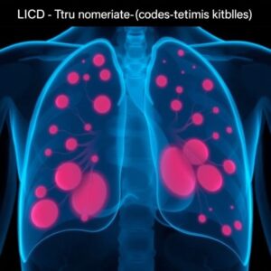
Chapter 5: Beyond the Solitary Nodule – Coding for Multiple Nodules and Specific Diagnoses
This is where coding becomes more complex and documentation becomes king.
The Challenge of Multiple Pulmonary Nodules
As indicated in the Index, multiple lung nodules are not coded with R91.1. The Index directs you to:
Nodule, lung > multiple > J98.4
Let’s verify in the Tabular List:
Chapter 10: Diseases of the respiratory system (J00-J99)
> J98 Other respiratory disorders
>> J98.4 Other disorders of lung
This is a very nonspecific code. Its parent code, J98, includes a variety of conditions like pulmonary collapse, disorders of the lung not elsewhere classified, and compensatory emphysema. J98.4 is essentially a catch-all for a lung condition that doesn’t fit a more specific category. While it can be used for multiple nodules, the coder must be cautious. If the physician provides any more specific context for the multiple nodules (e.g., “likely metastatic disease,” “suspicious for septic emboli”), a more specific code may be required.
Coding for a Confirmed Diagnosis (e.g., Malignancy, Infection, Benign Neoplasm)
When a diagnosis is confirmed, either by biopsy or, in some cases, by definitive clinical and radiological criteria, you abandon the “finding” code (R91.1) and use a definitive diagnosis code.
Scenario 1: Confirmed Primary Lung Cancer
The biopsy of the solitary nodule returns as “adenocarcinoma.”
-
Coding: You would use a code from category C34.- (Malignant neoplasm of bronchus and lung). The 4th character specifies the exact location (e.g., C34.11 – Malignant neoplasm of upper lobe, right bronchus or lung). A code from Chapter 2 (Neoplasms) always takes precedence.
Scenario 2: Nodule is a Metastasis from Colon Cancer
The patient has a history of colon cancer, and the lung nodule is biopsied, confirming it is metastatic adenocarcinoma consistent with a colorectal primary.
-
Coding: You would use a code for the secondary cancer in the lung: C78.01 (Secondary malignant neoplasm of right lung) or C78.02 (Secondary malignant neoplasm of left lung). The primary colon cancer (C18.9) would also be coded.
Scenario 3: Nodule is a Benign Hamartoma
A biopsy confirms the nodule is a benign hamartoma.
-
Coding: You would use a code from Chapter 2 for a benign neoplasm of the lung. The index path is: Neoplasm, neoplastic > lung > benign > D14.3. Verified in the Tabular List as D14.3 Benign neoplasm of bronchus and lung.
Scenario 4: Nodule is a Healed Granuloma from Histoplasmosis
The nodule is calcified and stable over two years, and the physician confidently documents it as a “healed granuloma due to remote histoplasmosis.”
-
Coding: You would code the underlying infection, B39.9 (Histoplasmosis, unspecified), as the physician has linked the nodule to a definitive, past disease process.
ICD-10-CM Coding Scenarios for Lung Nodules
| Clinical Scenario | Physician Documentation Key Phrases | Appropriate ICD-10-CM Code(s) | Rationale |
|---|---|---|---|
| Incidental Finding | “Incidental solitary pulmonary nodule, recommend follow-up CT.” | R91.1 | The nodule is an unconfirmed abnormal finding. |
| Multiple Nodules, Unspecified | “CT chest shows multiple subcentimeter pulmonary nodules, etiology unknown.” | J98.4 | Code for multiple nodules without a confirmed diagnosis. |
| Confirmed Primary Cancer | “Biopsy of RUL nodule: adenocarcinoma.” | C34.11 (Malignant neoplasm of upper lobe, right bronchus or lung) | Confirmed diagnosis from Chapter 2 (Neoplasms). |
| Metastatic Cancer to Lung | “History of breast cancer. Lung nodule biopsy consistent with metastatic breast carcinoma.” | C78.02 (Secondary malignant neoplasm of left lung) & C50.919 (Malignant neoplasm of unspecified site of female breast) | Code the secondary site and the primary cancer. |
| Benign Neoplasm | “Resected nodule confirmed as pulmonary hamartoma.” | D14.3 (Benign neoplasm of bronchus and lung) | Confirmed benign neoplasm from Chapter 2. |
| Infectious Cause | “Stable, calcified nodule consistent with prior histoplasmosis.” | B39.9 (Histoplasmosis, unspecified) | The nodule is a sequela of a confirmed infectious disease. |
Chapter 6: The Crucial Role of Documentation in Accurate Coding
The coder is entirely dependent on the quality of the physician’s documentation. Ambiguous documentation leads to ambiguous coding, which increases audit risk and potential denials.
What Coders Need from the Physician’s Note
For a lung nodule, the ideal documentation includes:
-
Number: Solitary or multiple.
-
Specificity: Is it a “nodule,” a “mass” (>3cm), or a “lesion”?
-
Laterality and Location: Right vs. left, and the specific lobe if known.
-
Clinical Context: Is it “incidental,” “suspicious for malignancy,” “stable from prior,” or “new”?
-
Final Diagnosis/Impression: The physician’s synthesis of the finding. (e.g., “Incidental solitary pulmonary nodule, likely benign, follow up in 12 months” vs. “Spiculated lung mass, highly suspicious for primary lung cancer.”).
The Radiologist’s Report: A Coder’s Best Friend
The radiology report is often the most detailed document. Coders should review both the “Findings” and the “Impression/Conclusion” sections. The “Impression” is the radiologist’s final summary and is the most reliable source for code assignment.
Querying the Physician: When and How
If the documentation is conflicting or unclear (e.g., the radiology report says “multiple nodules” but the pulmonologist’s note only mentions “a nodule”), the coder must initiate a physician query. A query is a formal, non-leading communication to clarify the record. For example: “Per the radiology report, there are multiple pulmonary nodules. Can you please clarify if the patient has a solitary nodule or multiple nodules for accurate coding?”
Chapter 7: Sequencing and Combination Coding – Putting It All Together
Often, a patient has more than one diagnosis. The order in which you list the codes (sequencing) is governed by official guidelines.
Principal Diagnosis vs. Secondary Diagnoses
The principal diagnosis is the condition established after study to be chiefly responsible for the patient’s admission/encounter. Secondary diagnoses are all coexisting conditions that affect the current care.
Case Study 1: Inpatient Admission
A patient is admitted to the hospital for worsening shortness of breath. Workup reveals bilateral pneumonia and also discovers an incidental solitary pulmonary nodule.
-
Principal Diagnosis: J18.9 (Pneumonia, unspecified organism) – This is the reason for the admission.
-
Secondary Diagnosis: R91.1 (Solitary pulmonary nodule) – This is an incidental finding that is being monitored but is not the reason for hospitalization.
Case Study 2: Outpatient Encounter
A patient sees a pulmonologist for the specific purpose of evaluating a solitary pulmonary nodule found on a recent CT. The pulmonologist reviews the images, discusses the findings, and orders a PET scan.
-
First-listed Diagnosis: R91.1 (Solitary pulmonary nodule) – This is the reason for the encounter.
Chapter 8: Common Pitfalls, Audit Risks, and How to Avoid Them
-
Pitfall: Using R91.1 for a confirmed diagnosis. This is the most significant error. Using an “abnormal finding” code when a definitive diagnosis exists (e.g., cancer) is incorrect and will be denied by payers.
-
Solution: Scrutinize the documentation for pathological results or definitive clinical statements.
-
-
Pitfall: Using R91.1 for multiple nodules.
-
Solution: Carefully review the radiology report. If “multiple” is stated, use J98.4 or a more specific code if the etiology is known.
-
-
Pitfall: Confusing “Suspected” vs. “Confirmed.” If a physician documents “suspicious for lung cancer,” you cannot code it as cancer. You code the reason for the study (e.g., the nodule, R91.1) and the signs/symptoms. The “rule-out” diagnosis is not coded in the outpatient setting.
-
Solution: Understand the ICD-10 guidelines for coding uncertain diagnoses in the inpatient vs. outpatient settings.
-
Chapter 9: The Future of Coding – ICD-11 and Beyond
The World Health Organization (WHO) has already released ICD-11, which offers a more modernized structure.
A Glimpse into ICD-11’s Structure for Respiratory Diseases
In ICD-11, a solitary pulmonary nodule would be found under:
CB41.0 Solitary pulmonary nodule
It remains under the chapter for “Symptoms, signs, or clinical findings, not elsewhere classified.” The structure is more logically aligned with current clinical practice and allows for easier electronic implementation.
The Impact of AI and Automated Coding Assistants
Artificial Intelligence (AI) and Natural Language Processing (NLP) are poised to revolutionize medical coding. These tools can read clinical documents and suggest codes, reducing manual effort. However, the human coder’s role will evolve to that of a auditor, validator, and clinical knowledge expert, ensuring the AI’s suggestions are contextually and clinically accurate. Understanding the nuances discussed in this article will remain a valuable skill.
Conclusion
Accurately coding a lung nodule in ICD-10-CM requires a careful, multi-step process: understanding the clinical picture, meticulously reviewing the physician’s documentation, navigating the ICD-10 manual from Index to Tabular List, and applying the correct code based on specificity and confirmation. The journey from a radiological finding (R91.1) to a definitive diagnosis code (e.g., C34.-, D14.3) is a critical pathway that ensures accurate reimbursement, valid health data, and, ultimately, high-quality patient care. Precision in coding is not an administrative task; it is a fundamental component of the modern healthcare ecosystem.
Frequently Asked Questions (FAQs)
Q1: What is the ICD-10 code for a lung nodule?
There isn’t one single code. The most common code is R91.1 for a solitary pulmonary nodule that is an unconfirmed finding. If there are multiple nodules, the code is typically J98.4. If the nodule is a confirmed cancer, benign tumor, or infection, you would use a different, more specific code from the relevant chapter.
Q2: Can I use R91.1 if the physician says the nodule is “suspicious for cancer”?
Yes, in most cases. “Suspicious for” is not a confirmed diagnosis. Until a biopsy or other definitive proof confirms malignancy, the condition is still classified as an abnormal finding, and R91.1 is appropriate. The reason for the encounter (e.g., evaluation of the nodule) is what you are coding.
Q3: What is the code for a lung mass (larger than 3 cm)?
A lung mass is coded differently. You would start in the Index under Mass, lung, which directs you to the Neoplasm Table. Unless a biopsy confirms it as benign, an unconfirmed lung mass is generally coded as a neoplasm of uncertain behavior (D49.1) or, more commonly in clinical practice, as a suspected malignancy, which is coded using the signs/symptoms that prompted the study. If it is confirmed as cancer, you would use the appropriate C34.- code.
Q4: The patient has lung cancer, and a new, separate nodule is found. What do I code?
You would code the confirmed lung cancer (C34.-). The new nodule could represent a metastasis, a new primary, or a benign process. Unless it is biopsied and confirmed, it is often considered part of the known cancer disease process. If the physician explicitly documents it as a “new primary,” then both cancers would be coded. Specific documentation is key.
Q5: Where can I find the official coding guidelines?
The official ICD-10-CM Official Guidelines for Coding and Reporting are published by the Centers for Disease Control and Prevention (CDC) and the Centers for Medicare & Medicaid Services (CMS). They are updated annually and are available for free on the CMS website.
Additional Resources
-
CDC ICD-10-CM Official Guidelines: https://www.cdc.gov/nchs/icd/icd-10-cm.htm (The primary source for all coding rules).
-
American Health Information Management Association (AHIMA): https://www.ahima.org (Provides education, certifications, and resources for medical coders).
-
American Academy of Professional Coders (AAPC): https://www.aapc.com (Another leading organization for coder education and certification).
-
Fleischner Society Guidelines: Publications Page (International, multidisciplinary guidelines for the management of incidental pulmonary nodules, essential for understanding clinical decision-making).
-
National Cancer Institute (NCI) – SEER Program: ICD-10-CM Neoplasm Coding Guidelines (Specific guidance on coding malignancies).
Date: October 11, 2025
Author: The Health Informatics Team
Disclaimer: The information contained in this article is for educational and informational purposes only and is not intended as a substitute for professional medical advice, diagnosis, or treatment. Always seek the advice of your physician or other qualified health provider with any questions you may have regarding a medical condition or treatment, and before undertaking a new health care regimen. Medical coding information should be verified with the most current official code sets and payer-specific guidelines.