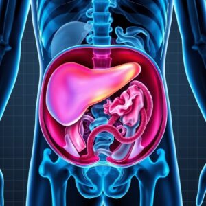
The human abdomen is a complex and vital region, a crowded metropolis of organs each performing functions essential to life. From the liver’s sophisticated detoxification plant to the pancreas’s precise endocrine and exocrine functions, from the kidneys’ intricate filtration systems to the labyrinthine networks of blood vessels, this space is both dynamic and delicate. For centuries, physicians relied on their hands, their ears, and their intuition to diagnose ailments hidden within this cavity. While valuable, these methods were often like trying to understand the plot of a movie by only listening to its soundtrack—you get the general idea but miss the crucial visual details.
The advent of medical imaging changed everything. It was like turning on the lights in a dark room. Among the most powerful of these illuminating technologies is Magnetic Resonance Imaging (MRI). Specifically, when a detailed, non-invasive, and radiation-free view of the soft tissues of the abdomen is required, an MRI with and without contrast, governed by the CPT code 74183, becomes an indispensable tool for modern medicine. This procedure provides a clarity and tissue characterization that other modalities often cannot match, allowing physicians to detect, diagnose, and monitor a vast array of conditions with unprecedented precision. This article serves as your ultimate guide to understanding this critical procedure—from the fundamental physics of the magnet to the complex nuances of medical billing, from the patient’s perspective on the table to the radiologist’s expert eye on the screen.
CPT Code 74183
2. Unpacking the CPT Code: What Does 74183 Really Mean?
In the world of U.S. healthcare, every medical procedure and service is assigned a specific code to standardize communication between healthcare providers, insurance companies, and regulators. These Current Procedural Terminology (CPT) codes are maintained by the American Medical Association (AMA). CPT code 74183 is precisely defined as:
“Magnetic resonance (e.g., proton) imaging, abdomen; without contrast material(s) followed by with contrast material(s) and further sequences.”
This definition is deceptively simple. Let’s break down its critical components:
-
“Magnetic resonance imaging, abdomen”: This specifies the modality (MRI) and the anatomical region (abdomen). The abdomen, for coding purposes, typically includes the liver, spleen, pancreas, kidneys, adrenals, and upper abdominal vasculature like the aorta and inferior vena cava. It generally does not include the pelvis (which has its own codes, 72195-72197).
-
“Without contrast material(s) followed by with contrast material(s)”: This is the core of the code. It mandates that the study must be performed in two distinct phases:
-
Non-Contrast Phase (Without): A complete set of images is acquired before any contrast agent is injected into the patient’s bloodstream. These “pre-contrast” images provide a baseline. They are excellent for identifying certain types of tissue characteristics, such as fat or blood products (e.g., in a hemorrhage), and for establishing the native appearance of structures before the contrast agent alters their appearance.
-
Contrast-Enhanced Phase (With): After the initial sequences, a gadolinium-based contrast agent (GBCA) is injected intravenously. The MRI scanner then acquires multiple additional sets of images at timed intervals—often referred to as dynamic phases. This allows radiologists to see how the contrast perfuses through the organs and any potential lesions over time.
-
-
“and further sequences”: This phrase is crucial. It implies that the post-contrast imaging is not just a single set of pictures. It involves multiple sequences performed after the contrast injection, capturing different phases of contrast enhancement (e.g., arterial, venous, delayed). This multi-phasic dynamic imaging is a key part of the value of 74183.
It is vital to distinguish 74183 from other related codes:
-
CPT 74181: MRI abdomen without contrast. This would be ordered if only a non-contrast study is deemed sufficient, such as for evaluating renal cysts or following known lesions that do not require contrast enhancement characterization.
-
CPT 74182: MRI abdomen with contrast. This is a rarely used code. It describes a study that is performed only with contrast. This would be inappropriate if any pre-contrast images were taken, which is almost always the case. If a study starts without contrast and then proceeds with contrast, it automatically becomes a 74183.
CPT Codes for Abdominal MRI
| CPT Code | Procedure Description | Common Use Cases |
|---|---|---|
| 74181 | MRI abdomen without contrast | Evaluating simple cysts, MRI urography without contrast, contraindication to contrast. |
| 74182 | MRI abdomen with contrast | Extremely rare. Only if no pre-contrast images are acquired. |
| 74183 | MRI abdomen without contrast followed by with contrast | Characterizing liver lesions, pancreatic mass evaluation, oncology staging, inflammatory conditions, vascular assessment. |
3. The Clinical Powerhouse: Indications for an MRI Abdomen w/wo Contrast
The dual-phase nature of 74183 makes it the gold standard for a wide range of clinical scenarios. Its ability to characterize tissue and visualize vascularity is unmatched.
A. Oncology: The Battle Against Cancer
-
Liver Lesion Characterization: This is one of the most common indications. When a potential mass is found on an ultrasound or CT scan, MRI w/wo contrast is the definitive test to determine if it is benign (e.g., a hemangioma or focal nodular hyperplasia) or malignant (e.g., hepatocellular carcinoma or metastasis). The dynamic contrast phases show how a lesion “enhances,” which is its fingerprint.
-
Staging Malignancies: For known cancers like liver, pancreatic, or renal cancer, 74183 is used to determine the tumor’s size, exact location, involvement of nearby vessels, and the presence of metastatic spread to other abdominal organs or lymph nodes. This information is critical for planning surgery, radiation, or other therapies.
-
Treatment Response Monitoring: During and after chemotherapy or other treatments, serial MRI scans are used to see if tumors are shrinking, staying stable, or growing.
B. Hepatobiliary and Pancreatic Disorders
-
Cirrhosis and Hepatocellular Carcinoma (HCC) Surveillance: In patients with chronic liver disease and cirrhosis, the risk of developing HCC is significantly elevated. MRI is a highly sensitive tool for screening these high-risk patients, often detecting small, early-stage cancers that are treatable.
-
Pancreatitis: MRI can excellently evaluate complications of pancreatitis, such as necrotizing pancreatitis, pseudocyst formation, and disconnected duct syndrome.
-
Pancreatic Cystic Lesions: MRI is superb at characterizing pancreatic cysts (e.g., IPMN, serous cystadenoma) and assessing for worrisome features that might indicate a pre-cancerous or cancerous change.
-
Cholangiocarcinoma: MRI with MR Cholangiopancreatography (MRCP) sequences is the best non-invasive test for diagnosing cancer of the bile ducts.
C. Renal and Adrenal Evaluation
-
Complex Renal Cysts: The Bosniak classification system for renal cysts relies heavily on contrast enhancement to categorize cysts and determine their risk of malignancy.
-
Renal Mass Characterization: Similar to liver masses, MRI can differentiate between benign renal oncocytomas and malignant renal cell carcinomas.
-
Adrenal Adenomas: MRI can use chemical shift imaging (a non-contrast technique) and contrast enhancement to identify benign adrenal adenomas, often avoiding the need for a biopsy.
D. Vascular Assessment
-
Aortic Pathology: MRI can accurately depict abdominal aortic aneurysms, aortic dissections, and signs of vasculitis without using ionizing radiation.
-
Mesenteric Ischemia: It can assess the patency of the celiac artery and superior mesenteric artery, looking for stenoses or occlusions that could cause intestinal ischemia.
-
Portal Venous System: It is excellent for evaluating portal vein thrombosis, cavernous transformation, and other portosystemic collateral vessels in patients with portal hypertension.
E. Inflammatory and Infectious Diseases
-
Inflammatory Bowel Disease (IBD): MRI enterography is a specialized form of abdominal MRI that is excellent for assessing the small bowel wall for inflammation, strictures, fistulas, and abscesses in patients with Crohn’s disease or ulcerative colitis.
-
Abscess Detection: MRI is highly sensitive for detecting abdominal and pelvic abscesses.
4. A Journey Through the Magnet: The Patient Experience from Start to Finish
Understanding what to expect can significantly reduce a patient’s anxiety.
A. Pre-Procedure Preparation
-
Scheduling and Instructions: The patient’s doctor’s office will schedule the exam, often at a hospital or an outpatient imaging center. The patient will receive specific instructions, which almost always include fasting for 4 to 6 hours prior to the exam. This reduces bowel motion and minimizes the risk of nausea or vomiting if contrast is used. Patients are usually encouraged to take their regular medications with small sips of water.
-
Screening Questionnaire: This is a critical safety step. The patient will fill out a detailed form asking about:
-
Metallic Implants: Pacemakers, implantable cardioverter-defibrillators (ICDs), aneurysm clips, cochlear implants, neurostimulators, and metal fragments in the eyes are often absolute or relative contraindications to MRI.
-
Allergies: Especially any history of allergy to gadolinium or other contrast agents.
-
Renal Function: History of kidney disease? A recent blood test (e.g., serum creatinine level) may be required to ensure the kidneys can safely filter out the gadolinium contrast.
-
Pregnancy: While MRI is generally preferred over CT in pregnancy if absolutely necessary, gadolinium contrast is typically avoided unless the diagnostic information is critical and cannot be obtained otherwise.
-
B. Day of the Exam: Arrival and Check-In
The patient will arrive, complete paperwork, and confirm their screening information with a technologist. They will be asked to change into a hospital gown to avoid any metal zippers or snaps on clothing interfering with the scan. A nurse or technologist will place an intravenous (IV) line, usually in the arm, for the contrast injection later.
C. Inside the MRI Suite
The MRI scanner is a large, tubular machine. The technologist will help the patient onto the scanning table and provide earplugs or headphones to protect hearing from the loud, repetitive knocking sounds the magnet makes during sequencing. They will place a device called a “coil” over the abdomen to help capture the signals. The table will then slide into the center of the magnet tunnel.
Key to the experience:
-
The Need for Stillness: The most important instruction for the patient is to remain as still as possible during each sequence, which can last from a few seconds to several minutes. Even slight movement can blur the images.
-
Communication: The patient will have a call button to squeeze if they feel uncomfortable or claustrophobic. The technologist can see and hear them at all times and will provide updates and breathing instructions (e.g., “Take a breath in, hold your breath, now breathe normally”).
D. The Scanning Process
-
Non-Contrast Sequences: The exam begins with a series of sequences performed without contrast. The patient will hear various sounds as different types of images (T1-weighted, T2-weighted, etc.) are acquired.
-
Contrast Injection: The technologist will remotely inject the gadolinium contrast agent through the IV line. The patient may feel a cool sensation, a metallic taste in the mouth, or a fleeting feeling that they need to urinate. These are normal and subside quickly.
-
Post-Contrast Sequences: Immediately after the injection, the scanner will acquire another rapid series of sequences, often asking the patient to hold their breath. This captures the “arterial phase.” This is followed by subsequent sequences after short delays (e.g., “portal venous phase,” “delayed phase”) to see how the contrast washes through the organs.
E. After the Scan
Once all sequences are complete, the table slides out, the IV is removed, and the patient is free to go. If they were fasting, they can now eat and drink normally. The gadolinium is eliminated by the kidneys over the next 24 hours. The images are sent to a picture archiving and communication system (PACS) for a radiologist to interpret.
5. The Science Behind the Image: How MRI Creates Detailed Pictures of the Abdomen
Unlike CT scans which use ionizing radiation, or ultrasound which uses sound waves, MRI utilizes a powerful magnetic field and radio waves to create images. The human body is mostly water. Water molecules (H₂O) contain hydrogen protons, which act like tiny magnets with a spin.
-
Alignment: When a patient enters the MRI scanner, the powerful main magnetic field (often 1.5 Tesla or 3.0 Tesla—tens of thousands of times stronger than Earth’s magnetic field) causes the randomly spinning hydrogen protons in the body to align with the field.
-
Excitation: A precisely tuned radiofrequency (RF) pulse is then applied, which knocks these aligned protons out of alignment.
-
Relaxation: When the RF pulse is turned off, the protons gradually return to their aligned state, releasing the energy they absorbed. This energy is emitted as a signal.
-
Signal Detection: The MRI coils placed around the patient detect these emitted signals.
-
Image Formation: A computer processes the complex signal data, using mathematical formulas (like the Fourier transform) to reconstruct a detailed, cross-sectional image of the inside of the body.
The timing of the RF pulses (repetition time, TR) and the time at which the signal is read (echo time, TE) can be altered to create different types of images that highlight different tissue properties:
-
T1-Weighted Images: Excellent for showing normal anatomy and fat-containing structures. Gadolinium contrast appears bright on T1 images.
-
T2-Weighted Images: Excellent for visualizing fluid, such as that found in cysts, edema, inflammation, and tumors. Simple fluid is very bright on T2 images.
Advanced techniques like diffusion-weighted imaging (DWI) can detect the random motion of water molecules within tissue. Restricted diffusion (where water movement is impeded) is often a feature of highly cellular tumors, making DWI a powerful tool for cancer detection.
6. The Role of Contrast: Gadolinium and Why It’s Sometimes Essential
A non-contrast MRI provides superb anatomical detail, but it’s like a detailed map of a city without any traffic information. The addition of a contrast agent shows the “traffic patterns”—the blood flow and vascularity—which is often the key to diagnosis.
What is Gadolinium?
Gadolinium is a rare earth metal. In its pure form, it is toxic. For medical use, it is chelated—bound to organic molecules—to create a stable, safe compound known as a gadolinium-based contrast agent (GBCA). This chelation allows it to be administered intravenously and safely excreted by the kidneys.
How It Works
Gadolinium is paramagnetic, meaning it interacts with the magnetic field of the MRI scanner. When injected into a vein, it circulates through the bloodstream and into the extracellular spaces of tissues. It does not cross intact cell membranes easily. Its presence locally distorts the magnetic field, causing protons nearby to relax more quickly. This shortens the T1 relaxation time of nearby water protons, making the areas where the contrast has pooled appear brilliantly bright on T1-weighted images.
The Diagnostic Power of Dynamic Imaging
The true power lies in observing how the contrast flows over time:
-
Arterial Phase (20-35 seconds post-injection): Contrast fills the arteries. Hypervascular tumors (like HCC or renal cell carcinoma) will show intense enhancement in this phase.
-
Portal Venous Phase (60-70 seconds post-injection): Contrast perfuses through the liver and other organs. This phase is optimal for visualizing most liver metastases and assessing overall organ enhancement.
-
Delayed/Hepatobiliary Phase (3+ minutes post-injection, depending on agent): Some contrast agents are taken up by liver cells (hepatocytes) and excreted into the bile. In this phase, normal liver tissue remains bright, while lesions that lack normal hepatocytes (like metastases) appear dark, creating a stark contrast.
This pattern of enhancement—when it starts, how intense it is, and how quickly it fades (a phenomenon known as “washout”)—provides a specific signature that allows radiologists to differentiate between benign and malignant lesions with high accuracy.
7. Interpreting the Unseen: A Radiologist’s Perspective on Reading the Scan
The radiologist is a physician specialized in interpreting medical images. Their analysis is a meticulous, multi-step process.
-
Systematic Review: They review all sequences—non-contrast T1, T2, DWI, etc.—in a systematic way, examining each organ in the abdomen: liver, spleen, pancreas, kidneys, adrenals, bowel, vasculature, lymph nodes, and bones.
-
Lesion Detection: On the non-contrast images, they identify any abnormalities: a dark lesion on T1, a bright lesion on T2, a bright lesion on DWI.
-
Lesion Characterization: This is where the contrast-enhanced sequences are pivotal. The radiologist toggles back and forth between the pre- and post-contrast images on their high-resolution monitor. They ask:
-
Does this lesion enhance? If not, it’s likely a simple cyst.
-
How does it enhance? Does it enhance intensely in the arterial phase (suggesting hypervascularity)? Does it show “washout” in the delayed phase (a classic feature of HCC)? Does it show progressive, slow filling (a classic feature of a hemangioma)?
-
-
Synthesis and Diagnosis: The radiologist synthesizes all this information: the lesion’s appearance on every sequence, its size, location, and enhancement pattern. They compare it to any prior exams to check for interval change. They then generate a structured report that describes the findings and provides a differential diagnosis or a definitive diagnosis based on established imaging criteria.
-
Communication: The final report is sent to the referring physician, who uses this information, combined with the patient’s clinical picture, to determine the next steps in care.
8. Navigating the Financial Landscape: Billing, Coding, and Reimbursement for 74183
The correct application of CPT code 74183 is essential for healthcare providers to receive appropriate reimbursement from insurance payers.
A. Medical Necessity: The Cornerstone of Reimbursement
The single most important factor is medical necessity. The referring physician must provide a clinical indication that justifies why a dual-phase contrast-enhanced MRI is required instead of a simpler, less expensive test like an ultrasound or non-contrast MRI. The diagnosis code (ICD-10 code) on the order must align with accepted guidelines for the procedure. For example:
-
ICD-10 code
C22.0(Liver cell carcinoma) justifies 74183. -
ICD-10 code
R93.5(Abnormal findings on diagnostic imaging of other abdominal regions, including retroperitoneum) may be denied as too vague without supporting clinical notes.
B. Bundling and Technical vs. Professional Components
-
Global Fee: The billing for 74183 can be submitted as a “global” fee, which includes both the technical component (cost of operating the scanner, contrast agent, technologist’s time, equipment) and the professional component (the radiologist’s interpretation and report).
-
Split Billing: Sometimes these are billed separately using modifiers:
-
-TC (Technical Component): Used by the facility that owns the scanner if they are not employing the radiologist.
-
-26 (Professional Component): Used by the radiologist who interprets the scan if they are billing separately from the facility.
-
C. Audits and Compliance
Coding for advanced imaging is subject to strict audits. “Unbundling” (billing separate codes for services that are included in 74183) or misrepresenting the service performed can lead to severe penalties, fines, and allegations of fraud. Documentation in the radiology report must clearly state that images were acquired both before and after contrast administration to support the use of 74183.
9. Safety First: Contraindications, Risks, and the Gadolinium Debate
MRI is a very safe modality overall, but specific, important risks must be managed.
A. Absolute Contraindications
-
Non-MRI Compatible Implanted Devices: Certain pacemakers, ICDs, and other electronic devices can malfunction or cause injury in the magnetic field. Special “MRI-conditional” devices are now common, but each requires specific protocols to be followed.
-
Ferromagnetic Aneurysm Clips: Certain older types of clips used in brain aneurysm surgery can torque in the magnetic field, causing catastrophic hemorrhage.
B. Relative Contraindications
-
Claustrophobia: Can often be managed with coaching, a head-first position, or in some cases, mild sedation.
-
First Trimester of Pregnancy: While no adverse effects have been proven, MRI is generally avoided in the first trimester unless the benefits overwhelmingly outweigh the theoretical risks. Gadolinium is avoided throughout pregnancy.
C. Risks Associated with Gadolinium
-
Allergic-Like Reactions: These are rare (<0.5%) and usually mild (nausea, hives). Severe anaphylactoid reactions are extremely rare.
-
Nephrogenic Systemic Fibrosis (NSF): This is a serious, rare condition that causes fibrosis of the skin and internal organs. It occurred in a small number of patients with severe renal impairment (GFR < 30) who received certain types of less stable (linear) GBCAs. This risk has been drastically reduced by:
-
Screening patients for kidney disease.
-
Using more stable (macrocyclic) agents in at-risk patients.
-
Avoiding GBCA administration in patients with acute renal failure or severe chronic kidney disease unless absolutely necessary.
-
-
Gadolinium Deposition: Recent studies have shown that tiny traces of gadolinium can be retained in the brain and bones years after administration, even in patients with normal kidney function. To date, no negative health effects have been linked to this deposition. However, as a precaution, the FDA and radiology societies advocate for using the lowest necessary dose of the most stable agent and avoiding unnecessary exams.
10. The Future is Bright: Technological Advancements in Abdominal MRI
The field of MRI is rapidly evolving, promising even greater speed, clarity, and functionality.
-
Artificial Intelligence (AI): AI algorithms are being developed to automate image acquisition, reduce image noise, and even assist radiologists by highlighting potential lesions or providing quantitative measurements of tumor response.
-
Higher Field Strengths: 7T (7 Tesla) scanners are moving into clinical research, offering potentially revolutionary signal-to-noise ratios and spatial resolution.
-
Compressed Sensing: This novel acquisition technique allows scanners to acquire data much faster, significantly reducing scan times and minimizing motion artifacts. This is particularly beneficial for patients who have difficulty holding their breath.
-
Hyperpolarized MRI: An emerging technique where contrast agents are “hyperpolarized” to dramatically increase their signal, allowing for real-time imaging of metabolic processes within the body.
11. Conclusion: The Indispensable Role of Advanced Imaging
CPT code 74183 represents far more than a billing number; it encapsulates a sophisticated, non-invasive diagnostic powerhouse that has revolutionized abdominal medicine. By providing exquisitely detailed, multi-phasic views of soft tissue and vascularity without ionizing radiation, it serves as an indispensable tool for detecting, characterizing, and monitoring a vast spectrum of diseases, from cancer to inflammation. As technology continues to advance with AI, faster sequences, and novel contrast mechanisms, the role of MRI abdomen with and without contrast will only grow, ensuring its place at the forefront of precision medicine and improved patient outcomes for years to come.
12. Frequently Asked Questions (FAQs)
Q1: How long does an MRI abdomen with contrast actually take?
A: While the actual time inside the scanner is typically between 30 to 45 minutes, you should plan for a total appointment time of 60 to 90 minutes. This allows for check-in, changing, screening, IV placement, the scan itself, and IV removal.
Q2: I have kidney problems. Can I still get gadolinium?
A: It depends on the severity. Your doctor will order a blood test to check your kidney function (eGFR). If you have severe kidney disease, the risks and benefits will be carefully weighed. If the MRI is essential, the radiologist will choose the safest type of gadolinium (a macrocyclic agent) and use the lowest possible dose. In some cases, the study may be performed without contrast.
Q3: Why do I have to hold my breath so many times during the scan?
A: Even small movements from breathing can blur the images of the abdomen, much like a camera taking a picture of a moving subject. By holding your breath for short periods (usually 10-20 seconds at a time), you “freeze” the motion of your diaphragm and liver, allowing the scanner to capture a crisp, clear image. The technologist will guide you precisely on when to hold and when to breathe.
Q4: What’s the difference between an MRI with contrast and a CT scan with contrast?
A: The key differences are:
-
Radiation: MRI uses magnetic fields and has no ionizing radiation. CT scans use X-rays and do involve radiation exposure.
-
Contrast Agent: MRI uses gadolinium. CT uses iodine-based contrast.
-
Tissue Detail: MRI generally provides superior soft tissue contrast, making it better for distinguishing between similar soft tissues (e.g., different types of liver lesions). CT is often faster and better for evaluating bone detail, calcifications, and lung tissue.
Your doctor chooses the best test based on the specific clinical question.
Q5: I’m claustrophobic. What are my options?
A: Inform your doctor and the imaging center beforehand. Options include:
-
Open MRI: These scanners are open on the sides but are generally lower strength, which can sometimes mean lower image quality for abdominal studies.
-
Wide-Bore MRI: Many modern “closed” scanners are shorter and have a wider opening (70 cm), which feels much less confining.
-
Prone Positioning: For some abdominal scans, you can lie on your stomach, allowing you to see out of the scanner.
-
Sedation: In some cases, your doctor can prescribe a mild oral sedative to help you relax during the procedure.
13. Additional Resources
-
RadiologyInfo.org: (A public website co-sponsored by the American College of Radiology (ACR) and the Radiological Society of North America (RSNA)) provides detailed, patient-friendly information on MRI procedures.
-
American College of Radiology (ACR) Appropriateness Criteria: Evidence-based guidelines to help doctors choose the most appropriate imaging exam for various clinical conditions.
-
American Medical Association (AMA) CPT Network: The official source for information on CPT codes (requires subscription).
-
FDA Safety Communication on Gadolinium-Based Contrast Agents: Provides the latest regulatory information and safety alerts.
Disclaimer
The information contained in this article is intended for educational and informational purposes only. It is not a substitute for professional medical advice, diagnosis, or treatment. Always seek the advice of your physician or other qualified health provider with any questions you may have regarding a medical condition, procedure, or billing code. The content regarding CPT codes is based on the American Medical Association’s proprietary codes and is subject to change. Always consult the most current, official AMA CPT code books for accurate billing and coding guidance. The author and publisher are not responsible for any errors or omissions or for any outcomes related to the use of this information.


