The human pelvis is a nexus of vital structures—a intricate basket of bones housing the reproductive organs, bladder, rectum, major blood vessels, and a complex network of muscles, nerves, and ligaments. When something goes wrong in this densely packed region, pain and dysfunction can be debilitating, yet the cause is often hidden from view, a mystery waiting to be solved. For decades, physicians relied on physical exams, basic X-rays, and invasive procedures to diagnose pelvic conditions. The advent of Magnetic Resonance Imaging (MRI) revolutionized this diagnostic process, offering an unprecedented, non-invasive window into the soft tissues of the body with exquisite detail.
However, the power of this technology is coupled with complexity, not just in its clinical application but also in its administration and billing. At the heart of this complexity for many patients, healthcare providers, and medical coders is a five-digit code: CPT 72198. This code, which signifies “Magnetic resonance (eg, proton) imaging, pelvis; with contrast material(s) and without contrast material(s),” is more than just a billing tool. It is a precise language that communicates a specific, sophisticated medical procedure to insurance companies, ensuring that providers are reimbursed appropriately and that patients understand the care they are receiving.
This article serves as the ultimate guide to CPT Code for MRI Pelvis with and without Contrast. We will embark on a detailed journey from the basic physics of MRI to the nuanced rules of medical coding. We will explore the anatomy of the pelvis, the reasons a doctor might order this advanced scan, and what a patient can expect from the moment the test is ordered to the moment they receive their results. Furthermore, we will demystify the financial and insurance landscape, providing clarity on costs and coverage. Whether you are a patient seeking to understand your upcoming procedure, a medical student, a healthcare administrator, or a coding professional, this comprehensive resource is designed to provide exclusive, in-depth knowledge, empowering you to navigate the world of pelvic MRI with confidence.
CPT Code for MRI Pelvis with and without Contrast
2. Understanding the Fundamentals: What is an MRI?
The Science Behind Magnetic Resonance Imaging
Unlike X-rays or CT scans which use ionizing radiation, MRI employs a powerful combination of magnetic fields and radio waves to create detailed images of the inside of the body. The core of an MRI scanner is a massive superconducting magnet, which creates an extremely strong, stable magnetic field. This field, typically measured in Tesla (T)—most clinical scanners are 1.5T or 3.0T—causes the protons in the hydrogen atoms within your body’s water and fat molecules to align with it.
When a specific radiofrequency pulse is applied, these protons are temporarily knocked out of alignment. The moment the pulse is turned off, the protons realign with the magnetic field, releasing energy in the form of a radio signal. It is this emitted signal that the scanner’s receivers detect. Different tissues (e.g., muscle, fat, fluid, tumor) have different densities of hydrogen atoms and varying relaxation times, meaning they emit uniquely identifiable signals. A sophisticated computer processes these myriad signals and translates them into exquisitely detailed cross-sectional images (slices) of the body in any plane: axial, sagittal, or coronal.
The primary advantage of MRI is its superior soft-tissue contrast. It can distinguish between subtle differences in tissues that appear identical on other imaging modalities, making it indispensable for evaluating muscles, ligaments, tendons, the brain, and organs.
Why Use Contrast? The Difference Between “With” and “Without”
A non-contrast MRI provides a superb anatomical map. However, the addition of a contrast agent can transform this static map into a dynamic functional one, revealing physiological information about blood flow, inflammation, and tissue viability.
The most common contrast agents used in MRI are gadolinium-based. Gadolinium is a rare earth metal with paramagnetic properties that, when safely chelated (bound to another molecule), shortens the relaxation time of protons nearby. This alteration in the magnetic properties causes the tissues that absorb the contrast agent to appear much brighter on T1-weighted images.
The protocol “with and without contrast” is deliberate and strategic:
-
“Without contrast” (non-contrast): Establishes a baseline anatomy. It excels at visualizing anatomy, detecting fat-containing lesions, fluid, hemorrhage, and bone marrow abnormalities.
-
“With contrast” (post-contrast): Administered intravenously during the scan, the contrast agent circulates and perfuses into tissues. Hypervascular tissues—those with rich blood supply, such as tumors, infections, and areas of active inflammation—will absorb more contrast and “enhance” or light up brightly. This allows radiologists to:
-
Detect lesions that are invisible on non-contrast images.
-
Characterize a known lesion (e.g., differentiating a benign cyst from a solid malignant tumor).
-
Define the extent of a disease process, such as mapping the precise borders of a cancer for surgical planning.
-
Monitor treatment (e.g., checking if a tumor is shrinking after chemotherapy by assessing its change in enhancement pattern).
-
The combination of both sets of images provides a comprehensive picture that is greater than the sum of its parts, offering both exquisite anatomical detail and crucial physiological data.
3. A Deep Dive into the Pelvis: Anatomy and Clinical Necessity
The Complex Landscape of the Male and Female Pelvis
The pelvis is a biomechanical and functional marvel. Its bony structure, composed of the sacrum, coccyx, and the two innominate bones (each made of the ilium, ischium, and pubis), provides attachment points for powerful muscles and protects the delicate internal organs.
-
In Females: The pelvis contains the uterus, cervix, ovaries, fallopian tubes, vagina, bladder, rectum, and urethra. It also houses a rich network of vasculature, nerves, and supporting ligaments like the broad and round ligaments of the uterus.
-
In Males: The pelvic cavity holds the bladder, rectum, prostate gland, seminal vesicles, and parts of the vas deferens. The prostate, in particular, is a common focus of pelvic MRI.
Surrounding these organs are muscles critical for stability and function, including the levator ani group that forms the pelvic floor, the obturator internus, the piriformis, and the large gluteal muscles.
Common Clinical Indications for a Pelvic MRI
A physician orders an MRI pelvis with and without contrast to answer specific, complex clinical questions that other tests cannot. Indications include:
-
Oncologic Staging and Surveillance: This is one of the most common reasons. MRI is the gold standard for local staging of cancers like:
-
Prostate Cancer: Determining if cancer is confined to the prostate gland or has spread beyond its capsule (extracapsular extension) or to the seminal vesicles.
-
Rectal Cancer: Precisely assessing the depth of tumor invasion, involvement of the mesorectal fascia (a key surgical boundary), and lymph node status.
-
Cervical, Endometrial, and Ovarian Cancers: Evaluating tumor size, location, invasion into adjacent structures (bladder, rectum), and identifying metastatic lymph nodes.
-
Bladder Cancer: Staging muscle-invasive disease.
-
-
Evaluation of Inflammatory and Infectious Processes:
-
Identifying abscesses (e.g., perirectal, tubo-ovarian).
-
Diagnosing osteomyelitis (bone infection) of the sacrum or iliac bones.
-
Evaluating inflammatory bowel disease (e.g., Crohn’s disease) for fistulas, sinus tracts, and abscesses.
-
-
Musculoskeletal and Pain Evaluation:
-
Assessing unexplained hip or pelvic pain.
-
Diagnosing occult (hidden) hip fractures not seen on X-ray.
-
Evaluating soft-tissue masses, tumors, or avascular necrosis of the femoral head.
-
Investigating endometriosis, a condition where uterine-like tissue grows outside the uterus, often requiring detailed MRI for surgical planning.
-
-
Congenital Anomalies: Evaluating developmental issues of the reproductive organs.
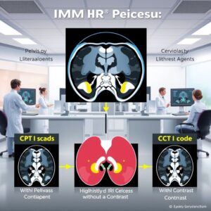
4. The Cornerstone of Billing: CPT Code 72198 Decoded
Code 72198: MRI Pelvis with and without Contrast
The American Medical Association’s (AMA) Current Procedural Terminology (CPT) code set is the universal language for describing medical, surgical, and diagnostic services. For pelvic MRI, there are three primary codes:
-
CPT 72195: Magnetic resonance (eg, proton) imaging, pelvis; without contrast material(s)
-
CPT 72196: Magnetic resonance (eg, proton) imaging, pelvis; with contrast material(s)
-
CPT 72198: Magnetic resonance (eg, proton) imaging, pelvis; with contrast material(s) and without contrast material(s)
Code 72198 is used only when both non-contrast and post-contrast imaging sequences are performed during the same session. It is a comprehensive code that encompasses the entire study. It is not appropriate to bill both 72195 and 72196 together for a single exam; doing so would be considered “unbundling” and is incorrect, leading to claim denials and potential audit flags.
Unbundling: The Critical Importance of Code Sequencing
The correct application of these codes is non-negotiable. If a provider performs a non-contrast MRI of the pelvis and, based on initial findings, decides to administer contrast for further evaluation, they must void the initial code (72195) and bill only the complete code, 72198. The rules are clear: the complete service supersedes the component services.
Modifiers: Adding Context to the Claim
Modifiers are two-character codes appended to a CPT code to provide additional information about the service without changing the code’s definition. Common modifiers used with 72198 include:
-
-26 (Professional Component): Used by the radiologist who interprets the images and writes the report, but does not own the equipment.
-
-TC (Technical Component): Used by the facility (hospital or imaging center) that owns the equipment, covers the technologist’s time, and supplies the contrast material.
-
-LT and -RT (Left Side, Right Side): Not typically used for a pelvis MRI as it is considered a single anatomic structure. However, if the study is focused on a specific joint like the hip, these may apply to different codes (e.g., hip MRI codes 73721-73723).
5. The Patient’s Journey: From Order to Results
Step 1: The Referral and Prior Authorization
The journey begins with a referral from a primary care physician, oncologist, urologist, gynecologist, or surgeon. The referring provider’s office then typically handles prior authorization (or pre-certification). This involves submitting clinical documentation (patient history, physical exam findings, results of prior tests) to the patient’s insurance company to prove the medical necessity of the scan. Without this authorization, the insurance company may deny the claim, leaving the patient with a significant financial responsibility.
Step 2: Preparation – What to Expect and How to Get Ready
Once authorized, the imaging facility contacts the patient to schedule the exam and provide preparation instructions, which are crucial for image quality and safety.
-
Safety Screening: The most critical step. The powerful magnet can turn metal objects into dangerous projectiles and interfere with implanted devices. Patients will be thoroughly screened for:
-
Absolute Contraindications: Certain aneurysm clips, cardiac pacemakers, implantable cardioverter-defibrillators (ICDs), cochlear implants, and metallic foreign bodies in the eye.
-
Relative Contraindications: Some older prosthetic heart valves, stents, joint replacements, and infusion pumps. These often require documentation of model and MR-compatibility.
-
-
Pregnancy and Breastfeeding: While gadolinium is generally avoided in pregnancy unless absolutely necessary, the MRI itself (without contrast) is considered safer than X-rays or CT. Gadolinium agents do pass into breast milk in tiny amounts; guidelines generally consider it safe to continue breastfeeding after contrast administration, though some mothers may choose to “pump and dump” for 12-24 hours as a precaution.
-
Kidney Function: For patients with known severe kidney disease (eGFR < 30 mL/min/1.73m²), gadolinium is often avoided due to a rare but serious risk called Nephrogenic Systemic Fibrosis (NSF). A recent blood test to check creatinine levels may be required.
-
Claustrophobia: Patients who are anxious about enclosed spaces should inform their doctor. Options include a mild oral sedative (arranging a ride home is necessary) or seeking out an imaging center with an “open-bore” MRI machine, which is less confining.
-
Fasting: Instructions vary. Often, no fasting is required for a pelvic MRI, but some facilities may ask for a few hours of fasting if contrast is planned to minimize potential nausea.
Step 3: The Day of the Exam – A Step-by-Step Walkthrough
-
Arrival and Check-in: The patient will fill out detailed safety screening forms and provide insurance information.
-
Changing and IV Placement: The patient will change into a gown without metal fasteners. A radiology nurse or technologist will then insert an intravenous (IV) line, usually in the arm or hand, for the contrast injection later in the exam.
-
Entering the Scan Room: The patient lies down on the motorized scanner table. Earplugs or headphones are provided to protect hearing from the loud knocking and buzzing sounds (up to 110 decibels) the magnet generates during sequencing.
-
The Scan Begins (Non-Contast Phase): The table slides into the magnet bore. The technologist, who operates the scanner from an adjacent room, provides instructions through an intercom. The patient must remain as still as possible, as motion blurs the images. Breathing instructions may be given. This first part acquires the “without contrast” images.
-
Contast Injection: The table slides out. The technologist connects the IV line to a syringe containing the gadolinium contrast agent. Using an automated injector, the contrast is administered. The patient may feel a cool sensation, a metallic taste in the mouth, or a fleeting feeling of needing to urinate. These are normal and subside quickly.
-
The Scan Continues (Post-Contrast Phase): The table re-enters the magnet, and a second set of sequences is run to capture the “with contrast” images.
-
Completion: The IV is removed, and the patient is free to get changed and leave. If sedated, they must have a driver.
Step 4: After the Scan – Image Processing and Radiologist Interpretation
The raw data from the scanner is processed by powerful computers to create hundreds of images. A specialized physician, a radiologist with expertise in body imaging, meticulously analyzes these images. They correlate the pre- and post-contrast sequences, comparing findings to any prior studies and the patient’s clinical history. They synthesize this analysis into a formal written report, which includes:
-
Technique: A description of the procedure performed.
-
Findings: A detailed, structured description of the normal and abnormal observations in all visualized organs and structures.
-
Impression/Conclusion: A summary of the most significant findings and a differential diagnosis, answering the clinical question that prompted the referral.
This report is sent to the referring physician, who discusses the results with the patient and determines the next steps in their care plan.
6. Financial Considerations and Insurance Navigation
Understanding Cost Variables and Pricing
The cost of an MRI pelvis with and without contrast is highly variable, ranging from $1,200 to over $5,000+ in the United States. Factors influencing cost include:
-
Geographic Location: Costs are higher in major metropolitan areas.
-
Facility Type: Hospital-based outpatient departments are almost always more expensive than free-standing independent imaging centers.
-
Insurance Negotiated Rates: The price an insurance company has agreed to pay is typically much lower than the facility’s “list price” or chargemaster rate.
The Intricate Dance of Insurance Coverage
Coverage is dictated by the patient’s specific insurance plan and the proven medical necessity of the procedure. The insurance company uses the CPT code (72198) and a corresponding diagnosis code (ICD-10 code, e.g., C61 for prostate cancer) to make this determination. If the diagnosis code does not align with the plan’s coverage policies for this advanced imaging, the claim will be denied.
Patient Responsibility: Deductibles, Co-pays, and Co-insurance
Even with authorization, the patient is often responsible for a portion of the cost:
-
Deductible: The amount the patient must pay out-of-pocket before insurance starts to pay.
-
Co-pay: A fixed fee (e.g., $100) for a specialized service like MRI.
-
Co-insurance: A percentage of the allowed amount (e.g., 20% of the $1,500 negotiated rate = $300 patient responsibility).
It is imperative for patients to contact their insurance provider before the exam to understand their specific financial obligations.
Summary of Pelvic MRI CPT Codes and Applications
| CPT Code | Procedure Description | Clinical Use Case | Key Billing Rule |
|---|---|---|---|
| 72195 | MRI Pelvis without contrast | Initial evaluation of pain, muscle injury, avascular necrosis. Establishing baseline anatomy. | Never bill with 72196. Bill only if NO contrast was given. |
| 72196 | MRI Pelvis with contrast | Follow-up exam of a known condition where only enhanced images are needed (rare). | Never bill with 72195. Bill only if contrast was given but NO non-contrast images were acquired. |
| 72198 | MRI Pelvis with and without contrast | Comprehensive evaluation for cancer staging, infection, inflammation. The gold standard for most indications. |
7. Advanced Applications and Future Directions
Beyond Anatomy: Functional MRI (fMRI) and Diffusion-Weighted Imaging (DWI)
Modern MRI is moving beyond pure anatomy. Techniques like Diffusion-Weighted Imaging (DWI) measure the random movement of water molecules within tissues. In highly cellular environments like tumors, water movement is restricted, making these areas appear bright on DWI. This is incredibly valuable in oncology for detecting small metastatic lymph nodes and monitoring early treatment response.
Artificial Intelligence in Musculoskeletal and Oncologic Imaging
AI and machine learning are poised to revolutionize radiology. AI algorithms are already being used as “second readers,” helping radiologists by:
-
Prioritizing urgent cases in a worklist.
-
Automatically measuring tumors to track growth over time.
-
Detecting subtle fractures or lesions that might escape the human eye.
-
Predicting tumor genetics and treatment response based on imaging patterns (“radiomics”).
8. Conclusion: Synthesizing Knowledge for Better Healthcare Outcomes
CPT code 72198 represents far more than a billing entry; it encapsulates a sophisticated diagnostic journey crucial for managing serious pelvic conditions. Its correct application ensures the financial viability of healthcare providers and clear communication across the care continuum. For patients, understanding this process—from the clinical need and safety preparations to the financial implications—demystifies a complex procedure and empowers them to be active participants in their own healthcare. As technology advances with functional techniques and AI, the value and precision of the pelvic MRI with and without contrast will only continue to grow, solidifying its role as an indispensable tool in modern medicine.
9. Frequently Asked Questions (FAQs)
Q1: How long does an MRI pelvis with and without contrast actually take?
A: The entire process, from check-in to leaving, can take 60 to 90 minutes. The actual time inside the scanner is typically 45 to 60 minutes, split between the non-contrast and post-contrast sequences.
Q2: I have a tattoo. Can I still have an MRI?
A: Most modern tattoos with FDA-approved inks are safe. However, some older tattoos, particularly those containing metallic pigments, may cause a burning sensation or skin irritation during the scan due to interaction with the magnetic field. Always inform your technologist about any tattoos so they can monitor you and, if possible, place a cold compress over the area to minimize discomfort.
Q3: What are the side effects of gadolinium contrast?
A: The vast majority of people experience no or very mild side effects, such as a temporary cold feeling, headache, or nausea. Severe allergic reactions are rare (approximately 0.01-0.2% of cases) and are similar to other iodine-based contrasts but often less severe. Facilities are equipped to handle such reactions immediately.
Q4: Why was my prior authorization for the MRI denied?
A: Denials usually occur due to a lack of proven medical necessity based on the insurer’s specific clinical guidelines. This could be because the referring physician did not provide enough supporting documentation, tried to order the test too early in the diagnostic workup (e.g., before trying an X-ray or ultrasound), or used a diagnosis code the plan does not cover for this advanced imaging. Your doctor’s office can appeal the denial with more information.
Q5: Is there an alternative to a traditional “closed” MRI machine for claustrophobic patients?
A: Yes. Options include:
-
Open-Bore MRI: These machines have magnets on the top and bottom with open sides. They are much less confining but may have a lower magnetic field strength (e.g., 1.0T vs. 1.5T/3.0T), which can sometimes result in slightly lower image resolution.
-
Wide-Bore MRI: A newer design that offers a shorter and wider tunnel than traditional scanners, providing more headroom and reducing the enclosed feeling, often without sacrificing magnetic field strength.
-
Sedation: Discussing mild oral anti-anxiety medication with your referring doctor is a common and effective solution.
10. Additional Resources
-
American College of Radiology (ACR): The premier organization for radiologists. Their “ACR Appropriateness Criteria” are evidence-based guidelines used by doctors to choose the right imaging exam for hundreds of clinical conditions.
-
RadiologyInfo.org: A public-facing website co-sponsored by the ACR and the Radiological Society of North America (RSNA). It provides easy-to-understand descriptions of every imaging procedure, including videos on what to expect.
-
American Medical Association (AMA) CPT Network: The official source for information on CPT codes (requires subscription for full access).
-
National Institutes of Health (NIH) – MedlinePlus: A reliable source for general health information, including details on medical procedures and conditions.
11. Disclaimer
This article is intended for informational and educational purposes only. It is not a substitute for professional medical advice, diagnosis, or treatment. Always seek the advice of your physician or other qualified health provider with any questions you may have regarding a medical condition, treatment, or procedure. Never disregard professional medical advice or delay in seeking it because of something you have read in this article. The information on medical coding and billing is provided as a general guide and may not reflect the most current updates or specific payer policies, which change frequently. Always consult current, official CPT codebooks and payer-specific guidelines for accurate coding and billing practices. The author and publisher disclaim any liability, loss, or risk incurred as a consequence, directly or indirectly, of the use and application of any of the contents of this article.


