In the intricate world of modern medicine, the ability to visualize the body’s hidden highways—the vast and complex network of arteries and veins—is nothing short of revolutionary. Angiography, the imaging of blood vessels, stands as a cornerstone of diagnostic and interventional cardiology, radiology, and neurology. It is the technique that allows physicians to peer into the living vasculature, identifying stenoses, occlusions, aneurysms, and malformations with remarkable clarity. From diagnosing life-threatening pulmonary embolisms to guiding the delicate placement of a stent in a coronary artery, angiography saves lives and improves patient outcomes every single day.
However, for the medical coder, this vital clinical procedure presents a formidable challenge. Translating the dynamic, contrast-filled images of an angiogram into the precise, alphanumeric language of ICD-10-PCS requires a deep and nuanced understanding of both medical terminology and the rigid logic of the coding system. A single misplaced character can mean the difference between accurate reimbursement and a costly denial, between a clear clinical picture in the data and a muddled one. This article is designed to be your definitive guide through this complex landscape. We will embark on a detailed journey, dissecting the ICD-10-PCS framework, unraveling the intent behind root operations, and applying these principles to real-world clinical scenarios. By the end, you will not only know how to code an angiography but understand why it is coded that way, empowering you with the knowledge and confidence to handle even the most complex vascular studies.
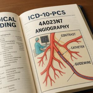
ICD-10-PCS Code for Angiography
2. Deconstructing the ICD-10-PCS Framework: The Seven-Character Code
Before we dive into the specifics of angiography, it is imperative to solidify our understanding of the ICD-10-PCS structure itself. Unlike its diagnosis-focused counterpart, ICD-10-CM, the Procedure Coding System (PCS) is entirely built on a multi-axial, seven-character alphanumeric code. Each character has a specific meaning and a defined set of values, and together, they create a highly specific description of a procedure.
Let’s break down the meaning of each character in the context of angiography:
-
Section (1st Character): This identifies the general section of the procedure. For all angiography procedures we will discuss, this will almost always be “Imaging”.
-
Body System (2nd Character): This specifies the general body system being imaged. For angiography, this is the “Anatomical Regions”.
-
Root Operation (3rd Character): This is the single most important character for angiography coding. It defines the objective or the intent of the procedure. The two primary root operations for angiography are “Measurement” and “Mapping”.
-
Body Part (4th Character): This character identifies the specific part of the anatomy that is the focus of the imaging procedure. For angiography in the Anatomical Regions body system, this refers to the specific vascular territory (e.g., coronary arteries, cerebral arteries, lower arteries).
-
Approach (5th Character): This describes the technique used to reach the site of the procedure. Common approaches for angiography include Percutaneous, Open, and Via Natural or Artificial Opening.
-
Device (6th Character): This character is used to specify a device that remains in or on the patient after the procedure is completed. For standalone diagnostic angiography, this is almost always “No Device”. However, if an imaging device like an Intracardiac Echocardiography (ICE) catheter is used and remains temporarily for guidance, it is specified here.
-
Qualifier (7th Character): This character provides additional information about the procedure. For angiography in the Anatomical Regions body system, this specifies the type of imaging modality or technique used. The most common qualifier for angiography is “Contrast”, indicating the use of an intravascular contrast agent. Other qualifiers include “Laser” or “No Qualifier,” but “Contrast” is the standard for angiographic studies.
This structured approach ensures that every coded procedure is described with a consistent and unambiguous level of detail. The following table provides a visual summary of this framework as it applies to a typical angiogram.
ICD-10-PAS Code Structure for Angiography in the “Imaging” Section
| Character Position | Definition | Example Value | Explanation for Angiography |
|---|---|---|---|
| 1st – Section | General Procedure Category | B | Imaging |
| 2nd – Body System | General Body System | 2 | Anatomical Regions |
| 3rd – Root Operation | Procedure’s Objective | 4 or B | Measurement (quantitative) or Mapping (visual road map) |
| 4th – Body Part | Specific Anatomical Site | D | Coronary Arteries |
| 5th – Approach | Technique to Reach Site | 3 | Percutaneous (through the skin via needle puncture) |
| 6th – Device | Device Remaining | Z | No Device (for standard diagnostic angio) |
| 7th – Qualifier | Additional Technique | X | Contrast (used to visualize vessels) |
| Sample Code | B24DZZX | Imaging of Anatomical Regions, Measurement, Coronary Arteries, Percutaneous Approach, No Device, Contrast |
3. The Cornerstone of Angiography Coding: Root Operations “Measurement and Mapping”
The selection of the root operation is the critical decision point in coding an angiography. It requires the coder to understand the clinical intent of the study as documented by the physician. The two options are “Measurement” and “Mapping,” and they are mutually exclusive.
3.1. Root Operation 4: Measurement – The Quantitative Assessment
The root operation Measurement is defined as: “Determining the level of a physiological or physical function at a point in time.” In the context of angiography, this translates to procedures where the primary goal is to obtain quantitative data about the blood vessels.
Key indicators for “Measurement” include:
-
Determining the percentage of stenosis (narrowing) in a vessel.
-
Measuring blood pressure gradients across a valve or a stenotic lesion.
-
Quantifying blood flow or velocity in a specific vessel.
-
Assessing the dimensions of a vessel, aneurysm, or defect.
When the physician’s report emphasizes the numerical assessment of a lesion’s severity or the hemodynamic significance of a finding, the root operation is “Measurement.” For example, a report stating “Left anterior descending artery showed a 90% proximal stenosis” or “A peak systolic gradient of 80 mmHg was measured across the aortic valve” clearly points toward a Measurement procedure.
3.2. Root Operation B: Mapping – The Roadmap of Vasculature
The root operation Mapping is defined as: “Determining the route or path of the conduction of electrical impulses or the functional location of a body part.” For angiography, this is interpreted as visualizing the anatomical course, patency, and connections of the blood vessels. It’s about creating a visual “road map.”
Key indicators for “Mapping” include:
-
Identifying the anatomical course and branches of a vessel.
-
Locating the source of bleeding (e.g., in a GI bleed).
-
Visualizing an arteriovenous malformation (AVM) and its feeding and draining vessels.
-
Assessing vessel patency (open vs. blocked) without specific quantitative measurements of stenosis.
-
Pre-operative planning to understand the vascular anatomy before a surgery.
If the report’s intent is to “define the anatomy,” “locate the bleed,” “visualize the AVM nidus,” or “assess runoff,” the root operation is likely “Mapping.”
3.3. Differentiating Between Measurement and Mapping in Clinical Practice
The distinction can be subtle. Consider a coronary angiography. If the report simply describes the vessels visualized and states there is “moderate disease in the circumflex artery,” it is a Mapping procedure. However, if the report specifically quantifies the disease as “a 70% lesion in the mid-circumflex artery,” it has crossed into Measurement territory.
Crucial Point: A single procedure can have only one root operation. If both Mapping and Measurement are performed during the same session on the same vascular territory, the root operation representing the more definitive procedure is assigned. In cases of doubt, the physician’s final impression and the clinical reason for the study (the indication) are the best guides. When a diagnostic angiogram (Mapping) immediately precedes and is used to guide an interventional procedure (e.g., a stent placement), the diagnostic angiogram is not coded separately, as the imaging is inherent to the catheter placement and the intervention.
4. The Access Point: Understanding the Approach (5th Character)
The approach describes the technique used to access the vascular system for the injection of contrast. The documentation must be reviewed carefully to identify the correct approach.
4.1. Percutaneous Approach (3)
This is the most common approach for angiography. It involves puncture of the skin with a needle to access a vessel, through which a catheter or sheath is placed. Common access sites include the femoral artery (common femoral artery), radial artery, and brachial artery. The term “Seldinger technique” almost always indicates a percutaneous approach. Code 3.
4.2. Open Approach (0)
An open approach involves cutting through the skin or mucous membrane and underlying tissues to directly expose the vessel for cannulation. This is less common but may be used in certain complex cases, re-operations, or in pediatric populations. Code 0.
4.3. Via Natural or Artificial Opening (7) and Via Natural or Artificial Opening Endoscopic (8)
These approaches are rarely used for standard angiography but can be relevant. For example, a procedure where a catheter is passed via the urethra (a natural opening) into the bladder and then into the ureter for a retrograde pyelogram would use approach 7. The endoscopic approach (8) would involve the use of an endoscope passed through a natural or artificial opening.
5. The Landscape of the Body: Anatomy of the Vasculature (4th Character)
The body part character specifies the vascular territory that is the focus of the imaging procedure. This is where detailed anatomical knowledge is essential. The “Imaging” section under “Anatomical Regions” has specific, pre-defined body part values for vascular systems.
5.1. The Arterial System: Upper and Lower Arteries, Head and Neck, Coronary, Pulmonary, and Aorta
-
Upper Arteries: This includes the arteries of the shoulder girdle and upper extremities (e.g., subclavian, axillary, brachial, radial, ulnar arteries). Body Part Character: R.
-
Lower Arteries: This includes the arteries of the pelvic region and lower extremities (e.g., common iliac, external iliac, femoral, popliteal, tibial, peroneal arteries). A “runoff” angiogram images this territory. Body Part Character: W.
-
Head and Neck Arteries: This includes the extracranial and intracranial cerebral circulation (e.g., carotid, vertebral, basilar, and cerebral arteries). Body Part Character: 2.
-
Coronary Arteries: This includes the left main, left anterior descending, circumflex, and right coronary arteries. Body Part Character: D.
-
Pulmonary Arteries: This includes the main, lobar, and segmental pulmonary arteries. Body Part Character: 4.
-
Aorta: This refers to imaging of the thoracic and/or abdominal aorta. Body Part Character: 4 (Note: The aorta shares the same body part character as Pulmonary Arteries in this table; the root operation and clinical context differentiate them).
5.2. The Venous System: Upper and Lower Veins, Head and Neck, and Portal Venous System
-
Upper Veins: Includes subclavian, axillary, brachial veins, etc. Body Part Character: 5.
-
Lower Veins: Includes iliac, femoral, popliteal veins, etc. Body Part Character: 6.
-
Head and Neck Veins: Includes jugular, cerebral veins, etc. Body Part Character: 3.
-
Portal Veins: Includes the portal vein, splenic vein, superior mesenteric vein. Body Part Character: P.
5.3. The Lymphatic System
Imaging of the lymphatic ducts and vessels. Body Part Character: 9.
6. The Tools of the Trade: Devices and Qualifiers (6th and 7th Characters)
6.1. The Crucial Role of Intracardiac Echocardiography (ICE)
For standard diagnostic angiography where no device remains, the 6th character is Z – No Device. However, an important exception occurs with the use of an Intracardiac Echocardiography (ICE) catheter. An ICE catheter is an ultrasound probe on a catheter that is advanced into the heart to provide real-time imaging guidance. If an ICE catheter is placed and remains in the heart during the procedure to guide the angiogram or an associated intervention, it is coded in the 6th character as C – Intracardiac Echocardiography. This is a common scenario in complex electrophysiology studies or structural heart procedures.
6.2. Imaging Guidance is Inherent
It is critical to remember that the imaging guidance required to perform the catheter placement (e.g., fluoroscopy, ultrasound for venous access) is considered inherent to the procedure and is not coded separately. The ICD-10-PCS code for the angiography itself encompasses all the imaging required to perform it.
6.3. Contrast Agents: The Unseen but Essential Tool
The 7th character, or Qualifier, specifies the imaging technique. For virtually all catheter-based angiograms, this will be X – Contrast. This indicates that an intravascular contrast agent was used to opacity the vessels. Other qualifiers like “Laser” or “Plain X-ray” are not applicable to standard angiography.
7. Clinical Coding Scenarios: From Report to Code
Let’s apply our knowledge to realistic clinical documentation.
7.1. Scenario 1: Diagnostic Coronary Angiography
Procedure Note: “After informed consent, the right common femoral artery was accessed percutaneously using the Seldinger technique. A 6-French sheath was placed. A Judkins right and left catheter were used to engage the coronary ostia. Contrast was injected, and multiple angiographic images were obtained of the left and right coronary systems. Findings: The left main coronary artery was normal. The left anterior descending artery had a 40% proximal lesion. The circumflex artery was normal. The right coronary artery was dominant and had a 30% mid lesion. Impression: Non-obstructive coronary artery disease.”
-
Analysis:
-
Intent: The primary goal was to visualize the coronary anatomy and quantify the stenosis (40% and 30%). This is a Measurement.
-
Body Part: The coronary arteries were the focus. D.
-
Approach: Percutaneous access of the femoral artery. 3.
-
Device/Qualifier: No device remained; contrast was used. Z, X.
-
-
ICD-10-PCS Code: B24D3ZX (Imaging, Anatomical Regions, Measurement, Coronary Arteries, Percutaneous, No Device, Contrast)
7.2. Scenario 2: Cerebral Angiogram for AVM Mapping
Procedure Note: “The patient was brought to the neuro-angiography suite. The right femoral artery was accessed percutaneously. A diagnostic catheter was advanced into the left internal carotid artery. Selective angiography was performed. The study revealed a 2 cm AVM nidus in the left parietal lobe, fed by branches of the left middle cerebral artery and drained by a single cortical vein into the superior sagittal sinus.”
-
Analysis:
-
Intent: The goal was to locate and define the anatomy of the AVM—its nidus, feeding arteries, and draining veins. This is creating a “map.” Mapping.
-
Body Part: The cerebral arteries (head and neck arteries) were the focus. 2.
-
Approach: Percutaneous. 3.
-
Device/Qualifier: No device; contrast used. Z, X.
-
-
ICD-10-PCS Code: B22?3ZX (Note: The exact character for Head and Neck Arteries in the Mapping table must be verified in the current year’s manual. For this example, we are using ‘2’ as per our earlier section).
7.3. Scenario 3: Lower Extremity Runoff Angiogram
Procedure Note: “Via a right common femoral artery access, an angiographic pigtail catheter was placed in the distal abdominal aorta. A power injection of contrast was performed, and serial images were obtained of the pelvis and lower extremities to assess for peripheral arterial disease. The study demonstrated a patent aorta and iliac systems. There was a focal 90% stenosis of the right superficial femoral artery. The runoff vessels were patent.”
-
Analysis:
-
Intent: The procedure was to assess for disease (Mapping), but a specific quantitative measurement (90% stenosis) was provided, making Measurement the correct root operation.
-
Body Part: The lower arteries were the focus. W.
-
Approach: Percutaneous. 3.
-
Device/Qualifier: No device; contrast used. Z, X.
-
-
ICD-10-PCS Code: B24W3ZX (Imaging, Anatomical Regions, Measurement, Lower Arteries, Percutaneous, No Device, Contrast)
8. Navigating the Nuances: Common Pitfalls and Expert Tips
-
Don’t Code the Catheter Placement Separately: The placement of the catheter to inject contrast is an inherent part of the imaging procedure in the “Imaging” section. Do not assign a separate code from the “Placement” section of ICD-10-PCS for the angiographic catheter.
-
Combined Procedures: If a diagnostic angiogram is performed and, based on its findings, an intervention (e.g., angioplasty, stent placement, embolization) is performed during the same session, you code only the intervention. The diagnostic imaging is bundled.
-
Multiple Vascular Territories: If multiple, distinct vascular territories are imaged (e.g., coronary arteries AND renal arteries), and separate, selective catheterizations and contrast injections are performed for each, multiple ICD-10-PCS codes may be warranted. However, if the renal arteries are visualized incidentally during an aortogram, a separate code is not assigned. The key is medical necessity and the work involved.
-
The Report is King: Always, always code based on the physician’s procedure report. Do not make assumptions based on the patient’s diagnosis or the name of the procedure in the schedule.
9. The Interventional Bridge: When Angiography Leads to Another Procedure
It is vital to understand the workflow in a catheterization lab. A diagnostic angiogram is often the first step. If it reveals a significant lesion, the physician may immediately proceed with a therapeutic intervention. In this case, the entire procedural session is defined by the intervention.
-
Example: A patient undergoes a coronary angiogram (which would be coded as B24D3ZX if standalone) that reveals a 99% lesion. The physician proceeds to perform a percutaneous coronary intervention (PCI) with stent placement.
-
Coding Action: You would not code the diagnostic angiogram. You would code the following:
-
The root operation Dilation for the angioplasty of the coronary artery.
-
The root operation Insertion for the placement of the coronary stent.
The imaging for the intervention is inherent to these procedure codes.
-
10. Conclusion: The Art and Science of Precision Coding
Accurate ICD-10-PCS coding for angiography hinges on a deep understanding of clinical intent, embodied by the root operations Measurement and Mapping. It demands precise identification of the vascular territory and the technical approach used. By methodically applying the seven-character framework and adhering strictly to the physician’s documentation, coders can ensure the integrity of medical data and the financial health of their organizations, ultimately supporting the vital work of clinical teams.
11. Frequently Asked Questions (FAQs)
Q1: What is the difference between an angiogram and an arteriogram?
A: Historically, “arteriogram” specified imaging of arteries, while “venogram” specified veins. “Angiogram” was a more general term. In modern clinical and coding practice, the terms are often used interchangeably, but the procedure is coded based on the actual vessels imaged (arterial vs. venous body part values in PCS).
Q2: How do I code a CT Angiogram (CTA) or MR Angiogram (MRA)?
A: CTA and MRA are coded differently from catheter-based angiograms. They are found in the “Imaging” section but under the body system “Anatomical Regions” for the specific area (e.g., Head, Chest, Abdomen) and use root operations like “Computerized Tomography” or “Magnetic Resonance Imaging.” The qualifier would indicate “High Osmolar Contrast,” “Low Osmolar Contrast,” or “Other Contrast.” They do not involve catheter placement.
Q3: What if the physician’s report does not specify a percentage of stenosis?
A: If the report uses qualitative terms like “mild,” “moderate,” or “severe” disease without quantification, or simply describes the anatomical course of the vessels, the root operation should be Mapping.
Q4: When do I use the “Device” character for an ICE catheter?
A: You use character C – Intracardiac Echocardiography only if the ICE catheter is used and it remains in place during the procedure. If it is removed immediately after use or not used at all, you would use Z – No Device.
Disclaimer: This article is intended for educational and informational purposes only. It is not a substitute for professional medical coding advice, official coding guidelines, or the current ICD-10-PCS code set. Medical coders must always consult the most current year’s official ICD-10-PCS manuals, Coding Clinic guidance, and payer-specific policies for accurate code assignment. The author and publisher disclaim any liability arising from the use or misuse of the information contained herein.
Date: November 17, 2025
Author: Medical Coding Intelligence Unit


