n the intricate ecosystem of modern healthcare, the seamless flow of information is as vital as the flow of blood. At the heart of this informational circulatory system lies medical coding, a discipline that translates complex clinical narratives into a standardized, alphanumeric language. For procedures involving the intricate landscape of the gastrointestinal tract, particularly gastroscopy (esophagogastroduodenoscopy or EGD), this translation is both an art and a science. The International Classification of Diseases, Tenth Revision, Procedure Coding System (ICD-10-PCS) provides the framework for this precise communication, moving far beyond the simplicity of its predecessor, ICD-9-CM, into a world of breathtaking specificity. A single, correctly built ICD-10-PCS code can communicate not just what was done, but where, how, why, and with what. This level of detail is paramount for reimbursement, quality reporting, public health tracking, and clinical research.
This article is designed to be the definitive guide for medical coders, health information management (HIM) professionals, and even clinicians seeking to understand the “why” behind the codes assigned to their work. We will embark on a detailed journey, dissecting the seven-character structure of ICD-10-PCS, applying it to the vast array of gastroscopic procedures, and illuminating the path to accurate, defensible coding. We will move from the foundational principles of the system’s architecture to the nuanced application required for complex, multi-procedural encounters. By the end of this guide, you will not only know how to build a code; you will understand the clinical rationale behind each character, empowering you to navigate even the most challenging operative reports with confidence and precision.
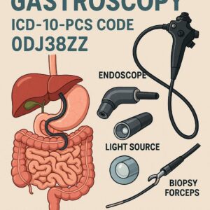
ICD-10-PCS Code for Gastroscopy
2. Deconstructing the Gastroscopic Procedure: A Primer for Coders
Before a coder can accurately translate a procedure, they must first understand it. A gastroscopy, formally an esophagogastroduodenoscopy (EGD), is a minimally invasive endoscopic procedure that allows a gastroenterologist or surgeon to visually examine the upper gastrointestinal (GI) tract. This includes the esophagus, stomach, and duodenum (the first part of the small intestine).
The procedure is typically performed with the patient under sedation. A gastroscope—a long, thin, flexible tube with a high-definition camera and a light at its tip—is inserted through the mouth and gently advanced down the esophagus. The camera transmits real-time video to a monitor, allowing the physician to assess the mucosa (lining) for abnormalities such as inflammation, ulcers, bleeding, tumors, or strictures.
Crucially, the gastroscope contains a working channel. This allows the physician to pass specialized instruments through the scope to perform therapeutic interventions without making any external incisions. These instruments can include:
-
Biopsy Forceps: To take small tissue samples (biopsies) for pathological analysis.
-
Snares: To remove polyps or other growths.
-
Needles: To inject medications (e.g., epinephrine for a bleeding ulcer).
-
Cautery Probes: To burn (cauterize) bleeding vessels or ablate abnormal tissue.
-
Balloons or Dilation Catheters: To stretch open narrowed areas (strictures).
-
Clips: To mechanically close a bleeding site or a perforation.
This dual nature—diagnostic visualization and therapeutic intervention—is the central concept that drives ICD-10-PCS coding. A single EGD encounter can transition from a simple inspection to a life-saving hemostatic procedure in moments, and the coding must reflect every clinically significant action taken.
3. The Architecture of ICD-10-PCS: A Seven-Character System
ICD-10-PCS is built on a logical, multi-axial structure. Each code is composed of seven characters, each representing a specific aspect of the procedure. Unlike ICD-10-CM diagnosis codes, there are no decimals. The meaning of each character is position-dependent.
Here is the breakdown of the seven characters:
-
Character 1: Section – The broadest category (e.g., Medical and Surgical, Obstetrics, Imaging).
-
Character 2: Body System – The general physiological system involved (e.g., Gastrointestinal System, Hepatobiliary System).
-
Character 3: Root Operation – The objective of the procedure; the definitive, core action performed.
-
Character 4: Body Part – The specific anatomical site where the root operation was performed.
-
Character 5: Approach – The technique used to reach the operative site.
-
Character 6: Device – Any device that remains in or on the patient after the procedure is completed.
-
Character 7: Qualifier – Adds additional information about the procedure that is not captured in the other characters.
For gastroscopy procedures, the vast majority of codes will fall within the Medical and Surgical section (character 1). Our focus will be exclusively on this section.
4. The Foundation: Section and Body System (Characters 1 & 2)
For virtually all gastroscopic procedures, the first two characters are fixed.
-
Character 1: Section = 0 (Medical and Surgical)
-
Character 2: Body System = D (Gastrointestinal System)
The GI system in PCS includes the mouth and pharynx, esophagus, stomach, intestines, and anus. Therefore, our codes for EGD will always begin with 0D.
5. The Core of the Code: The Root Operation (Character 3)
The root operation is the most critical and often the most challenging character to determine. It requires a careful reading of the operative report to identify the principal objective of each distinct procedure. A single EGD report may document multiple root operations, each requiring its own code.
Inspection: The Diagnostic Cornerstone
-
PCS Definition: “Visually and/or manually exploring a body part.”
-
Clinical Correlation: This is the purely diagnostic part of the EGD. The physician is looking at the anatomy without performing any therapeutic intervention. Crucially, a biopsy alone does not change the root operation from Inspection. Taking a biopsy is considered a part of the diagnostic inspection.
-
When to Use: When the procedure is solely diagnostic, even if biopsies are taken. The report might state: “EGD performed for evaluation of dyspepsia. Exam revealed mild gastritis. Biopsies taken from gastric antrum and body.” The primary goal was to visually explore and sample for diagnosis.
-
Key Point: If only an inspection with biopsy is performed, this is the only root operation code you need.
Excision, Resection, and Extraction: Taking Tissue
These three root operations are often confused but have distinct definitions.
-
Excision (B):
-
PCS Definition: “Cutting out or off, without replacement, a portion of a body part.”
-
Clinical Correlation: This is used for the removal of a portion of a body part, not the entire thing. The most common example is a polypectomy, where a polyp is snipped off from the stomach or duodenal wall. The polyp is a portion of the wall. Another example is an endoscopic mucosal resection (EMR), where a piece of abnormal mucosa is removed.
-
-
Resection (T):
-
PCS Definition: “Cutting out or off, without replacement, all of a body part.”
-
Clinical Correlation: This is rarely used in routine gastroscopy. It would apply if the entire stomach were removed endoscopically, which is not a standard procedure. Resection is more common in open or laparoscopic surgery (e.g., gastrectomy).
-
-
Extraction (D):
-
PCS Definition: “Pulling or stripping out or off all of a body part or any portion of a body part by the use of force.”
-
Clinical Correlation: The key differentiator from Excision is the “use of force” on the body part itself. This is used for the removal of foreign bodies. For example, if a food bolus or a accidentally swallowed object is lodged in the esophagus and is grasped and pulled out with forceps or a snare, the root operation is Extraction.
-
Destruction: Eradicating Tissue
-
PCS Definition: “Physical eradication of all or a portion of a body part by the direct use of energy, force, or a destructive agent.”
-
Clinical Correlation: The tissue is not taken out; it is ablated or eradicated in situ. Common methods in gastroscopy include:
-
Argon Plasma Coagulation (APC): Using ionized argon gas to deliver high-frequency current to ablate tissue.
-
Laser: Using light energy to vaporize tissue.
-
Heater Probe: Using thermal energy to coagulate and destroy tissue.
-
-
When to Use: For ablation of bleeding lesions (e.g., arteriovenous malformations), ablation of Barrett’s esophagus, or destruction of small, flat polyps that are not excised.
Dilation: Opening a Pathway
-
PCS Definition: “Expanding an orifice or the lumen of a tubular body part.”
-
Clinical Correlation: This is used for stretching open a narrowed (stenotic) area. This can be done with:
-
Balloon Dilation: A deflated balloon is passed through the scope’s channel, positioned across the stricture, and inflated.
-
Bougienage: Passing a series of progressively wider rigid dilators.
-
-
When to Use: For dilation of an esophageal stricture, pyloric stenosis, or an anastomotic stricture.
Bypass, Alteration, and Restriction: Changing Anatomy
-
Bypass (1):
-
PCS Definition: “Altering the route of passage of the contents of a tubular body part.”
-
Clinical Correlation: In gastroscopy, this is most commonly associated with the placement of a duodenal stent. The stent bypasses a malignant obstruction in the duodenum, allowing stomach contents to pass through. It can also refer to the creation of a gastroenteric anastomosis surgically, but that is not an endoscopic procedure.
-
-
Restriction (V):
-
PCS Definition: “Partially closing an orifice or the lumen of a tubular body part.”
-
Clinical Correlation: This is used for procedures like endoscopic sleeve gastroplasty (ESG), where sutures are placed in the stomach to reduce its volume and restrict food intake for weight loss.
-
-
Alteration (S):
-
PCS Definition: “Modifying the natural anatomic structure of a body part without affecting the function of the body part.”
-
Clinical Correlation: Primarily used for cosmetic or reconstructive procedures. It is less common in gastroscopy but could apply to a procedure that changes the shape of the stomach for a non-functional reason.
-
Introduction and Removal: Putting In and Taking Out
-
Introduction (3):
-
PCS Definition: “Putting in or on a therapeutic, diagnostic, nutritional, physiological, or prophylactic substance except blood or blood products.”
-
Clinical Correlation: This is a very specific root operation. It is used for the instillation of a substance that is not a device. The most common example in EGD is the injection of a substance (e.g., epinephrine or a sclerosing agent) into a bleeding ulcer to achieve hemostasis. Note: The injection of a substance for hemostasis is coded separately from any Destruction or Control of bleeding performed at the same site.
-
-
Removal (P):
-
PCS Definition: “Taking out or off a device from a body part.”
-
Clinical Correlation: Used when a previously placed device is taken out. For example, the removal of a gastrostomy tube (PEG tube) via endoscopy, or the removal of a migrated stent.
-
Repair and Transplantation: Fixing and Replacing
-
Repair (Q):
-
PCS Definition: “Restoring, to the extent possible, a body part to its normal anatomic structure and function.”
-
Clinical Correlation: Used for closing a perforation or a tear. The most common application is the placement of hemostatic clips (e.g., through-the-scope clips) to close a bleeding vessel or a mucosal defect. Note: If the clips are placed for control of bleeding, the root operation is the one defined for controlling bleeding (see Qualifier section), not Repair. Repair is for closing a defect.
-
-
Transplantation (Y):
-
PCS Definition: “Putting in or on all or a portion of a living body part taken from another individual or animal to physically take the place and/or function of all or a portion of a similar body part.”
-
Clinical Correlation: This is not applicable to gastroscopy.
-
6. Navigating the Body Part (Character 4)
The body part character specifies the exact anatomical site where the root operation was performed. The choices are found in the ICD-10-PCS tables under the Gastrointestinal System (0D). Accurate identification is key.
Common Body Part Values for EGD:
-
Esophagus:
D -
Upper Esophagus:
0 -
Middle Esophagus:
1 -
Lower Esophagus:
2 -
Esophagogastric Junction:
3 -
Stomach:
4 -
Stomach, Pylorus:
5 -
Duodenum:
6 -
Duodenum, First Portion:
7 -
Duodenum, Second Portion:
8 -
Duodenum, Third Portion:
9 -
Duodenum, Fourth Portion:
B
Critical Concept: The body part is specific to the root operation. If an inspection is performed of the entire upper GI tract, the body part is the Esophagus, Stomach, and Duodenum, which has a specific value (C). However, if a polyp is excised from the stomach and an ulcer in the duodenum is cauterized, you have two separate codes:
-
Excision of stomach polyp: Body Part =
4 Stomach -
Destruction of duodenal ulcer: Body Part =
6 Duodenum
You would not use the general C value for these therapeutic procedures.
7. The Surgical Approach (Character 5)
The approach describes how the surgeon reached the site of the procedure. For gastroscopy, this is almost always:
-
Via Natural or Artificial Opening (7): The scope is passed through a natural opening (the mouth).
-
Via Natural or Artificial Opening Endoscopic (8): This is the most precise and correct approach for a gastroscopy. The “Endoscopic” qualifier specifies that the procedure was performed using an endoscope as the instrument. This is the default and should be used for all standard EGDs.
Other approaches like Open (0) or Percutaneous (3) are used for surgical or radiologically guided procedures and are not applicable to standard gastroscopy.
8. Device and Qualifier (Characters 6 & 7)
These final two characters add layers of specificity.
Character 6: Device
This character is used only if a device remains after the procedure is completed. If no device remains, the value is Z (No Device).
-
Examples:
-
Bypass with Stent Placement: Device =
C (Intraluminal Device) -
Control of Bleeding with Clip Placement: Device =
C (Intraluminal Device)(the clip is the device) -
Placement of a PEG Tube: This is a different root operation (Change), but the Device would be
J (Gastrointestinal Tube)
-
Character 7: Qualifier
The qualifier provides the final piece of information. Its meaning changes depending on the root operation.
-
For Inspection: Qualifier =
X (Diagnostic) -
For Excision: Qualifier =
X (Diagnostic)if the tissue is sent to pathology, otherwiseZ (No Qualifier). It is almost alwaysX. -
For Control of Bleeding: This is a special case. There is no specific “Control” root operation. Instead, you use the root operation that was performed to achieve control (e.g., Destruction, Occlusion via clipping, etc.) and the Qualifier is
3 (Control).-
Example: Cauterizing a bleeding ulcer with APC: Root Operation =
Destruction, Qualifier =3 (Control). -
Example: Placing a clip on a bleeding vessel: Root Operation =
Occlusion (L), Qualifier =3 (Control).
-
9. Practical Application: Building Codes from Complex Reports
Let’s apply our knowledge to real-world operative reports.
Scenario 1: Diagnostic EGD with Biopsies
-
Report: “EGD performed for iron deficiency anemia. Scope passed without difficulty. Exam revealed mild erythema in the gastric antrum. No active bleeding, varices, or masses seen. Biopsies taken from gastric antrum and duodenum.”
-
Analysis: The sole objective was visual examination and sampling for diagnosis. One root operation covers it all.
-
Code Build:
-
Section: 0 (Medical and Surgical)
-
Body System: D (Gastrointestinal)
-
Root Operation: J (Inspection)
-
Body Part: C (Esophagus, Stomach, and Duodenum) – The inspection and biopsies were of the entire tract.
-
Approach: 8 (Via Natural or Artificial Opening Endoscopic)
-
Device: Z (No Device)
-
Qualifier: X (Diagnostic)
-
-
Final ICD-10-PCS Code: 0DJC8ZX
Scenario 2: Complex Therapeutic EGD
-
Report: “EGD performed for food impaction. A large meat bolus was identified in the mid-esophagus. It was successfully extracted with a Roth net. Upon removal, a bleeding superficial ulcer was noted at 30cm. The bleeding was controlled with the injection of 5cc of 1:10,000 epinephrine in 4 quadrants around the vessel. A through-the-scope clip was then deployed for definitive hemostasis. Finally, a 2cm pedunculated polyp was identified in the gastric body and was removed with a cold snare. The polyp was retrieved and sent to pathology.”
-
Analysis: This report describes four distinct procedural objectives, requiring four separate codes.
-
Extraction of the foreign body.
-
Introduction of a substance (epinephrine) to control bleeding.
-
Occlusion of the vessel (with a clip) to control bleeding.
-
Excision of the polyp.
-
-
Code 1: Extraction of Foreign Body
-
0 (Med/Surg) + D (GI System) + D (Extraction) + 1 (Middle Esophagus) + 8 (Endoscopic) + Z (No Device) + Z (No Qualifier)
-
Code: 0DD18ZZ
-
-
Code 2: Introduction of Substance for Control
-
0 (Med/Surg) + D (GI System) + 3 (Introduction) + 1 (Middle Esophagus) + 8 (Endoscopic) + Z (No Device) + 5 (Qualifier for “Other Substance” for hemostasis – check table)
-
Code: 0D318Z5
-
-
Code 3: Occlusion of Vessel for Control
-
0 (Med/Surg) + D (GI System) + L (Occlusion) + 1 (Middle Esophagus) + 8 (Endoscopic) + C (Intraluminal Device – the clip) + 3 (Control)
-
Code: 0DL18C3
-
-
Code 4: Excision of Polyp
-
0 (Med/Surg) + D (GI System) + B (Excision) + 4 (Stomach) + 8 (Endoscopic) + Z (No Device) + X (Diagnostic)
-
Code: 0DB48ZX
-
Common Gastroscopy Root Operations and Their Codes
| Procedural Goal | Root Operation | Body Part Example | Approach | Device | Qualifier | Example Code | Clinical Example |
|---|---|---|---|---|---|---|---|
| Diagnostic Exam | Inspection (J) | C (Esoph, Stom, Duod) | 8 | Z | X | 0DJC8ZX | EGD with biopsies for dyspepsia |
| Polyp Removal | Excision (B) | 4 (Stomach) | 8 | Z | X | 0DB48ZX | Cold snare polypectomy |
| Foreign Body Removal | Extraction (D) | 2 (Lower Esophagus) | 8 | Z | Z | 0DD28ZZ | Removal of impacted food bolus |
| Ablate Bleeding Ulcer | Destruction (5) | 6 (Duodenum) | 8 | Z | 3 (Control) | 0D568Z3 | APC to a bleeding duodenal ulcer |
| Dilate Stricture | Dilation (7) | 2 (Lower Esophagus) | 8 | Z | Z | 0D728ZZ | Balloon dilation of peptic stricture |
| Place Stent | Bypass (1) | 6 (Duodenum) | 8 | C (Stent) | Z | 0D168CZ | Duodenal stent for malignancy |
| Inject for Hemostasis | Introduction (3) | 4 (Stomach) | 8 | Z | 5 (Substance) | 0D348Z5 | Epinephrine injection around ulcer |
| Clip a Vessel | Occlusion (L) | 1 (Middle Esophagus) | 8 | C (Clip) | 3 (Control) | 0DL18C3 | Clip placement on bleeding Mallory-Weiss tear |
| Remove PEG Tube | Removal (P) | 4 (Stomach) | 8 | Z | Z | 0DP48ZZ | Endoscopic removal of old gastrostomy tube |
10. Common Pitfalls and Pro Tips for Flawless Coding
-
Confusing Biopsy with Excision: Remember, a biopsy is part of the Inspection root operation. You only use Excision when a lesion (like a polyp) is cut out. Taking multiple random biopsies of flat mucosa is Inspection.
-
Missing the “Control” Qualifier: A very common error is coding the destruction or occlusion of a bleeding site without the
3qualifier. This changes the clinical meaning and is often necessary for accurate reimbursement. -
Incorrect Body Part for Inspection: Using a general body part value like
C (Esophagus, Stomach, Duodenum)for a therapeutic procedure. This value is only for the Inspection root operation when the entire tract is examined. Therapeutic procedures must be coded to the specific body part treated. -
Not Coding All Root Operations: A single operative report is a narrative of the encounter. You must identify every distinct objective the physician accomplished. If they do five things, you may need five codes.
-
Overthinking the Approach: For standard gastroscopy, the approach is almost always
8 (Via Natural or Artificial Opening Endoscopic).
11. The Role of Documentation: A Partnership with Providers
Accurate coding is impossible without precise documentation. Coders rely entirely on the physician’s operative report. Incomplete or vague documentation leads to queries, delayed billing, and potential down-coding.
What Coders Need to See in a Report:
-
Indication: Why was the procedure performed?
-
Findings: A detailed description of what was seen in each segment (esophagus, stomach, duodenum).
-
Techniques: Exactly what was done. “Bleeding controlled” is not enough. How was it controlled? “Injection,” “cautery,” “clip placement”?
-
Location: The specific anatomical site of every intervention (e.g., “gastric antrum,” “first portion of the duodenum”).
-
Specimens: What was sent to pathology and from where.
Engaging in a collaborative relationship with providers to educate them on the elements of a “coder-friendly” report is a best practice in any HIM department.
12. Conclusion
Mastering ICD-10-PCS for gastroscopy requires a methodical understanding of its seven-character logic and the clinical procedures it represents. By anchoring your coding in the precise definitions of root operations, meticulously identifying body parts, and leveraging the specificity of devices and qualifiers, you can ensure complete and accurate translation of clinical care into data. This precision is the bedrock of a compliant and efficient revenue cycle, robust quality metrics, and a true reflection of the sophisticated work performed by gastroenterologists every day. Continuous learning and a strong partnership with clinical documentation integrity specialists are the keys to ongoing success in this dynamic field.
13. Frequently Asked Questions (FAQs)
Q1: If multiple biopsies are taken from different areas (esophagus, stomach, duodenum) during a diagnostic EGD, do I need multiple codes?
A: No. A single Inspection code with the body part value “C” (Esophagus, Stomach, and Duodenum) suffices. The act of taking biopsies is inherent to the diagnostic inspection.
Q2: How do I code a screening EGD on an asymptomatic patient?
A: This is still coded with the Inspection root operation (0DJC8ZX). The “screening” intent is conveyed by the ICD-10-CM diagnosis code (e.g., Z12.81), not the PCS procedure code.
Q3: What is the difference between Occlusion with a clip and Repair with a clip?
A: It depends on the intent.
-
Occlusion: The goal is to block a tubular body part (e.g., a blood vessel). Used for controlling bleeding. Qualifier is
3 (Control). -
Repair: The goal is to close a physical defect or restore anatomy (e.g., closing a mucosal tear or a perforation). The qualifier would be specific to the technique, like
Y (Other Device)for a clip.
Q4: An EGD was performed, but the scope could not be advanced beyond the esophagus due to a tight stricture. How is this coded?
A: You would code the Inspection of the body parts that were successfully examined (e.g., the esophagus). If an attempt was made to dilate the stricture but was unsuccessful, you would still code the Dilation procedure that was attempted.
Q5: Where can I find the official ICD-10-PCS tables and guidelines?
A: The Centers for Medicare & Medicaid Services (CMS) and the National Center for Health Statistics (NCHS) are the official publishers. These resources are available on the CMS ICD-10 website.
Date: November 24, 2025
Author: Gastrointestinal Coding Specialist
Disclaimer: The information contained in this article is for educational and informational purposes only and does not constitute medical or coding advice. It is essential to consult the official ICD-10-PCS code set, payer-specific guidelines, and the patient’s medical record for accurate coding. The author and publisher are not responsible for any errors or omissions or for any outcomes related to the use of this information.


