Imagine two sheets of slick plastic wrap laid gently against one another. They can slide smoothly, but when pulled apart, they resist separation and may cling back together. This is akin to the visceral and parietal pleura—two thin, glistening membranes that line the lungs and the inside of the chest wall, respectively. The potential space between them, the pleural space, is minimal, containing only a tiny amount of lubricating fluid. This elegant design allows the lungs to expand and contract with minimal friction during the vital act of breathing. However, when air, blood, or other substances invade this potential space, it becomes a very real and problematic cavity, leading to a collapse of the lung—a condition known as a pneumothorax.
The pneumothorax is more than a medical term; it is a potentially life-threatening event that can arise spontaneously in tall, thin young men (primary spontaneous pneumothorax) or as a complication of underlying lung diseases like emphysema (secondary spontaneous pneumothorax). It can also result from trauma. The symptoms—sudden, sharp chest pain and shortness of breath—are dramatic and debilitating. While a small, first-time pneumothorax may be managed with observation or simple aspiration, the specter of recurrence looms large. For patients experiencing recurrent episodes, or for those with persistent air leaks (such as in complicated emphysema or certain post-surgical states), a temporary chest tube is merely a stopgap measure. The fundamental question becomes: How can we eliminate the pleural space to prevent the lung from collapsing again? The answer lies in a procedure that has evolved over decades, marrying mechanical intervention with biological healing: mechanical pleurodesis.
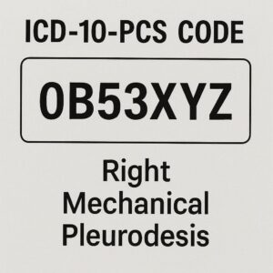
icd 10 pcs code for right mechanical pleurodesis
2. Defining the Solution: What is Pleurodesis?
Pleurodesis is a definitive surgical procedure designed to create adhesions, or scar tissue, between the parietal and visceral pleural layers. The goal is not to repair the pleura to its original, slick state but rather to intentionally cause a controlled, inflammatory response that results in the two layers permanently fusing together. By obliterating the pleural space, we eliminate the cavity where air or fluid can accumulate, thereby preventing recurrence of pneumothorax or malignant pleural effusion.
The concept is ancient in principle, but modern in execution. The history of pleurodesis dates back over a century, with early attempts involving the insufflation of irritating substances like silver nitrate or talc into the chest. Over time, the methodology has refined into three primary categories, each with its own mechanism of action:
-
Chemical Pleurodesis: Introduction of an irritant (e.g., talc slurry, doxycycline) via a chest tube to provoke inflammation.
-
Surgical Pleurodesis (Pleurectomy): The physical stripping of the parietal pleura, leaving a raw, bleeding surface that heals by adhering to the visceral pleura.
-
Mechanical Pleurodesis: The physical abrasion of the pleural surfaces using dry gauze, a specialized scratch pad, or a laser to create a diffuse area of injury that leads to adhesion formation.
This article will focus intently on the last of these—mechanical pleurodesis—and specifically, its application on the right side of the chest, as captured in the precise language of modern medical coding: ICD-10-PCS code 0BJL4ZZ.
3. The Nuts and Bolts of Medical Coding: A Primer on ICD-10-PCS
Before we dissect the specific code, it is essential to understand the system it belongs to. The International Classification of Diseases, 10th Revision, Procedure Coding System (ICD-10-PCS) is a complex, multi-axial system used in hospital settings in the United States to report inpatient procedures. Unlike its diagnosis counterpart (ICD-10-CM) or the CPT codes used for physician services, ICD-10-PCS is built on a structure of seven alphanumeric characters. Each character represents a specific aspect of the procedure, providing an astonishing level of detail. This granularity allows for precise tracking of medical interventions, which is critical for reimbursement, healthcare statistics, epidemiological research, and quality assessment.
The seven characters of an ICD-10-PCS code represent the following:
-
Section: The broad category (e.g., Medical and Surgical, Obstetrics, Imaging).
-
Body System: The general physiological system involved.
-
Root Operation: The objective of the procedure (e.g., cutting, altering, removing).
-
Body Part: The specific anatomical site.
-
Approach: How the surgeon accessed the site (e.g., open, percutaneous, via natural orifice).
-
Device: Any device that remains after the procedure.
-
Qualifier: An additional detail for further specificity.
This structure transforms a narrative operative report into a standardized, machine-readable code. It is a language of precision, and mastery of it is essential for health information management professionals.
4. A Deep Dive into the Code: Deciphering 0BJL4ZZ (Right Mechanical Pleurodesis)
Let us now apply this framework to our primary code of interest: 0BJL4ZZ. This code is not a random string of characters; it is a complete procedural description.
-
Character 1: 0 – Section – Medical and Surgical
This immediately tells us the procedure is classified under the largest section of ICD-10-PCS, encompassing all invasive procedures performed on the body’s anatomical structures. -
Character 2: B – Body System – Pleural Cavity
This character specifies the body system as the Pleural Cavity. This is distinct from the Respiratory System or the Lung. ICD-10-PCS meticulously distinguishes between operating on the lung parenchyma (Body System = Lungs) and operating within the pleural space (Body System = Pleural Cavity). Mechanical pleurodesis is an intervention directed at the lining of that space, hence the correct designation is B. -
Character 3: J – Root Operation – Alteration
This is the conceptual heart of the code. The ICD-10-PCS defines Alteration as “Modifying the natural anatomic structure of a body part without affecting the function of the body part.” The official guideline further clarifies that the purpose of an Alteration is to improve appearance. At first glance, this seems paradoxical—pleurodesis is done for functional reasons, not cosmetic ones. However, in the specific logic of PCS, pleurodesis is classified under Alteration because it changes the anatomical structure (fusing the pleural layers) without restoring a function to a body part that is malfunctioning. It is not repairing a defect (Repair), it is not taking out something (Excision), it is altering the anatomy to achieve a clinical goal. This is a critical nuance that coders must understand. -
Character 4: L – Body Part – Pleura, Right
This character provides laterality and specificity. “L” corresponds to the Pleura, Right. This explicitly codes for a procedure performed on the right hemithorax. A left mechanical pleurodesis would have a different character in this position (K). -
Character 5: 4 – Approach – Thoracoscopic
The approach is 4, which stands for Thoracoscopic. This indicates the procedure was performed using video-assisted thoracoscopic surgery (VATS). This is a minimally invasive approach where small incisions (ports) are made in the chest wall, a camera is inserted, and long, thin instruments are used to perform the operation. Other possible approaches for pleurodesis could be Open (0) or Percutaneous (3, for chemical pleurodesis via tube), but for modern mechanical abrasion, thoracoscopic is most common. -
Character 6: Z – Device – No Device
No device is used in a standard mechanical pleurodesis and left in the body. The abrasive material (e.g., gauze) is introduced, used, and removed. Therefore, this character is Z for No Device. -
Character 7: Z – Qualifier – No Qualifier
There is no additional qualifier needed for this specific procedure, so the final character is also Z.
Thus, 0BJL4ZZ translates to: Medical and Surgical procedure on the Pleural Cavity, involving an Alteration of the Right Pleura, performed via a Thoracoscopic approach, with no device left in place. This single code encapsulates a vast amount of clinical and technical information.
5. The Clinical Pathway: Indications and Patient Selection for Mechanical Pleurodesis
Not every pneumothorax warrants a pleurodesis. The decision to proceed is a careful balance of clinical judgment, patient factors, and the risk of recurrence. The primary indications for mechanical pleurodesis include:
-
Recurrent Spontaneous Pneumothorax: After a second ipsilateral (same-side) pneumothorax, the risk of a third exceeds 60%, making definitive intervention the standard of care.
-
Persistent Air Leak: When a chest tube fails to resolve the pneumothorax after 5-7 days (in primary spontaneous pneumothorax) or longer in secondary cases.
-
Secondary Spontaneous Pneumothorax in High-Risk Patients: Especially in patients with chronic obstructive pulmonary disease (COPD), where each episode carries significant morbidity and mortality.
-
Contralateral Pneumothorax: A history of pneumothorax on the opposite side increases the risk on the untreated side.
-
Certain Professions or Avocations: “Professionals at risk” such as pilots, divers, and those living in remote areas may opt for definitive surgery after a first event.
-
Failed Previous Chemical Pleurodesis: Mechanical pleurodesis can be a next-step intervention.
Patient selection is paramount. A thorough preoperative workup includes high-resolution CT scanning to identify underlying blebs or bullae (which may also be resected during the same procedure), pulmonary function tests to assess respiratory reserve, and a careful evaluation of comorbidities. The patient must be fit enough to tolerate single-lung ventilation, which is required for VATS.
6. Inside the Operating Room: A Step-by-Step Walkthrough of the Procedure
Understanding the code requires understanding the action it describes. Here is a detailed narrative of a typical right mechanical pleurodesis via VATS:
-
General Anesthesia & Positioning: The patient is placed under general anesthesia. A double-lumen endotracheal tube is positioned to allow the anesthesiologist to selectively ventilate the left lung and collapse the right lung, providing space and visibility within the right pleural cavity. The patient is positioned in a full left lateral decubitus position (on their left side).
-
Incision and Access: Three small incisions (approximately 1-2 cm each) are made in strategic locations on the right lateral chest wall. A trocar is inserted, and the thoracoscope (camera) is introduced into the chest. The other ports are placed under direct visualization for instruments.
-
Exploration and Identification: The surgeon inspects the entire right hemithorax. The lung is carefully examined for the source of the air leak—often an apical bleb or bulla. The condition of the pleural surfaces is assessed.
-
Resection of Blebs (if present): Using a mechanical stapler introduced through one of the ports, any identified blebs or bullae at the apex of the lung are resected. This addresses the immediate cause of the pneumothorax.
-
The Mechanical Abrasion: This is the core of the pleurodesis. The surgeon introduces a piece of dry, sterile surgical gauze (mounted on a ring forceps) or a dedicated pleural abrader (a scratch pad-like device on an extended handle) through a port. Under direct vision, the parietal pleura—particularly over the apex and upper ribs, the lateral chest wall, and the diaphragm—is vigorously abraded. The goal is to create a uniform, diffuse area of bleeding and inflammation without damaging underlying structures. The visceral pleura over the lung surface may also be gently abraded.
-
Irrigation and Inspection: The chest is irrigated with warm saline solution to remove debris and blood clots. The surgical field is inspected for hemostasis.
-
Chest Tube Placement and Closure: One or two chest tubes are positioned under direct vision, typically at the apex and base. The lung is re-inflated by the anesthesia team. The instruments and camera are removed, and the small incisions are closed with sutures.
7. The Thoracoscopic Revolution: Minimally Invasive Approach (VATS)
The approach character “4” in our code represents a monumental shift in thoracic surgery. Prior to VATS, mechanical pleurodesis required a posterolateral thoracotomy—a large, muscle-cutting incision that spread the ribs. This resulted in significant postoperative pain, a longer recovery, and greater morbidity. The Video-Assisted Thoracoscopic Surgery (VATS) approach has transformed the procedure. Benefits include:
-
Markedly reduced postoperative pain
-
Shorter hospital stay (often 1-3 days)
-
Faster return to normal activities
-
Improved cosmetic results
-
Equivalent, if not superior, efficacy in preventing recurrence
This minimally invasive triumph is precisely what is captured by the fifth character of the ICD-10-PCS code, highlighting the system’s ability to document technological progress in care delivery.
8. The Tools of Adhesion: Mechanical Abrasion and Its Variants
The “mechanical” in mechanical pleurodesis refers to the method of inducing inflammation. The classic technique involves dry gauze abrasion. Alternatives include:
-
Pleural Abraders: Commercially available disposable devices with an abrasive surface.
-
Laser Pleurodesis: Using a laser beam to create pleural injury (less common).
-
Talc Poudrage: While talc is a chemical irritant, its application via VATS as a powder (poudrage) is sometimes grouped with mechanical methods but is coded differently in PCS as an Inspection of the pleural cavity with Introduction of a substance.
The choice often depends on surgeon preference and institutional protocols. Studies have shown that mechanical abrasion and talc poudrage have similar high success rates (>90%) for preventing pneumothorax recurrence.
9. Weighing the Options: Mechanical vs. Chemical vs. Surgical Pleurodesis
Comparison of Pleurodesis Modalities
| Feature | Mechanical Pleurodesis (via VATS) | Chemical Pleurodesis (via Chest Tube) | Surgical Pleurodesis (Pleurectomy) |
|---|---|---|---|
| Primary Indication | Recurrent/Persistent Pneumothorax | Malignant Effusion; Pneumothorax in poor surgical candidates | Recurrent Pneumothorax; Failed other methods |
| Mechanism | Physical abrasion of pleural surfaces | Inflammatory reaction to irritant (e.g., talc, doxycycline) | Physical removal of parietal pleura |
| Invasiveness | Minimally invasive (VATS) | Least invasive (tube bedside) | Most invasive (thoracotomy or large VATS) |
| Analgesia Needs | Moderate (post-op) | Low (pleuritic pain during instillation) | High (significant post-op pain) |
| Hospital Stay | Short (1-3 days) | Variable (depends on underlying condition) | Longer (3-7 days) |
| Efficacy | Very High (>90%) | High for effusion; Moderate for pneumothorax | Highest (>95%) but with more morbidity |
| Common Complications | Prolonged air leak, bleeding, infection | Fever, pain, ARDS (rare with talc), failure | Hemorrhage, nerve injury, persistent pain |
| ICD-10-PCS Root Op | Alteration (J) | Introduction (3) of substance into pleural cavity | Resection (T) of the pleura |
10. The Road to Recovery: Postoperative Management and Expected Outcomes
Postoperatively, the patient is monitored in a recovery unit. Pain is managed with a multimodal approach (e.g., regional nerve blocks, IV and oral analgesics). The chest tube is connected to a suction system and monitored for air leak and drainage. A chest X-ray confirms lung expansion. The tube is removed once the air leak has ceased and drainage is minimal. Patients are encouraged to mobilize early and perform deep breathing exercises to promote lung expansion. Most can return to light activities within a week and full activities, including strenuous exercise, within 4-6 weeks. The success rate for preventing recurrent pneumothorax is excellent, exceeding 90-95% when combined with bleb resection.
11. Navigating Complications: What Can Go Wrong?
While safe, mechanical pleurodesis carries potential risks:
-
Prolonged Air Leak (>5 days): The most common complication, often managed with continued chest tube drainage.
-
Hemorrhage: From the abrasion sites; usually minor.
-
Infection: Empyema (pleural space infection) is a serious but rare complication.
-
Failure & Recurrence: Occurs in a small percentage, potentially requiring re-intervention.
-
Complications of VATS/Ane sthesia: Such as injury to intercostal vessels/nerves, atelectasis, or arrhythmias.
12. The Critical Link: Why Accurate ICD-10-PCS Coding Matters
Accurate assignment of 0BJL4ZZ is not a clerical afterthought; it is a vital component of patient care and healthcare system integrity.
-
Reimbursement: The code determines the Diagnosis-Related Group (DRG), which directly impacts hospital payment. Incorrect coding can lead to underpayment or audit-related takebacks.
-
Quality & Outcomes Tracking: Accurate data allows hospitals to track recurrence rates, complication rates, and the success of their surgical programs.
-
Research & Epidemiology: Aggregated, accurate codes enable large-scale research on the effectiveness of different pleurodesis techniques.
-
Clinical Decision Support: Coded data feeds algorithms that can help identify best practices and improve patient care pathways.
A coder must carefully review the operative report to ensure all seven characters are supported: Was it truly an Alteration? Was it on the right pleural cavity? Was the approach definitively thoracoscopic? This diligence is the backbone of data integrity.
13. Beyond 0BJL4ZZ: Related Codes and Clinical Scenarios
Our focus code exists within a family. Other related ICD-10-PCS codes include:
-
0BJK4ZZ: Alteration of Left Pleura, Thoracoscopic.
-
0BJ04ZZ: Alteration of Pleura, Right, Open Approach.
-
0B954ZZ: Resection of Right Pleura (for pleurectomy), Thoracoscopic.
-
0B938ZZ: Inspection of Pleural Cavity, Right, Thoracoscopic (often used as the root operation for VATS exploration or talc poudrage, with a separate code for the talc introduction).
A common scenario is a VATS procedure where the surgeon performs a wedge resection of a right apical bleb followed by a right mechanical pleurodesis. This would require two codes:
-
0BTG4ZZ: Excision of Right Lung, wedge resection, Thoracoscopic.
-
0BJL4ZZ: Alteration of Right Pleura, Thoracoscopic.
This demonstrates how multiple codes paint a complete picture of a multifaceted surgical intervention.
14. The Future of Pleural Management: Emerging Trends and Technologies
The field continues to evolve. Trends include:
-
Uniportal VATS: Performing the entire procedure through a single, slightly larger incision, potentially reducing pain further.
-
Non-intubated VATS: Performing VATS under sedation and regional anesthesia without general endotracheal intubation, reducing anesthesia-related side effects.
-
Enhanced Recovery After Surgery (ERAS) Protocols: Standardized pathways for perioperative care to optimize recovery.
-
Biologic Sealants: Investigational use of fibrin glues or other agents to augment pleurodesis or seal air leaks.
-
Refinements in Abrasion Tools: Development of more effective and controlled mechanical abrasion devices.
15. Conclusion
Mechanical pleurodesis, elegantly captured by ICD-10-PCS code 0BJL4ZZ, stands as a definitive and highly effective solution for recurrent pneumothorax, blending precise surgical technique with the body’s innate healing response. Its evolution toward minimally invasive VATS has significantly improved patient outcomes and recovery. Mastering the clinical details of this procedure and the precise logic of its corresponding code is essential for thoracic surgeons, coders, and healthcare administrators alike, ensuring optimal patient care, accurate data representation, and the efficient functioning of the modern healthcare system.
16. Frequently Asked Questions (FAQs)
Q1: What is the difference between ICD-10-PCS code 0BJL4ZZ and a CPT code for the same procedure?
A: ICD-10-PCS (0BJL4ZZ) is used for reporting inpatient hospital procedures in the U.S. CPT codes (e.g., 32650 for thoracoscopic pleurodesis) are used for billing professional physician services. They are two separate systems used for different purposes.
Q2: If a patient has a mechanical pleurodesis performed on both the right and left sides during the same operation, how is it coded?
A: You would assign two codes: 0BJL4ZZ (Alteration of Right Pleura) and 0BJK4ZZ (Alteration of Left Pleura). ICD-10-PCS requires laterality specificity.
Q3: Why is mechanical pleurodesis coded as an “Alteration” and not a “Repair”?
A: Per ICD-10-PCS guidelines, “Repair” is used when restoring a body part to its normal function. Pleurodesis does not restore the pleura to its original, sliding function; it intentionally alters its anatomy to prevent a space from forming. Hence, “Alteration” is the correct root operation.
Q4: How long does the effect of a mechanical pleurodesis last?
A: It is intended to be permanent. The adhesions formed are scar tissue, providing a lifelong fusion of the pleural layers in the majority of cases.
Q5: Can a patient live a normal life after a pleurodesis?
A: Yes. Once fully recovered, most patients can return to all normal activities, including sports. There may be a slight, often imperceptible, reduction in lung volume on the operated side, but it rarely impacts overall pulmonary function significantly.
Disclaimer: This article is for informational and educational purposes only. It is not intended as medical advice, coding advice, or a substitute for professional clinical or coding consultation. Always rely on the patient’s specific medical record, physician documentation, and current official coding guidelines for all healthcare and coding decisions. The author and publisher disclaim any liability arising directly or indirectly from the use of this information.
Date: December 09, 2025
Author: Clinical Coding & Surgical Insights Team


