Imagine the steady, rhythmic drumbeat of a healthy heart—a coordinated symphony of electrical impulses triggering mechanical contractions that sustain life. Now, imagine that symphony devolving into a chaotic, disorganized crackle—a relentless electrical storm within the upper chambers of the heart. This is the reality for millions living with atrial fibrillation (AFib), the most common sustained cardiac arrhythmia worldwide. AFib is not merely a palpitation; it is a formidable contributor to stroke, heart failure, dementia, and diminished quality of life. For decades, management relied on medications that often proved insufficient or intolerable. The quest for a definitive treatment led to a revolutionary insight: the chaotic electrical waves of AFib often originate from specific points within the heart’s own venous system—the pulmonary veins. From this discovery was born a transformative procedure: Pulmonary Vein Isolation (PVI).
PVI represents a cornerstone of modern interventional cardiac electrophysiology. It is a deliberate, precise therapeutic intervention designed to quell the electrical storm by creating circumferential zones of non-conductive scar tissue around the ostia (openings) of the pulmonary veins, thereby blocking the errant impulses from propagating into the left atrium. This article serves as an exhaustive treatise on PVI, crafted to engage clinicians, healthcare administrators, medical coders, and curious patients alike. We will journey from the fundamental anatomy to the high-tech electrophysiology lab, and crucially, into the intricate world of medical coding. Accurate coding, specifically within the ICD-10-PCS (Procedure Coding System), is not merely an administrative task; it is the language that translates complex clinical work into data that drives research, quality metrics, and appropriate reimbursement. Mastering this language is essential for capturing the full scope and technological nuance of this life-changing procedure., filled with professional detail, illustrative graphics, and definitive coding guidance.
icd 10 pcs code for pulmonary vein isolation
2. Anatomy and Electrophysiology: The Foundation of Atrial Fibrillation
2.1 The Normal Cardiac Conduction System
The heart’s innate pacemaker, the sinoatrial (SA) node, generates an electrical impulse that spreads uniformly across the right and left atria, causing them to contract and push blood into the ventricles. The impulse then converges at the atrioventricular (AV) node, is briefly delayed, and then races down the Bundle of His and Purkinje fibers to synchronously activate the ventricles. This elegant system ensures efficient, sequential pumping.
2.2 The Pulmonary Veins: From Passive Conduits to Arrhythmic Triggers
Anatomically, the pulmonary veins (typically four: left superior, left inferior, right superior, right inferior) are vessels responsible for carrying oxygenated blood from the lungs to the left atrium. They were long considered passive conduits. However, groundbreaking work in the late 1990s revealed that the myocardial sleeve—an extension of the left atrial heart muscle—often extends several centimeters into the pulmonary veins. Within these sleeves, ectopic foci (abnormal pacemaker cells) can become electrically active, firing rapid, disorganized impulses that bombard the left atrium and initiate or perpetuate AFib. These veins, therefore, are not just plumbing; they are often the “trigger points” for the arrhythmia.
2.3 The Pathophysiology of Atrial Fibrillation
AFib is characterized by a “trigger” and a “substrate.” The trigger is often the rapid firing from these pulmonary vein foci. The substrate is the abnormal atrial tissue itself—often remodeled by age, hypertension, heart failure, or other conditions—that allows multiple, self-sustaining wavelets of electricity to circulate chaotically (the “multiple wavelet hypothesis”). PVI primarily addresses the trigger, but extensive ablation can also modify the substrate. The result is the electrocardiographic hallmark of AFib: an irregularly irregular rhythm with no discernible P-waves, replaced by chaotic “fibrillatory waves.”
(Illustrative graphic would be inserted here showing normal sinus rhythm vs. AFib on ECG, and an anatomical model highlighting the pulmonary veins and ectopic foci.)
3. Evolution of a Cure: The History of Pulmonary Vein Isolation
The history of PVI is a testament to medical innovation. The first catheter-based ablation for AFib, pioneered in the 1990s, targeted the AV node itself (requiring a permanent pacemaker) or attempted to create linear lesions mimicking the surgical Cox-Maze procedure—both cumbersome and with limited success. The seminal shift came with the work of Dr. Michel Haïssaguerre and colleagues in 1998, who meticulously mapped and demonstrated that the initiating triggers for AFib overwhelmingly originated in the pulmonary veins. The first PVIs involved focal ablation inside the veins, but this carried a high risk of pulmonary vein stenosis (dangerous narrowing). The procedure evolved to the current standard: wide-area circumferential ablation at the antrum (the funnel-shaped region of the left atrium surrounding the vein ostia). This approach effectively isolates the triggers while minimizing stenosis risk. The technology has paralleled this evolution, moving from simple fluoroscopy-guided catheters to sophisticated 3D mapping and novel energy sources.
4. The Modern PVI Armamentarium: Technologies and Energy Sources
The core principle of PVI—creating a continuous, transmural (full-thickness) scar—can be achieved through various energy modalities, each with distinct mechanisms and profiles.
4.1 Radiofrequency Ablation (RFA): The Workhorse
RFA uses a catheter-tipped electrode to deliver high-frequency alternating current. This current generates resistive heating at the tissue-contact interface, causing cellular necrosis and coagulative tissue death, forming a scar. Modern RFA uses irrigated-tip catheters that circulate fluid at the tip to cool the surface, preventing char formation and allowing for deeper, more controlled lesion creation. It is highly effective and allows for flexible, point-by-point lesion placement, but requires significant operator skill to create contiguous lesions.
4.2 Cryoballoon Ablation: The Single-Shot Revolution
Introduced in the mid-2000s, cryoballoon technology uses a catheter with an inflatable balloon at its tip. Upon inflation at the pulmonary vein ostium, the balloon is filled with nitrous oxide, which expands and undergoes rapid Joule-Thomson cooling, dropping temperatures to -50°C to -60°C. This cryothermal energy freezes tissue, causing apoptosis (programmed cell death) and forming a homogeneous, contiguous circumferential lesion in a single application. Its advantages include shorter procedure times, a potentially gentler tissue effect, and a faster learning curve. The key challenge is achieving complete occlusion of the vein with the balloon for effective freezing.
4.3 Pulsed Field Ablation (PFA): The Emerging Paradigm
PFA represents the most significant recent advance. Instead of thermal energy (heat or cold), PFA delivers trains of ultra-rapid, high-voltage electrical pulses. These pulses create irreversible electroporation—punching nanoscale holes in the cell membranes of cardiomyocytes (heart muscle cells), leading to cell death, while theoretically sparing adjacent structures like the esophagus, nerves, and blood vessels due to their different electrical properties. This tissue selectivity promises a dramatically improved safety profile, particularly for avoiding esophageal injury (atrio-esophageal fistula) and pulmonary vein stenosis. Early data show excellent efficacy and safety.
4.4 Laser, Microwave, and Other Modalities
Other energies like laser (focused light energy) and microwave have been explored but have not achieved widespread adoption for PVI due to technical challenges or safety profiles. RFA and cryoballoon remain dominant, with PFA rapidly gaining ground.
5. Inside the Electrophysiology Lab: A Step-by-Step Walkthrough of a PVI Procedure
5.1 Pre-Procedural Planning and Patient Preparation
Planning begins with imaging: a cardiac CT scan or MRI to precisely define pulmonary vein anatomy, left atrial size, and rule out thrombus. Anticoagulation is managed carefully. The procedure is performed under moderate sedation or general anesthesia. Sterile preparation is paramount.
5.2 Vascular Access and Transseptal Puncture
Catheters are introduced via femoral vein access. A diagnostic catheter is advanced to the right atrium. A critical step is the transseptal puncture: using a specialized needle under fluoroscopic and often intracardiac echocardiography (ICE) guidance, the physician creates a controlled puncture through the interatrial septum to gain access to the left atrium.
5.3 3D Electroanatomic Mapping: Creating a Digital Heart
A mapping catheter is used in conjunction with a system (e.g., CARTO, Ensite) to create a real-time, color-coded 3D geometry of the left atrium and pulmonary veins. This map integrates with pre-procedural CT scans and displays voltage information (scar vs. healthy tissue) and catheter location with millimeter accuracy, minimizing reliance on fluoroscopy.
5.4 The Isolation Itself: Catheter Navigation and Lesion Delivery
Depending on the technology:
-
For RFA: An irrigated RF ablation catheter is navigated point-by-point around each vein ostium. Each lesion is delivered for 20-40 seconds, guided by contact force sensing to ensure adequate tissue contact.
-
For Cryoballoon: The cryoballoon catheter is positioned at each vein ostium, inflated, and occlusion is confirmed. A freeze cycle (typically 3-4 minutes) is then applied.
-
For PFA: A PFA-specific catheter (often basket or flower-shaped) is positioned, and a series of pulsed field applications are delivered.
5.5 Confirmation of Isolation: The Endpoint of Success
After the initial set of lesions, a circular mapping catheter (lasso catheter) is placed within each vein. The absence of any electrical signals inside the vein (electrical silence) or the demonstration of entrance block (no electrical conduction from the atrium into the vein) confirms successful isolation. Any gaps are targeted for additional ablation.
5.6 Post-Procedure Care and Immediate Follow-up
Sheaths are removed, and hemostasis is achieved. The patient is monitored for complications (bleeding, pericardial effusion). A period of post-procedural anticoagulation is mandatory. A “blanking period” of 3 months is recognized, during early arrhythmia recurrence is common due to inflammation and does not indicate procedural failure.
6. Decoding the Procedure: The Critical Realm of ICD-10-PCS Coding for PVI
Accurate procedural coding is foundational to healthcare data integrity. For inpatient procedures in the U.S., the ICD-10-PCS system is used.
6.1 Understanding the ICD-10-PCS Structure
ICD-10-PCS codes are alphanumeric, seven characters long. Each character specifies a particular aspect of the procedure:
-
Section (e.g., Medical and Surgical)
-
Body System
-
Root Operation (the objective of the procedure)
-
Body Part
-
Approach
-
Device
-
Qualifier
6.2 Deconstructing the PVI Code: Character by Character
For PVI, we are always in the Medical and Surgical section (character 1: 0). The key decision points are in the root operation and body part.
6.3 The Master Table: ICD-10-PCS Code Building for PVI
| ICD-10-PCS Character | Character Position & Meaning | Options for PVI | Notes & Clinical Correlation |
|---|---|---|---|
| 1 | Section: Medical and Surgical | 0 | Always “0” for PVI. |
| 2 | Body System: Heart and Great Vessels | 2 | The procedure is performed on the heart/pulmonary vein interface. |
| 3 | Root Operation | B5: Destruction L5: Extirpation |
B5: Destruction is the most common and appropriate. It is defined as “physical eradication of all or a portion of a body part by the direct use of energy, force, or a destructive agent.” Ablation uses energy (thermal, cryo, electrical) to eradicate conductive tissue. L5: Extirpation (taking or cutting out solid matter from a body part) is less common and typically used only if a discrete focus is mapped and ablated inside the pulmonary vein itself, not for standard circumferential antrum isolation. |
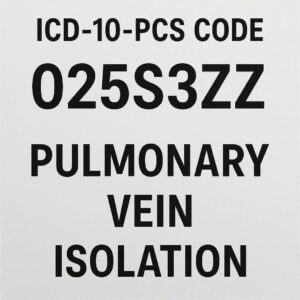
| 4 | Body Part | 3: Pulmonary Vein, Right 4: Pulmonary Vein, Left M: Pulmonary Vein, Right Superior N: Pulmonary Vein, Right Inferior T: Pulmonary Vein, Left Superior V: Pulmonary Vein, Left Inferior |
Crucial: You must code EACH pulmonary vein that is isolated. If all four are isolated (the standard procedure), you will report four separate codes. Using the less specific “3” or “4” is acceptable if the operative report does not specify superior/inferior. However, greater specificity is preferred if documented. |
| 5 | Approach | 3: Percutaneous 7: Via Natural or Artificial Opening |
3: Percutaneous is standard for catheter-based ablation via femoral venous access. 7 would be used only in exceedingly rare endoscopic surgical approaches. |
| 6 | Device | Z: No Device | Character 6 is always Z (No Device) for ablation. The catheter is a tool, not a device left in place. |
| 7 | Qualifier | Z: No Qualifier X: Diagnostic |
Z: No Qualifier is used for therapeutic ablation. X: Diagnostic is used only if the procedure is solely for diagnostic mapping with no therapeutic ablation delivered. |
Example Codes:
-
Isolation of all four pulmonary veins via cryoballoon ablation:
-
02583ZZ Destruction of Pulmonary Vein, Right Superior, Percutaneous Approach
-
02584ZZ Destruction of Pulmonary Vein, Right Inferior, Percutaneous Approach
-
0258TZZ Destruction of Pulmonary Vein, Left Superior, Percutaneous Approach
-
0258VZZ Destruction of Pulmonary Vein, Left Inferior, Percutaneous Approach
-
6.4 Common Coding Scenarios and Complexities
-
Additional Ablation: If lesions are also placed in the left atrial posterior wall, mitral isthmus, or for cavotricuspid isthmus ablation (for flutter), separate codes from body parts 5: Atrium, Right or 7: Atrium, Left would be added.
-
Failed/Partial Isolation: Code only the veins on which the procedure was actually performed, as documented. Do not code for intention.
-
Repeat Procedures: Coding is identical; the code reflects the procedure performed, not whether it’s a first or redo.
6.5 Avoiding Pitfalls: Distinguishing PVI from Other Ablations
-
Atrial Flutter Ablation: Root operation is the same (Destruction), but Body Part is typically 5: Atrium, Right or 7: Atrium, Left, with a Qualifier sometimes specifying the cavotricuspid isthmus.
-
AV Node Ablation: Body Part is H: Atrioventricular Node, often with a Device character for a pacemaker lead if performed concomitantly.
7. Outcomes, Risks, and the Future of PVI
7.1 Efficacy and Success Rates
Success is typically defined as freedom from atrial arrhythmias off antiarrhythmic drugs. For paroxysmal AFib, single-procedure success rates are ~70-80%, rising to ~85-90% with a repeat procedure. Success rates are lower for persistent and long-standing persistent AFib (~50-70%), often requiring more extensive ablation.
7.2 Complications: Recognition and Management
Major complications occur in 2-5% of cases:
-
Cardiac Tamponade (0.5-1%): Emergency pericardiocentesis required.
-
Stroke/TIA (<1%): Mitigated by pre-procedural imaging and anticoagulation.
-
Pulmonary Vein Stenosis (<1% with modern techniques): Managed with dilation/stenting.
-
Phrenic Nerve Palsy (more common with cryoballoon on the right side): Usually transient.
-
Atrio-Esophageal Fistula (0.1-0.2%): A rare but often fatal complication thermal ablation; PFA may eliminate this risk.
7.3 The Horizon: AI, Robotics, and Personalized Ablation Strategies
The future lies in precision ablation. Artificial intelligence will analyze ECGs and imaging to predict optimal ablation targets. Robotic catheter navigation may enhance stability and precision. PFA technology will mature. The goal is moving from a “one-size-fits-all” circumferential PVI to patient-specific ablation strategies targeting individual arrhythmia substrates.
8. Conclusion
Pulmonary Vein Isolation stands as a testament to the convergence of anatomical insight, technological innovation, and procedural mastery in curing atrial fibrillation. From its historical roots to the advent of pulsed field ablation, PVI continues to evolve, offering patients freedom from arrhythmia and its associated risks. Understanding this procedure in its full depth—from the electrophysiology lab to the precise language of ICD-10-PCS coding—is essential for all stakeholders in cardiovascular care to ensure optimal patient outcomes, accurate data representation, and the continued advancement of the field.
9. Frequently Asked Questions (FAQs)
Q1: Is PVI a cure for atrial fibrillation?
A: It is considered a potentially curative procedure, especially for paroxysmal AFib. Success rates are high, but some patients may require more than one procedure or may continue to need medication. It is the most effective intervention available for reducing AFib burden.
Q2: What is the recovery time after a PVI?
A: Most patients stay overnight for observation and go home the next day. Light activities can be resumed in a few days, but strenuous exercise should be avoided for 1-2 weeks. The heart undergoes healing and remodeling for up to 3 months (the “blanking period”).
Q3: How do I choose between Radiofrequency and Cryoballoon ablation?
A: The choice is made by your electrophysiologist based on your specific anatomy, type of AFib, and their expertise. Both are highly effective. Cryoballoon can be faster, while RF allows for more tailored lesion sets. Discuss the pros and cons for your individual case.
Q4: In ICD-10-PCS, why do I need four codes for one PVI procedure?
A: Because each pulmonary vein is a distinct, separate body part in the coding system. The procedure of Destruction (ablation) is performed on each individual anatomical structure. Reporting four codes accurately reflects the full work performed.
Q5: What is the single most important documentation tip for accurate PVI coding?
A: The operative report must explicitly name each pulmonary vein that was successfully isolated (e.g., “entrance block was confirmed in all four pulmonary veins” or “the right superior and right inferior veins were isolated, but isolation of the left veins could not be achieved”). Specificity is key.
10. Additional Resources
-
Heart Rhythm Society (HRS): www.hrsonline.org – Provides patient education materials and clinical practice guidelines for AFib ablation.
-
American College of Cardiology (ACC): www.acc.org – Offers extensive clinical resources and latest research in electrophysiology.
-
CDC – Atrial Fibrillation: www.cdc.gov/heartdisease/atrial_fibrillation.htm – Public health information on AFib.
Disclaimer
Date: December 13, 2025
Author:Medical Content Specialist
The information contained in this article is for educational and informational purposes only and is not intended as a substitute for professional medical advice, diagnosis, or treatment. Always seek the advice of your physician or other qualified health provider with any questions you may have regarding a medical condition or procedure. The coding information provided is for illustrative educational purposes and should be verified against the official ICD-10-PCS coding manuals and payer-specific guidelines for accurate procedural coding and billing. The author and publisher are not responsible for any errors or omissions or for any consequences from application of the information herein.


