In the high-stakes world of modern healthcare, a hip fracture is more than a medical diagnosis; it is a life-altering event. For the elderly population, it carries a sobering one-year mortality rate of up to 30%. For the healthcare system, it represents a significant clinical and financial challenge, with costs running into billions of dollars annually. At the very heart of bridging the clinical event with the administrative process lies a seemingly cryptic language: the International Classification of Diseases, Tenth Revision, Procedure Coding System (ICD-10-PCS). For the procedure that has become the gold standard for stabilizing one of the most common hip fractures—the intertrochanteric fracture—the correct application of this language is not merely an administrative task. It is a critical function that ensures accurate reimbursement, drives quality metrics, facilitates vital research, and ultimately, reflects the true complexity of the care provided. This article is a deep dive into the universe of ICD-10-PCS coding for intertrochanteric hip nailing, a journey that will take us from the intricate anatomy of the femur to the precise logic of procedural coding, empowering coders, clinicians, and healthcare administrators alike.
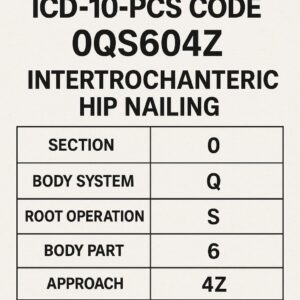
ICD-10-PCS code for intertrochanteric hip nailing
2. Anatomy of a Fracture: Deconstructing the Intertrochanteric Region
To accurately code a procedure, one must first understand the anatomy it addresses. The intertrochanteric region is not just a “part of the hip”; it is a dynamic and mechanically critical junction.
The Hip Biomechanics: A Marvel of Engineering
The hip is a classic ball-and-socket synovial joint, designed for both mobility and remarkable stability. The femoral head (the “ball”) articulates with the acetabulum of the pelvis (the “socket”). Connecting the femoral head to the long, straight femoral shaft are two vital bony prominences: the greater and lesser trochanters. These serve as the attachment levers for the powerful gluteal and iliopsoas muscles, which control hip movement. The weight-bearing forces from the upper body are transmitted down the femoral neck to the shaft, and the intertrochanteric line is where these forces converge and are redirected.
The Intertrochanteric Line: A Zone of High Stress
The intertrochanteric region is the area of bone found between the greater and lesser trochanters. It is composed primarily of cancellous (spongy) bone, which is highly vascular and excellent at healing but is mechanically weaker than the cortical (compact) bone of the shaft. In young, healthy bone, this area can withstand immense forces. However, with age and conditions like osteoporosis, the cancellous bone becomes porous and fragile. A simple fall from a standing height can then generate forces sufficient to cause a catastrophic break through this region.
Classifying the Break: The AO/OTA Foundation for Coding
Not all intertrochanteric fractures are the same. The AO Foundation/Orthopaedic Trauma Association (AO/OTA) classification is the universal language surgeons use to describe these injuries, and it has direct implications for coding complexity. The classification is as follows:
-
31-A1: Stable Pertrochanteric Fracture: A simple, non-comminuted (two-part) fracture line. The posteromedial cortex is intact, making it inherently stable.
-
31-A2: Unstable Pertrochanteric Fracture with Posteromedial Comminution: The fracture line extends, with multiple fragments (comminution) in the critical posteromedial area, which includes the lesser trochanter. This compromises stability.
-
31-A3: Intertrochanteric Fractures Involving the Lateral Cortex: This includes reverse obliquity and transverse fractures. The primary fracture line runs laterally, making them highly unstable and particularly suited for intramedullary nailing, as sliding hip screws are prone to failure.
* AO/OTA Classification of Intertrochanteric Fractures and Coding Implications*
| AO/OTA Class | Description | Stability | Preferred Surgical Method | Coding Complexity |
|---|---|---|---|---|
| 31-A1 | Simple, two-part fracture | Stable | Sliding Hip Screw or Cephalomedullary Nail | Low |
| 31-A2 | Comminuted fracture with posteromedial involvement | Unstable | Cephalomedullary Nail | Moderate to High (may involve additional procedures like fragment fixation) |
| 31-A3 | Reverse obliquity or transverse fracture | Very Unstable | Cephalomedullary Nail | High (often requires precise reduction and locking) |
Understanding this classification allows the coder to anticipate the procedural details they will encounter in the operative report. An A3 fracture, for instance, almost guarantees the use of an intramedullary nail and may involve more complex reduction maneuvers.
3. The Surgical Solution: Intertrochanteric Nailing Unveiled
The evolution from traction and prolonged bed rest to modern internal fixation represents one of orthopaedic surgery’s great successes. The intertrochanteric nail is the pinnacle of this evolution for unstable fracture patterns.
Philosophy of Intramedullary Fixation: Why a Nail?
The principle is one of load-sharing rather than load-bearing. An intramedullary nail is inserted into the hollow medullary canal of the femur, acting as an internal splint. It aligns the fragments and bears a significant portion of the mechanical load, allowing for immediate post-operative weight-bearing in many cases. This is in contrast to extramedullary devices like the sliding hip screw, which act as a side plate and are subject to higher bending forces, leading to a higher risk of failure in unstable fractures.
The Cephalomedullary Nail: The Modern Workhorse
The term “cephalomedullary” literally means “head” and “marrow cavity.” This describes the implant perfectly. It consists of:
-
The Main Nail: A sturdy metal rod that sits in the femoral shaft’s medullary canal.
-
The Lag Screw (or Blade): A large screw that passes through a proximal hole in the nail, up the femoral neck, and into the femoral head. This screw is designed to “collapse” or slide within the nail, allowing for impaction at the fracture site as healing occurs—a crucial feature to prevent cut-out of the screw from the head.
-
Distal Locking Screws: One or two screws placed through holes at the distal end of the nail, traversing the femoral cortex. These prevent the nail from rotating or sliding distally within the canal, providing rotational stability.
Step-by-Step Surgical Walkthrough: From Incision to Closure
A detailed operative report will describe these steps, which form the basis for coding:
-
Positioning and Preparation: The patient is placed on a fracture table, and the leg is positioned and placed in traction to achieve a preliminary reduction under fluoroscopy.
-
Incision: A small lateral incision is made at the hip, proximal to the greater trochanter.
-
Entry Point and Canal Preparation: The surgeon enters the medullary canal, typically at the tip of the greater trochanter or through a piriformis fossa approach. A guide wire is passed down the canal, followed by sequential reamers to widen the canal to accommodate the nail.
-
Nail Insertion: The appropriately sized nail is attached to its insertion jig and manually tapped into position down the medullary canal.
-
Proximal Locking (Cephalic Component): Using the jig, a guide wire is passed across the femoral neck into the head. Its position is verified on live X-ray (fluoroscopy). The lag screw is then drilled and tapped over this guide wire and inserted.
-
Distal Locking: Using either a jig-guided or free-hand fluoroscopic technique, the distal locking screws are placed through stab incisions.
-
Closure: The incisions are irrigated, and the deep tissues, fascia, and skin are closed in layers.
(Image: A detailed anatomical illustration showing a cephalomedullary nail in place within a femur, with labels for the nail, lag screw, distal locking screws, greater trochanter, and femoral head.)
4. ICD-10-PCS Demystified: The Language of Procedural Coding
ICD-10-PCS is a multi-axial system where each character has a specific meaning, and the combination of these characters defines a unique procedure.
The 7-Character Alphanumeric System: A Logical Framework
-
1st Character: Section – Broad category (e.g., Medical and Surgical = 0).
-
2nd Character: Body System – The general physiological system (e.g., Lower Joints = Q).
-
3rd Character: Root Operation – The objective of the procedure (e.g., Reposition = S).
-
4th Character: Body Part – The specific anatomical site (e.g., Femoral Shaft = 7).
-
5th Character: Approach – The technique used to reach the site (e.g., Open = 0).
-
6th Character: Device – Any device left in place (e.g., No Device = Z).
-
7th Character: Qualifier – An additional attribute for further specificity (e.g., No Qualifier = Z).
The Importance of the Objective: Why “Reposition” is Rarely Correct
The root operation is the cornerstone of ICD-10-PCS coding. For fracture care, the three most common root operations are:
-
Reposition (S): Moving to its normal location, or other suitable location, all or a portion of a body part. The key is that the procedure is focused solely on moving the body part. No device is coded with Reposition.
-
Insertion (H): Putting in a non-biological device that remains in the body after the procedure. If the primary objective is to put in a device, and any moving of body parts is incidental, this is the correct root operation.
-
Supplement (U): A device is used to physically reinforce and/or augment the function of a body part. This was a common miscode for this procedure in the past.
The official ICD-10-PCS guidelines provide the definitive answer. Guideline B3.3 states that if a procedure is performed to treat a fracture, the root operation Reposition is used. The device used to maintain the reduction (the nail and screws) is not coded separately. The procedure of “open reduction internal fixation” (ORIF) is, in the language of PCS, a Reposition.
5. Building the Code: A Character-by-Character Deconstruction of 0QS70ZZ
Let’s build the primary code for a standard open intertrochanteric nailing procedure.
-
Section: 0 – Medical and Surgical
-
This is the correct section for the vast majority of invasive, therapeutic procedures performed in an operating room.
-
-
Body System: Q – Lower Joints
-
The Lower Joints body system includes the anatomical joints of the lower extremities. Crucially, the PCS table includes not just the joint articulation itself but also the adjoining bones. The hip joint body part values include the femoral head and neck. However, for a fracture that extends into the shaft, the specific body part must be carefully considered.
-
-
Root Operation: S – Reposition
-
As per the official guideline, the objective of the procedure is to “move the femoral fracture fragments to their normal anatomic location.” The insertion of the nail and screws is the method used to maintain this repositioning; it is not the primary objective.
-
-
Body Part: 7 – Femoral Shaft
-
This is the most critical and often misunderstood character. The PCS body part for a hip procedure is not “Hip.” The choices are:
-
Femoral Head (3)
-
Femoral Neck (4)
-
Femoral Shaft (7)
-
-
An intertrochanteric fracture, by definition, occurs between the trochanters. The lesser trochanter is part of the proximal femoral shaft. Therefore, a fracture that involves the intertrochanteric region and the lesser trochanter is coded to the Femoral Shaft (7). This aligns with the AO/OTA classification, where involvement of the lesser trochanter (A2, A3) defines the fracture as involving the shaft. For a pure, simple intertrochanteric fracture without shaft involvement, the body part would be Femoral Neck (4). However, in clinical practice, most fractures treated with a nail have some degree of comminution extending towards the shaft, making Femoral Shaft (7) the most frequently used value.
-
-
Approach: 0 – Open
-
An “Open” approach is defined as cutting through the skin or mucous membrane and any other body layers necessary to expose the site of the procedure. The initial lateral hip incision, dissection down to the bone, and creation of the entry portal into the medullary canal qualify as an open approach. The subsequent placement of the distal locking screws through separate stab incisions is considered part of the same overall open procedure.
-
-
Device: Z – No Device
-
This is a direct consequence of selecting the root operation Reposition. The guideline is explicit: the device used to maintain the repositioning is not coded.
-
-
Qualifier: Z – No Qualifier
-
There is no additional qualifier needed for this specific procedure in the Lower Joints body system.
-
Therefore, the primary code for an open reduction and internal fixation of an intertrochanteric femur fracture using an intramedullary nail is 0QS70ZZ: Reposition of Femoral Shaft, Open Approach.
6. The Device Dilemma: Coding the Implant with 0QSH0Z?
A common point of confusion arises from the existence of a code like 0QSH0Z? (Insertion of Intramedullary Fixation Device into Femoral Shaft, Open Approach). Why isn’t this the correct code?
The answer lies in the hierarchy of objectives defined by the official guidelines. The coder must identify the root operation that most accurately describes the principal objective of the procedure.
-
Scenario A (Principal Objective: Fix Fracture): The patient has a painful, deformed leg from a traumatic fracture. The surgeon’s goal is to put the bone back together so it can heal. The nail is the means to achieve this goal. Root Operation = Reposition.
-
Scenario B (Principal Objective: Insert Device): The patient has a non-union of a fracture that is already in acceptable alignment. The surgeon’s goal is to insert a new, more robust nail to stimulate healing. The bone is not being “moved,” it is being “reinforced.” Root Operation = Insertion.
In the vast majority of acute intertrochanteric fracture cases, the scenario is “A.” The fracture is the problem, and repositioning is the solution. The insertion of the nail is an integral part of the repositioning procedure and is not coded separately.
7. Navigating Nuances and Complex Scenarios
Coding is rarely about a single, perfect code. Real-world cases are messy and complex.
Bilateral Fractures: Duplicate Procedures or Distinct Encounters?
If a patient falls and sustains simultaneous bilateral intertrochanteric fractures, and both are fixed in the same operative session, you would assign the same code (0QS70ZZ) twice. Modifiers (like the LT/RT modifiers used in CPT) are not used in ICD-10-PCS. The PCS system itself does not specify laterality in the code; the fact that two identical procedures are coded indicates that they were performed on two separate body parts (the left and right femurs).
Hybrid Procedures: When Reposition is Paired with Other Operations
Sometimes, the surgeon must perform additional, distinct procedures.
-
Cerclage Wiring: If a large, separate fragment (e.g., a lesser trochanteric fragment) is wrapped with a wire or cable to secure it, this is a separate root operation: Supplement (0QU00?Z). The device would be the cable (qualifier would need to be determined from the table).
-
Bone Grafting: If bone graft is placed at the fracture site to aid healing, this is coded as Supplement (0QU00J9) if it’s autograft or allograft, representing the biological material.
The Unstable Fracture: Addressing the Lesser Trochanter
As discussed, involvement of the lesser trochanter shifts the body part to the Femoral Shaft. The coder must read the operative report’s “Pre-operative Diagnosis” and “Description of Procedure” to confirm the exact anatomical location of the fracture.
Percutaneous Approach: Is it Ever Applicable?
While the main part of the procedure is open, one might wonder if the distal locking screws, placed through stab incisions, constitute a percutaneous approach. The official guideline A9 states that if multiple approaches are used to accomplish the same root operation, the approach value is coded to the most invasive approach. Since the nail insertion requires an open approach, the entire procedure is coded as Open (0).
Treatment of Non-Union and Malunion: A Different Objective
For a non-union (the bone has not healed) or a malunion (the bone has healed in a bad position), the objective changes. The surgeon may need to perform a Reposition again if the fragments are moved. However, they might also perform an Excision of the fibrous non-union tissue, an Insertion of a new nail, or a Supplement with a bone graft. The operative report must be analyzed to determine the principal objective of the current procedure.
8. Coding for Associated Procedures: A Holistic Patient Record
A patient’s encounter involves more than just the main procedure.
-
Diagnostic Arthroscopy (0UJ68ZZ): Rarely done in acute trauma, but if the surgeon scopes the hip joint to assess for concomitant labral or cartilage damage, this would be an additional code.
-
Bone Grafting (0QU00J9): As mentioned, this is a separate procedure for Supplement using autologous tissue substitute.
-
Wound Debridement (0JQ60ZZ): If the fracture was open (broken skin), the surgeon would extensively debride non-viable tissue from the skin, muscle, and bone before fixation. This is coded separately using the root operation Excision from the “Skin,” “Muscles,” and “Bones” body systems as appropriate.
-
Blood Transfusions: Administered blood products are coded from the Administration section (3).
9. The Clinical Documentation Improvement (CDI) Imperative
The coder can only code what the document says. Clear and precise documentation is paramount.
What Coders Need from Surgeons: A Detailed Breakdown
An ideal operative report for an intertrochanteric nailing should clearly state:
-
Pre-operative Diagnosis: e.g., “Left unstable intertrochanteric femur fracture, AO/OTA 31-A2”
-
Post-operative Diagnosis: (Should be the same).
-
Procedure Performed: e.g., “Open reduction and internal fixation of left intertrochanteric femur fracture with cephalomedullary nail.”
-
Description:
-
Confirmation that the fracture was manipulated and reduced.
-
Specifics of the approach: “A standard lateral approach to the proximal femur was made.”
-
Anatomical landmarks: “The fracture was noted to extend from the greater trochanter to the lesser trochanter.”
-
Implant details (manufacturer, size) are less critical for ICD-10-PCS but are good practice.
-
Any additional procedures performed (e.g., “A cerclage cable was placed around a large, displaced lesser trochanteric fragment.”).
-
Query Opportunities: Clarifying Ambiguity
If the report is vague, a coder or CDI specialist may need to query the physician:
-
“The report describes an ‘intertrochanteric fracture,’ but the procedure details mention addressing the ‘lesser trochanteric fragment.’ Can you clarify if the fracture involves the lesser trochanter, confirming the body part as the femoral shaft?”
-
“For the cerclage wiring, was the objective to re-approximate a fragment (Reposition) or to reinforce the bone (Supplement)?”
10. Case Studies: From Operative Report to Final Code
Case Study 1: Standard Intertrochanteric Fracture Fixation
-
Report Snippet: “An 80-year-old female sustained a left hip fracture after a fall. Diagnosis: Left intertrochanteric femur fracture. Procedure: The patient was positioned on a fracture table. A lateral incision was made over the proximal femur. The fracture was identified and reduced under fluoroscopic guidance. The medullary canal was reamed, and a 10mm x 240mm cephalomedullary nail was inserted. A lag screw was placed in the center-center position of the femoral head. Distal locking was performed with one screw. Closure was performed in layers.”
-
Coding Analysis:
-
The objective is to fix a fracture: Root Operation = Reposition (S).
-
The fracture is intertrochanteric, a region that includes the proximal shaft: Body Part = Femoral Shaft (7).
-
A lateral incision was made: Approach = Open (0).
-
No separate device is coded.
-
-
Final Code: 0QS70ZZ
Case Study 2: Complex Fracture with Cerclage Wiring
-
Report Snippet: “A 65-year-old male with a comminuted left intertrochanteric fracture with a large, displaced lesser trochanteric fragment (AO/OTA 31-A2). After preliminary reduction and nail insertion, the lesser trochanteric fragment was noted to be unstable. It was reduced and secured with two cerclage cables.”
-
Coding Analysis:
-
Primary Procedure: Reposition of Femoral Shaft with Nail = 0QS70ZZ
-
Secondary Procedure: The cerclage cabling is a distinct objective. The cables are physical devices that reinforce the bone. Root Operation = Supplement (U). The body part is still the Femoral Shaft (7). The device is an Internal Fixation Device in the Lower Joints (Qualifier = J).
-
-
Final Codes: 0QS70ZZ and 0QU70JZ
Case Study 3: Revision for Failed Extramedullary Device
-
Report Snippet: “The patient presents with a failed sliding hip screw for an intertrochanteric fracture, with cut-out of the lag screw and varus collapse. Procedure: Removal of the failed sliding hip screw implant. Open reduction of the malunited fracture. Insertion of a new cephalomedullary nail.”
-
Coding Analysis:
-
This is a complex case. The principal objective is to revise the failed fixation. This involves:
-
Removal of old device: 0QPN0ZZ (Removal of Device from Femoral Shaft, Open)
-
Reposition of the malunited bone: 0QS70ZZ (Reposition of Femoral Shaft, Open)
-
Insertion of the new nail: Since the bone is being repositioned and the new nail is the method to hold it, the Insertion is not coded separately per Guideline B3.3. The Reposition is the definitive procedure.
-
-
-
Final Codes: 0QPN0ZZ and 0QS70ZZ
11. Conclusion: Synthesizing Knowledge for Coding Excellence
Mastering the ICD-10-PCS code for intertrochanteric hip nailing requires a synthesis of anatomical knowledge, surgical understanding, and strict adherence to coding guidelines. The principal code, 0QS70ZZ, is built on the foundation that the objective is Reposition of the Femoral Shaft via an Open approach, with the nail serving as an uncoded methodological component. Navigating complex scenarios demands a meticulous analysis of the operative report to identify distinct procedural objectives. Ultimately, precise coding is not an abstract exercise; it is the critical link that faithfully translates a surgeon’s skill into accurate data, ensuring the integrity of the patient record, the financial health of the institution, and the advancement of orthopaedic science.
12. Frequently Asked Questions (FAQs)
Q1: Why is the code for inserting a large metal nail “No Device”?
A: This is the most common point of confusion. In ICD-10-PCS logic, the device is not coded when its insertion is an integral part of accomplishing the root operation. For fracture care, the root operation is “Reposition.” The nail and screws are the means to hold the repositioned bone; they are not the objective. The objective was to move the bone back into place.
Q2: When would I use the “Insertion of Intramedullary Fixation Device” (0QSH0Z?) code?
A: You would use this code when the primary goal is to put in the device itself, and no significant “repositioning” is occurring. A classic example is an exchange nailing for a non-union where the fracture is already in acceptable alignment but has failed to heal. The surgeon is removing a broken or ineffective nail and inserting a new, larger one to provide more stability. The objective is “Insertion,” not “Reposition.”
Q3: How do I know if the body part is Femoral Neck (4) or Femoral Shaft (7)?
A: You must rely on the physician’s documentation. If the report specifies an “intertrochanteric fracture” and mentions the “lesser trochanter,” it is almost always involving the shaft. A pure “femoral neck fracture” is intracapsular and occurs further up, near the femoral head. If the documentation is unclear, a query is necessary.
Q4: The surgeon made a small incision for the nail and used stab incisions for the screws. Why isn’t this a Percutaneous approach?
A: The ICD-10-PCS guideline A9 states that if multiple approaches are used to accomplish the same objective, the procedure is coded to the most invasive approach. Creating an entry portal into the medullary canal through an incision that cuts down to the bone is considered an “Open” approach. The percutaneous portions are part of the same overall procedure and do not change the primary approach code.
Date: November 26, 2025
Author: Dr. Jonathan Thorne, MD, CCS-P
Disclaimer: *This article is intended for educational purposes and to illustrate professional coding principles. It is not a substitute for the official ICD-10-PCS guidelines, coding manuals, or professional medical advice. Medical coders must always rely on the most current year’s official resources and facility-specific protocols for final coding decisions.*


