
The human body is a map of hidden wires, and few dental procedures trigger as much anxiety about “hitting a nerve” as wisdom tooth extraction. Patients sit in the consultation chair, gripping the armrests, asking variations of the same question: “If you pull this tooth, will it paralyze my face?” This fear stems from a fundamental confusion about what lives inside a tooth versus what surrounds it. The question “Are there nerves in your wisdom teeth?” requires a precise anatomical answer, because the truth sits at the intersection of living pulp tissue and the major sensory highways of the jaw.
Yes, wisdom teeth contain nerves. Every healthy tooth does. The internal chamber of a tooth houses a delicate, jelly-like tissue called the dental pulp, which is rich with nerve fibers, blood vessels, and connective tissue. These nerves give the tooth its ability to feel hot coffee and cold ice cream. However, the nerve that keeps you awake at night before surgery is not inside the tooth. It is the inferior alveolar nerve, a major trunk that often snakes intimately around the roots of the lower wisdom tooth. This article dissects the internal innervation of the third molar, distinguishes it from the catastrophic nerve injury risk, and maps the exact anatomy that governs surgical consent forms.
The Living Core: Dental Pulp Under the Enamel
Let us drill down, literally, through the layers. The outer shell of a wisdom tooth is enamel—acellular, dead, crystalline. Beneath that lies dentin, a microtubular living tissue that transmits fluid shifts. At the very center sits the pulp chamber, extending down the roots as root canals. This pulp is a highly innervated organ.
The nerve fibers entering the apex of the wisdom tooth are branches of the trigeminal nerve (Cranial Nerve V), specifically the maxillary division (V2) for upper wisdoms and the mandibular division (V3) for lower wisdoms. These fibers are predominantly A-delta (sharp, piercing pain) and C-fibers (dull, throbbing, inflammatory pain). They enter the tiny apical foramen at the tip of the root and fan out into a plexus called the Raschkow’s plexus, right beneath the odontoblast cell layer. This is the biological alarm system. When decay breaches the enamel and hits the dentin, these nerve endings fire. When the pulp becomes irreversibly inflamed (pulpitis), the throbbing pain keeps you awake because the C-fibers are exquisitely sensitive to the increased internal pressure inside the rigid pulp chamber. Yes, your wisdom tooth has a rich nerve supply, and it can scream just as loudly as any other tooth.
Why This Matters for “Dead” Wisdom Teeth
A common patient assumption is that a wisdom tooth that isn’t hurting has no nerve. This is biologically false. A tooth can have a necrotic pulp—the nerve tissue inside has liquefied—but the nerve supply at the root apex is still alive and can still transmit pain from the surrounding infected bone. More critically, the tooth remains connected to the body’s nervous system via the periodontal ligament. The PDL is a collagenous sling that cushions the tooth in the socket, and it is packed with proprioceptive and nociceptive nerve endings. Even a “dead” tooth provides sensory feedback when you chew or when a dentist elevates it during extraction. You cannot escape the neural network. The tooth is a plug in a socket woven with sensory wiring.
The Inferior Alveolar Nerve: The Real Source of Fear
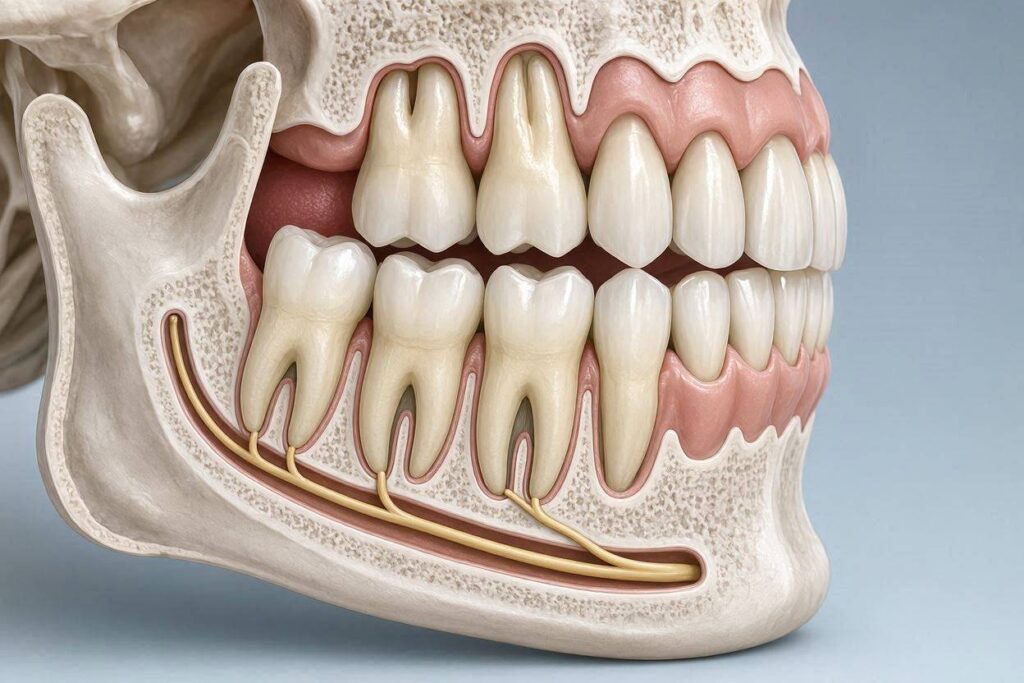
When a patient asks about “the nerve,” they rarely care about the microscopic pulp inside the crown. They are asking about the Inferior Alveolar Nerve (IAN), the large, macroscopic sensory trunk that runs in a bony canal just beneath the roots of the lower molars. This nerve provides sensation to the lower lip, chin, and lower teeth on that side.
The wisdom tooth, especially the lower third molar, is famous for having roots that dance dangerously close to this canal. A panoramic X-ray often shows the dark shadow of the roots superimposing over the white lines of the IAN canal. The proximity is the entire game of lower wisdom tooth extraction. If the nerve is merely touching the root, a careful extraction often leaves it intact. If the nerve is wrapped around the root or perforates it, the risk of damage skyrockets. This is not a risk from the nerve inside the tooth; it is a risk from the nerve hosting the tooth. The distinction must be made crystal clear: extracting the tooth removes the pulp nerves, which is irrelevant because the tooth is being discarded. But injuring the IAN removes sensation to the face, which can be permanent and life-altering.
The Lingual Nerve: The Hidden Danger
While everyone fixates on the IAN, the lingual nerve is the silent victim of wisdom tooth surgery. This nerve runs along the inner (lingual) side of the lower jaw, supplying taste sensation to the anterior two-thirds of the tongue and general sensation to the floor of the mouth and gums. It is not inside the tooth, but it sits just under the gum tissue on the tongue side of the third molar.
A sloppy surgical flap that extends too far lingually, or an elevator that slips, can sever or crush the lingual nerve. Unlike the IAN, which is protected inside a bony canal, the lingual nerve is soft, naked tissue in many areas. When patients report losing taste or feeling a burning, electric sensation on their tongue after wisdom tooth surgery, the lingual nerve was damaged. This is a pure external nerve injury; it has nothing to do with nerves in the tooth itself. But because the terms blur together in informed consent discussions, understanding the geography is vital. The tooth nerve is inside. The lip nerve is below. The tongue nerve is inside the gum to the tongue side.
Radiographic Signs: When the Roots Wrap the Nerve
Oral surgeons look for specific radiographic warning signs that the IAN is not just near the tooth but potentially intertwined with it. The 3D Cone Beam CT scan has revolutionized this diagnosis, replacing the flat, shadowy 2D panoramic X-ray for high-risk cases. Warning signs of true interposition include:
- Darkening of the root: The black line of the canal crosses the root shadow, suggesting the root has a groove or notch that the nerve sits in.
- Narrowing of the canal: The canal appears squeezed as it passes the root, indicating the nerve is physically displaced.
- Interruption of the lamina dura: The thin white line of the canal wall disappears where it touches the root, implying the nerve is no longer separated from the root by bone.
If these signs are present, the nerve is not just “close”—it may be attached to the root surface. This is a surgical nightmare. The tooth itself is not the nerve, but the nerve has essentially melded with the root’s external cementum. When this occurs, pulling the tooth may literally avulse a segment of the nerve. This is where the intra-tooth nerve (the dying pulp) and the extra-tooth nerve (the IAN) become pathologically linked. Chronic inflammation from the wisdom tooth can cause the nerves to become “friendly” at a microscopic level, making separation impossible.
The “Nerve Inside” vs. “Nerve Outside” Table
To eliminate confusion permanently, here is the comparative anatomy of the neural structures relevant to a wisdom tooth extraction.
| Structure | Location | Function | Fate During Extraction | Injury Consequence |
|---|---|---|---|---|
| Dental Pulp Nerves | Inside the tooth (crown and roots). | Hot/cold, sharp pain. | Removed with the tooth. | None (tooth is discarded). |
| Periodontal Ligament Nerves | Lining the socket wall. | Pressure, proprioception. | Stretched and severed. | Temporary local socket soreness. |
| Inferior Alveolar Nerve (IAN) | Mandibular canal below the roots. | Sensation to lower lip/chin. | Compressed, severed, or stretched. | Paresthesia (numbness), paralysis. |
| Lingual Nerve | Soft tissue lingual to the molar. | Taste and sensation to tongue. | Severed or crushed. | Loss of taste, tongue numbness. |
This table is the essential clinical map. The internal nerve is a sacrifice; the external nerves are the surgical gauntlet.
Paresthesia: The Persistent Numbness
When the IAN or lingual nerve is damaged, the result is paresthesia—a partial or complete loss of sensation that may be temporary or permanent. The statistics vary by study, but roughly 0.5% to 2% of lower third molar extractions result in some degree of temporary IAN paresthesia, with permanent damage (beyond 6–12 months) estimated at 0.1% to 0.5%.
A numb lip is not just an inconvenience. It means you cannot feel if soup is burning your skin. You drool without knowing it. You bite your lower lip repeatedly, causing traumatic ulcers. Kissing feels asymmetrical. The psychological burden of a “dead” face segment is immense. This is the risk that generates the lawsuits. The nerve inside the wisdom tooth dies a painless death in the surgical waste bin. The nerve below the wisdom tooth, if injured, lives on as a dysfunctional sensory ghost, sending either zero signals or false “pins and needles” signals for decades. The patient who asks, “Are there nerves in wisdom teeth?” deserves to know that the dangerous nerve is the one they cannot feel in the tooth, but the one they will feel missing in the world.
Maxillary Wisdom Teeth: The Sinus Nerve Connection
Upper wisdom teeth present a different neural landscape. The IAN does not exist in the maxilla. Instead, the posterior superior alveolar nerve supplies the upper molars. The risk here is not sensory loss to the face but a nerve-driven headache phenomenon. The roots of the upper wisdom tooth often protrude into the maxillary sinus. The sinus membrane itself is innervated by branches of the trigeminal nerve.
If an upper wisdom tooth extraction is traumatic or creates an oro-antral communication (a hole into the sinus), the patient can suffer from chronic neuropathic sinus pain. The nerve pain inside the tooth is gone, but the nerve pain in the sinus lingers. This is often misdiagnosed as a sinus infection. The nerve anatomy here is diffuse and less catastrophic than the IAN, but it explains why some patients trade a toothache for a “ghost headache” after extraction. The interconnectedness of the maxillary dentition and the sinus nerves is a testament to the head’s complex wiring.
Coronectomy: Leaving the Nerves Undisturbed
Because the nerve outside the tooth is the priority, modern surgery has a clever trick: the coronectomy. If the wisdom tooth roots are deeply wrapped around the IAN, the surgeon cuts off the crown (the visible part) and leaves the roots intentionally buried in the jawbone. The pulp nerves inside those roots are severed from the crown, so they die. The IAN remains untouched. The risk of paresthesia drops to nearly zero.
This procedure acknowledges that the tooth nerve is a non-issue, but the IAN nerve is everything. The retained root fragment eventually stabilizes or is encased in bone. The trade-off is a very small risk of late infection requiring a second surgery. For the patient, the peace of mind is worth it. The decision matrix is simple: lose the crown and the internal nerve (via coronectomy) or lose the tooth and risk taking a piece of the external nerve with it. The existence of this procedure proves that oral surgeons care about one nerve and one nerve only during extraction—and it is not the one inside the tooth.
Important Note: The Staged Extraction Protocol
In certain extremely high-risk cases where the IAN is encircling the root, the surgeon may recommend orthodontic extrusion before extraction. An orthodontist places a bracket on the wisdom tooth and slowly pulls it away from the nerve over 2–3 months. The tooth nerve is still alive and screaming (braces on a wisdom tooth hurt), but the IAN nerve is slowly decompressed as the roots are teased away from the canal. This is the ultimate demonstration of nerve hierarchy: we will torture the tooth’s own nerve to protect the lip nerve.
Trigeminal Neuralgia and Phantom Tooth Pain
We must address the dark side of dental neurology. Sometimes the nerve inside the tooth is not the problem; the nerve ganglion itself is diseased. Trigeminal neuralgia is a lightning-strike pain condition affecting Cranial Nerve V. Patients often mistakenly believe a specific wisdom tooth is the culprit. They demand extraction. The tooth is pulled, and the pain remains—because the pain was never in the tooth; it was in the nerve trunk at the skull base.
This is phantom tooth pain, or atypical odontalgia. The pulp nerves in the extracted tooth are sitting in a medical waste bin, but the patient still feels the toothache. This phenomenon is the cruelest proof that pain is a brain construct, not a tooth event. The nerve inside the tooth was just the messenger. The true source was the hyper-excitable nerve center. For a small percentage of patients asking this question, their wisdom tooth nerve is perfectly healthy, but their trigeminal ganglion is firing randomly. Removing the tooth removes a healthy messenger, leaving the culprit intact. This is why a thorough neurological history is essential before elective third molar extraction.
The Inflammatory Mediator Bridge
There is a fascinating biological bridge between the nerve inside the tooth and the nerve outside. If the wisdom tooth has a chronic periapical infection (an abscess pocket at the root tip), the inflammatory soup—cytokines like TNF-alpha, prostaglandins, and nerve growth factor—can chemically sensitize the IAN.
In these cases, the patient’s lip might already tingle or feel “weird” before surgery because the infected tooth nerve is spewing inflammatory waste onto the nearby IAN. The two nerves are chemically coupled. When the surgeon removes the tooth, the IAN often recovers because the source of chemical irritation is gone. This is why some pre-operative paresthesia resolves post-extraction. The tooth nerve infected the main nerve; removing the patient zero stopped the spill. However, during surgery, the already-inflamed IAN is fragile and more prone to physical injury. The nerve-to-nerve relationship is not just anatomical; it is biochemical. A hot, infected socket is a toxic environment for the IAN, making the timing of extraction a neurological balancing act.
Regeneration Research: Can the Tooth Nerve Be Saved?
Neuroscience is pushing boundaries on whether the pulp nerve can be regenerated. Stem cell research focuses on implanting dental pulp stem cells into a cleaned canal to regrow a living nerve. For wisdom teeth specifically, this research is an ironic footnote. Wisdom teeth are often extracted and thrown away, but they are also a goldmine of mesenchymal stem cells for banking.
Companies now offer to cryopreserve your extracted wisdom tooth pulp because the nerves and stem cells inside are young, pluripotent, and potentially useful for future regenerative medicine. In this light, the nerves in your wisdom teeth are not just sensory alarms; they are a biological resource. The nerve tissue you never think about until it aches might one day repair a spinal cord or a cardiac muscle. This scientific horizon recontextualizes the question completely. Yes, there are nerves in your wisdom teeth, and they are precious enough that some families pay hundreds of dollars to store them in liquid nitrogen.
Conclusion
Wisdom teeth are fully innervated organs, containing a living pulp with A-delta and C-nerve fibers that register pain, temperature, and pressure, making them biologically identical to any other tooth in your mouth. However, the nerve that dominates surgical consent—the inferior alveolar nerve—is not inside the tooth but runs beneath it in a bony canal, and injury to this external structure can cause permanent numbness of the lip and chin. The distinction between the internal, disposable pulp nerve and the external, functionally vital sensory nerve is the single most important anatomical fact any patient must grasp before signing a surgical release.
FAQ
Q: If the wisdom tooth nerve dies, why do I still need anesthesia for extraction?
A: Even if the pulp inside the tooth is necrotic (dead), the periodontal ligament anchoring the tooth to the bone is richly innervated and fully alive. You will feel the pressure and the stretching of those fibers during extraction. More importantly, the surrounding gum tissue, bone, and the inferior alveolar nerve itself must be anesthetized. The internal tooth nerve is a tiny fraction of the neural network involved in pulling a tooth.
Q: Can the nerve grow back if it’s damaged during extraction?
A: Peripheral nerves can regenerate at a rate of approximately 1 millimeter per day, but only if the injury is a crush (axonotmesis) and the nerve sheath (endoneurium) is intact. If the nerve is completely severed (neurotmesis) and the ends are not surgically approximated, regeneration is haphazard and often results in a painful neuroma—a tangled ball of disorganized nerve scar tissue that fires spontaneously. This is why microsurgical nerve repair is sometimes indicated if a lingual nerve is cut cleanly during surgery.
Q: Are upper wisdom teeth safer regarding nerves?
A: Generally, yes, because the inferior alveolar nerve does not exist in the maxilla. However, the posterior superior alveolar nerve can be bruised, and the palate can be injured if the greater palatine nerve is compressed by an improperly positioned surgical instrument. The sensory loss is much less noticeable to the patient (small area of inner gum), so it is rarely litigated, but it is a nerve injury nonetheless.
Additional Resource
For detailed medical illustrations and 3D interactive models of the trigeminal nerve pathways relevant to third molars, visit the anatomical resources provided by the American Association of Clinical Anatomists at www.clinical-anatomy.org.