If you have been told you need a detailed look at your liver, your doctor has likely ordered an MRI. It is one of the most advanced tools we have for seeing what is happening inside the abdomen. But when you look at the paperwork, you might see a confusing string of numbers and abbreviations: CPT code MRI liver w/wo contrast.
It looks like a puzzle. But it is really just a specific language that doctors and insurance companies use to make sure everyone is on the same page. Understanding this language is important. It helps you know what you are paying for and why the test is necessary.
In this guide, we will break down everything you need to know about liver MRI coding. We will cover the difference between a scan with contrast versus without, the specific CPT codes used, how much you might expect to pay, and how to prepare for the exam. Whether you are a patient trying to understand a bill or a medical coder looking for clarity, this article is designed to help.
CPT Code MRI Liver w wo Contrast
What is a Liver MRI?
Before we dive into the numbers, let us talk about the test itself. An MRI, or Magnetic Resonance Imaging, is a type of scan that uses powerful magnets and radio waves to create detailed pictures of the organs inside your body. Unlike X-rays or CT scans, MRIs do not use radiation.
When a doctor orders an MRI of the liver, they are looking for a very clear, three-dimensional view. The liver is a complex organ. It handles hundreds of vital functions, from filtering toxins to helping with digestion. An MRI can help identify:
-
Tumors (both benign and malignant)
-
Scarring (cirrhosis)
-
Fatty liver disease
-
Abscesses or infections
-
Blockages in the bile ducts
-
Damage from trauma
The detail provided by an MRI is often superior to other imaging methods. This makes it the go-to choice for surgeons and oncologists who need to make critical decisions about treatment.
Understanding “With and Without Contrast”
When you see the phrase “w/wo contrast,” it refers to whether a special dye is used during the scan. This is a crucial distinction, and it directly affects the CPT code used.
MRI Liver Without Contrast
In this scenario, you simply lie on the table, and the machine takes images of your liver in its natural state. This is often done first to establish a baseline. It shows the structure and size of the organ.
MRI Liver With Contrast
This involves injecting a contrast agent, usually gadolinium, into a vein in your arm. This dye travels through the bloodstream and into the liver. It highlights specific areas.
Why Do Both?
You might wonder why doctors often order both phases. The reason is accuracy. By taking images before the contrast is injected, and then immediately after, radiologists can see how blood flows through the liver.
Tumors often have different blood flow patterns than healthy tissue. They might “light up” with contrast in a way that makes them visible. A cancerous lesion might absorb the dye differently than a benign cyst. By comparing the “without” images to the “with” images, the radiologist gets a dynamic view of the liver’s health.
Important Note: If you have kidney problems or a known allergy to contrast materials, it is vital to tell your doctor before the exam. Contrast is generally safe, but your medical team needs to know your history.
The Specific CPT Codes for Liver MRI
CPT stands for Current Procedural Terminology. These codes are maintained by the American Medical Association (AMA). They are used to standardize medical billing across the United States.
For an MRI of the liver, there are three specific codes. They are often confused, so let us clarify them.
CPT 74181: MRI Abdomen Without Contrast
This code is used when the MRI is performed on the abdomen without any contrast material. While it covers the abdomen, it is often used for liver imaging when a doctor just needs a basic structural view. However, if the order specifies “liver w/wo,” this is usually not the final code used for billing.
CPT 74182: MRI Abdomen With Contrast
This code covers an MRI of the abdomen where contrast material is administered. Again, this is a broad abdominal code. If the scan only involves the contrast phase and no pre-contrast images, this might be used. However, for the specific “with and without” protocol, we look to the next code.
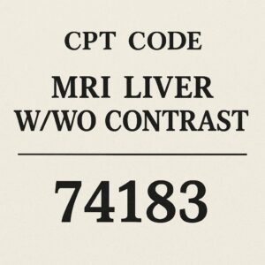
CPT 74183: MRI Abdomen Without Contrast Followed by With Contrast
This is the code you are looking for. CPT 74183 is the specific code for a magnetic resonance imaging procedure of the abdomen that includes both the non-contrast and contrast phases.
When a doctor orders a “CPT code MRI liver w/wo contrast,” they are almost always referring to 74183.
This code covers the entire process:
-
The initial scan without the dye.
-
The injection of the contrast agent.
-
The subsequent scans after the injection.
It is a comprehensive code that recognizes the extra time, skill, and resources required to perform a dynamic study of the liver.
Comparative Table: CPT 74181 vs 74182 vs 74183
To make the differences crystal clear, here is a simple breakdown of the three codes you might encounter.
| CPT Code | Description | What It Includes | Typical Use Case |
|---|---|---|---|
| 74181 | MRI Abdomen w/o contrast | Only non-contrast images | Screening for large masses, basic anatomy check, or if patient has kidney failure. |
| 74182 | MRI Abdomen w/ contrast | Only contrast-enhanced images | Follow-up on a known finding; less common for initial liver diagnosis. |
| 74183 | MRI Abdomen w/o & w/ contrast | Both non-contrast and contrast phases | The standard for liver lesions, tumors, and characterization. This is the code for a comprehensive liver exam. |
As you can see, 74183 is the most comprehensive option. It provides the radiologist with the full picture needed to make an accurate diagnosis.
How CPT 74183 Applies to Liver Imaging
When a patient presents for a liver MRI, the protocol is almost always designed to justify the use of 74183. The radiologist is not just looking at the liver; they are looking at the behavior of the liver.
Let us look at a practical scenario.
Imagine a patient has a spot on their liver found during a routine ultrasound.
The doctor orders an MRI liver w/wo contrast.
The technologist performs the scan.
They start with 74181 (without contrast) to see the spot in its natural state.
Then, they inject the contrast and perform dynamic imaging.
This second half falls under 74182 (with contrast).
However, because the two are performed together in a single session for a single diagnostic purpose, they are bundled into 74183.
The Importance of Contrast in Liver Diagnosis
Contrast is essential for characterizing liver lesions. There are many types of spots that can appear on the liver.
-
Hemangiomas: These are benign blood vessel tumors. They have a very specific “fill-in” pattern with contrast.
-
Focal Nodular Hyperplasia (FNH): Another benign lesion that has a distinct “central scar” that lights up with contrast.
-
Hepatocellular Carcinoma (HCC): A primary liver cancer. It has a specific pattern of washing out contrast quickly.
Without the “with and without” comparison provided by code 74183, it is very difficult for a radiologist to tell the difference between these conditions. This is why the CPT code matters. It ensures the imaging center is paid for the complex protocol required to give you an accurate answer.
Financial Considerations: Cost and Insurance
One of the biggest concerns for patients is the cost. An MRI is an expensive test, and the addition of contrast adds to the complexity and price.
Factors Affecting Cost
The price for CPT 74183 can vary wildly based on several factors:
-
Geographic Location: Costs are generally higher in large metropolitan areas compared to rural clinics.
-
Facility Type: A hospital-based outpatient department will usually charge more than an independent imaging center.
-
Insurance Negotiations: Insurance companies negotiate rates with providers. The “list price” is rarely what is actually paid.
-
Deductibles: If you have a high-deductible health plan, you may be responsible for the negotiated rate until your deductible is met.
Average Cost Estimates
It is difficult to give a fixed price, but here is a general range for CPT 74183 in the United States.
-
Without Insurance (Cash Price): $1,500 to $4,000+
-
With Insurance (Negotiated Rate): $400 to $1,200 (depending on plan)
-
Medicare: Medicare has a set fee schedule. For 2026, the facility rate for 74183 is typically between $300 and $600.
Important Note: Always ask for the CPT code before scheduling your appointment. You can call your insurance company and give them the code (74183) to get an accurate estimate of your out-of-pocket cost. Do not rely on verbal descriptions like “liver MRI.” Use the code.
How to Reduce Your Bill
If you are concerned about costs, you have options.
-
Shop Around: Independent imaging centers often offer lower rates than hospitals.
-
Ask for the Cash Price: Many facilities offer a discount if you pay upfront without using insurance.
-
Financial Assistance: Non-profit hospitals are required to offer financial assistance programs. Ask about their policy.
Preparing for Your Liver MRI
Preparation is key to a successful scan. If you move during the scan or if there is interference, the images become blurry, and you might have to do it again.
Food and Drink
For a liver MRI using CPT 74183, you will likely be asked to fast.
-
Nothing to eat or drink for 4 to 6 hours before the exam.
-
This is important because food in the stomach or intestines can create artifacts (blurring) in the images.
-
It also ensures that the gallbladder is full, which helps the radiologist see it clearly.
Medications
You can usually take your regular medications with a small sip of water unless your doctor tells you otherwise. If you are diabetic, talk to your doctor about your insulin dose, as fasting may affect your blood sugar levels.
What to Wear
-
Wear comfortable, loose-fitting clothing.
-
Avoid metal: You will be asked to remove jewelry, watches, glasses, and hairpins.
-
You will likely be given a hospital gown to wear to ensure no metal is on your clothing.
Safety Screening
Before entering the MRI room, you will fill out a safety questionnaire. The magnet is incredibly strong. You must tell the technologist if you have:
-
Pacemaker or implantable defibrillator
-
Aneurysm clips
-
Cochlear implants
-
Metal fragments in your eyes
-
Certain types of stents or artificial joints (most are safe, but they need to know)
What to Expect During the Procedure
If you have never had an MRI before, the process can seem intimidating. Knowing what will happen can help ease your anxiety.
Step 1: Arrival and Check-In
You will arrive, check in, and verify your insurance information. You will sign consent forms acknowledging the use of contrast.
Step 2: IV Placement
A nurse or technologist will place a small IV line into a vein in your hand or arm. This is for the contrast injection.
Step 3: The Scan Begins
You will lie on a padded table that slides into the large, donut-shaped magnet.
-
The Noise: The machine makes loud knocking and buzzing noises. You will be given earplugs or headphones.
-
Holding Still: This is the hardest part. You must hold your breath for short periods (usually 10 to 20 seconds at a time). The technologist will give you instructions over an intercom: “Breathe in, breathe out, and hold your breath.”
Step 4: The Contrast Injection
Partway through the scan, the technologist will inject the contrast through your IV.
-
Sensation: You might feel a cool sensation rushing through your arm. Some people report a metallic taste in their mouth. This is normal.
-
The Scan Resumes: The machine will take another series of images immediately after the injection. This captures the contrast as it flows through the liver arteries, portal veins, and out into the tissue.
Step 5: Completion
The entire exam typically lasts 30 to 45 minutes. After the scan, the technologist will remove your IV. You can go back to your normal activities immediately.
Interpreting the Results: What the Radiologist Looks For
After the scan, a radiologist—a doctor specializing in medical imaging—will review the images. They will write a report and send it to your primary doctor or specialist.
When looking at a CPT 74183 study, the radiologist focuses on several key things.
Liver Morphology
They look at the size and shape of the liver. Is it enlarged? Is the surface smooth or bumpy? A bumpy surface often suggests cirrhosis.
Signal Intensity
On the images, tissues appear in shades of black, white, and gray. The radiologist looks for “lesions” that have a different signal than the surrounding liver.
Contrast Kinetics
This is the most valuable part of the “with and without” study.
-
Arterial Phase: Images taken seconds after contrast. Arteries are bright. Tumors that have high blood supply (like HCC) will “light up” here.
-
Portal Venous Phase: A few seconds later. The contrast flows into the liver tissue.
-
Delayed Phase: Minutes later. Benign lesions often hold onto contrast longer than malignant ones.
By comparing these phases, the radiologist can classify a lesion with high confidence.
Common Findings
-
Simple Cyst: A fluid-filled sac. It will appear black on all sequences. Usually benign and no follow-up needed.
-
Hemangioma: A bright spot that fills in from the edges. Benign.
-
Hepatocellular Carcinoma (HCC): A lesion that is bright in the arterial phase but “washes out” in the delayed phase.
Conclusion
Understanding the specifics of your medical imaging can feel overwhelming, but it does not have to be. The CPT code MRI liver w/wo contrast, specifically 74183, represents a sophisticated diagnostic tool designed to provide the clearest possible picture of your liver health. It distinguishes itself from simpler scans by combining both non-contrast and contrast phases, allowing radiologists to characterize lesions with remarkable accuracy. Whether you are navigating insurance costs or simply preparing for the procedure, knowing the difference between these codes empowers you to take control of your healthcare journey.
Frequently Asked Questions (FAQ)
1. Is CPT 74183 the same as an MRI of the liver?
Yes, in the vast majority of cases. While 74183 technically codes for an MRI of the abdomen, it is the specific code used when a physician orders a comprehensive liver study that requires both pre-contrast and post-contrast imaging.
2. Will I feel sick after the contrast injection?
Most patients tolerate gadolinium-based contrast very well. Some may experience a mild headache, nausea, or a cold sensation at the injection site. Severe allergic reactions are rare. If you have a history of allergies, inform your technologist beforehand.
3. How long does it take to get results?
Results are typically available within 24 to 72 hours. The radiologist must review the images, compare the phases, and dictate a formal report. Your ordering physician will then discuss the findings with you.
4. Can I have an MRI with contrast if I am pregnant?
MRIs are generally avoided in the first trimester unless absolutely necessary. Contrast agents are rarely given to pregnant patients. If you are pregnant or suspect you might be, you must inform your doctor and the MRI facility before the exam.
5. What if I am claustrophobic?
It is common to feel anxious in the MRI machine. You should tell your doctor ahead of time. They can prescribe a mild sedative to help you relax during the scan. Many facilities also offer open-bore MRI machines, which are less confining.
Additional Resources
For more detailed information on MRI safety, contrast agents, and liver disease, you can visit the Radiological Society of North America (RSNA) website. They offer patient-friendly information sheets and video explanations of common imaging procedures.
Link: RadiologyInfo.org – MRI of the Body
Disclaimer: This article is for informational purposes only and does not constitute medical advice. CPT codes and pricing are subject to change. Always consult with your healthcare provider for medical guidance and your insurance provider for specific billing information.


