
You peer into the mirror after a tooth extraction, and your heart skips a beat. The socket where your tooth used to be has taken on a whitish, grayish, or yellowish appearance. Panic sets in as you wonder whether this means infection, a dry socket, or some other complication. You reach for your phone and start searching for answers.
The truth is that a white appearance at an extraction site is often completely normal and actually signals that healing is progressing as it should. However, in some cases, white tissue can indicate a problem that needs attention. Knowing how to distinguish between normal healing tissue and signs of trouble empowers you to care for your extraction site properly and know when to call your dentist. This guide covers everything about the color changes that occur during socket healing, from the first day through complete recovery.
Understanding the Normal Healing Process
When a tooth is removed, your body immediately launches a complex cascade of healing events. The extraction socket goes through several distinct phases, each with its own characteristic appearance. Understanding this timeline helps you interpret what you see in the mirror.
The First 24 Hours: Blood Clot Formation
Immediately after the tooth comes out, blood fills the socket. This is essential. The blood coagulates into a clot that serves multiple critical functions. It stops the bleeding, seals off the underlying bone and nerve endings from the oral environment, and provides the scaffold upon which new tissue will grow.
During this first day, the clot appears dark red. Your dentist places gauze over the site, and you bite down to apply pressure. Once the bleeding stops completely, the clot should remain undisturbed in the socket. You must avoid actions that could dislodge it, such as using a straw, spitting forcefully, or smoking.
Days Two to Three: The White Appearance Begins
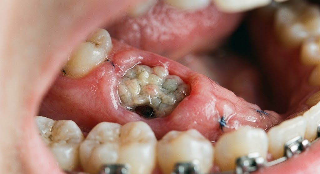
Around forty-eight to seventy-two hours after extraction, you may notice a white or yellowish film forming over the clot. This is granulation tissue, and its appearance is a positive sign. Granulation tissue consists of new tiny blood vessels, collagen-producing cells, and immune cells that are rebuilding the area. The whitish color comes from the fibrin and cellular debris that accumulate as part of the healing process.
This tissue looks somewhat like a thin, whitish membrane or a soft, pale covering over the socket. It may appear slightly raised compared to the surrounding gum tissue. Many patients mistake this normal tissue for pus or infection, but the two look distinctly different.
Days Four to Seven: Granulation Tissue Matures
The granulation tissue continues to develop and fill the socket from the bottom upward. The white or yellowish appearance may persist or gradually transition to a pinkish hue as new blood vessels proliferate and the tissue thickens. By the end of the first week, the socket typically appears partially or mostly filled with this new tissue, and the edges of the gum begin to close inward.
Weeks Two to Four: Epithelialization
The gum tissue, or epithelium, begins to grow over the granulation tissue, closing the socket from the edges inward. The white appearance fades and is replaced by normal pink gum tissue. The socket depression becomes shallower and less noticeable. By the end of the first month, the site looks much like the surrounding gum, though the contour may still feel different to your tongue.
Months One to Six: Bone Remodeling
Beneath the gum surface, new bone fills in the socket. This process continues for several months after the extraction. The gum tissue itself looks normal during this phase, though the ridge where the tooth used to be gradually changes shape as the bone remodels.
Normal White Tissue vs. Problematic White Tissue
Not all white appearances are created equal. Learning to distinguish between normal healing tissue and signs of complications helps you respond appropriately.
Characteristics of Normal Healing Tissue
Normal granulation tissue has a consistent appearance. It looks like a thin, whitish or pale yellowish film that sits within the socket, not extending significantly above the surrounding gum. It does not cause increasing pain. In fact, pain typically decreases steadily after the first few days. The tissue adheres to the socket walls and clot; it does not wipe away easily.
There is no foul odor or taste associated with normal healing tissue. A slight metallic taste from trace amounts of blood is normal, but a truly unpleasant, persistent bad taste suggests a problem. The tissue does not bleed when you gently rinse or when you touch it lightly with a clean finger.
Characteristics of Problematic White Tissue
Pus or purulent drainage looks different from granulation tissue. Pus is thicker, often more opaque, and may appear as a distinct collection rather than a uniform film. It may ooze from the socket or form a pimple-like bump on the gum adjacent to the extraction site. Pus usually accompanies increasing pain, swelling, redness, and sometimes fever.
A dry socket presents differently. Instead of seeing tissue filling the socket, you see an empty or partially empty hole where the bone is visible. The pain of a dry socket is its hallmark: severe, throbbing, radiating to the ear or temple, and not controlled by over-the-counter pain medications. The exposed bone may appear white or grayish, but it is hard and bare, not soft tissue.
Food debris trapped in the socket can also appear white. Bits of bread, rice, or other soft foods can lodge in the socket and take on a whitish, sodden appearance. Unlike granulation tissue, food debris often comes out with gentle rinsing or irrigation.
Oral thrush, or candidiasis, can cause white patches on the gums, tongue, and extraction site. These patches often look like cottage cheese and can be wiped away, leaving a red, bleeding surface underneath. Thrush typically occurs in people who have taken antibiotics, use steroid inhalers, or have compromised immune systems.
| Appearance | Normal Granulation Tissue | Pus/Infection | Dry Socket | Food Debris | Oral Thrush |
|---|---|---|---|---|---|
| Color | White-yellow film | Thick yellow/white | Bare white/gray bone | Variable white | Bright white patches |
| Pain Level | Decreasing | Increasing | Severe, throbbing | Mild discomfort | Mild to moderate |
| Removal | Cannot wipe away | May drain | Not applicable | Rinses out easily | Wipes off, bleeds |
| Odor | None to mild | Foul | Foul breath possible | May have food odor | Mild or none |
| Timing | 2-3 days after extraction | 3-7 days after | 2-5 days after | Anytime after eating | Anytime, especially after antibiotics |
Day-by-Day Healing Timeline and Appearance
Knowing what to expect each day after your extraction helps you recognize when things are on track and when they deviate from the normal course.
Day of Extraction
The socket appears red and filled with a dark blood clot. Some oozing of blood is normal, and the gauze placed by your dentist may show pink staining. Swelling begins and increases over the first few hours. Apply ice packs to the outside of your cheek to minimize swelling. Take prescribed or recommended pain medication before the local anesthetic wears off completely.
Day One Post-Extraction
The clot should be firmly in place. You may notice a whitish film beginning to form on the surface of the clot. This is the earliest granulation tissue. Swelling peaks around this time and then begins to subside. Bruising may appear on the cheek or jaw. Continue with soft foods and gentle salt water rinses starting twenty-four hours after the extraction, if your dentist approves.
Day Two to Three Post-Extraction
The white or yellowish granulation tissue becomes more apparent and may cover most of the visible clot surface. Pain should be decreasing noticeably. Swelling continues to subside. You can begin warm salt water rinses more regularly, especially after eating, to keep the area clean. The white tissue should not cause alarm; it is a sign of healing.
Day Four to Seven Post-Extraction
The socket continues to fill with granulation tissue. The white appearance may persist or gradually transition to pink. Any remaining discomfort should be mild. You can begin to reintroduce more normal foods, though you should still avoid chewing directly on the extraction site. Continue rinsing gently.
Week Two and Beyond
The gum tissue closes over the socket, and the white tissue is no longer visible. The site appears pink like the surrounding gum. A small indentation may remain where the socket was, but it should be clean and not tender. You can resume normal eating and oral hygiene routines, though you may want to be gentle around the site for another week or two.
How to Care for the Extraction Site
Proper care of the extraction site prevents complications and supports the formation of healthy granulation tissue. The white healing tissue needs gentle handling to avoid disruption.
The First 24 Hours: Critical Do’s and Don’ts
Do bite firmly on the gauze your dentist provides, changing it as directed. The pressure helps the clot form and stabilize. Do rest with your head elevated, even while sleeping, to reduce bleeding and swelling. Do apply ice packs in twenty-minute intervals to the outside of your face.
Do not rinse your mouth at all during the first twenty-four hours. The suction action of swishing can dislodge the clot. Do not spit; let saliva dribble out if necessary. Do not use a straw, as the negative pressure is one of the most common causes of clot dislodgement. Do not smoke; the chemicals in tobacco impair healing, and the sucking action risks the clot. Do not engage in strenuous exercise, as increased blood pressure can restart bleeding.
Days Two Through Seven: Gentle Cleaning
After the first twenty-four hours, begin gentle salt water rinses. Mix half a teaspoon of salt in a cup of warm water. Do not swish vigorously. Instead, gently move your head to roll the water around your mouth, then let it fall from your mouth without spitting forcefully. Rinse after each meal and before bed.
Brush your other teeth normally, but avoid the extraction site. Keep your toothbrush at least one tooth away from the socket. You can use a soft, damp cloth or gauze to very gently wipe the teeth adjacent to the extraction site if they feel fuzzy.
Avoid commercial mouthwashes during the first week unless your dentist specifically instructs otherwise. Many contain alcohol or other ingredients that irritate the healing tissue.
Dietary Recommendations
Stick to soft, cool, or lukewarm foods for the first several days. Yogurt, pudding, applesauce, mashed potatoes, smooth soups (not hot), scrambled eggs, and smoothies all provide nutrition without traumatizing the socket. Avoid anything with seeds, small grains, or particles that could lodge in the socket.
Gradually reintroduce firmer foods as the socket heals and becomes less tender. Chew on the opposite side of your mouth. Avoid crunchy, sharp, or sticky foods that could disturb the healing tissue. Stay well-hydrated with water, but remember not to use a straw.
Complications That Alter the Healing Appearance
While most extractions heal uneventfully, certain complications produce distinct visual and sensory changes that warrant attention.
Dry Socket (Alveolar Osteitis)
Dry socket occurs when the blood clot dislodges or dissolves prematurely, leaving the underlying bone and nerve endings exposed. The condition affects between two and five percent of routine extractions, with higher rates for lower wisdom teeth and in smokers.
The primary symptom is severe pain that begins two to five days after the extraction. The pain often radiates to the ear, temple, or neck on the same side. Over-the-counter pain relievers provide little relief. When you look at the socket, you see an empty or partially empty hole. The bone may be visible as a whitish-gray surface. There is no soft tissue covering.
Dry socket requires professional treatment. Your dentist will clean the socket and place a medicated dressing that provides rapid pain relief. The dressing may need to be changed every few days until the socket begins to fill with granulation tissue. Do not attempt to treat dry socket at home.
Surgical Site Infection
Infection at an extraction site is uncommon in healthy individuals but can occur. Signs include increasing pain after the initial post-operative period, swelling that worsens rather than improves after the first two days, redness and warmth of the surrounding gum and cheek, pus or purulent drainage from the socket, a foul taste or odor, and sometimes fever and general malaise.
If you suspect infection, contact your dentist immediately. Treatment typically involves drainage of any pus, irrigation of the socket, and a course of antibiotics. Do not delay, as untreated infections can spread to deeper tissues.
Sharp Bony Edges (Spicules)
As the socket heals, small fragments of bone may work their way to the surface. These bone spicules can appear as hard, white, sharp points poking through the gum. They may cause discomfort and feel rough to your tongue. Small spicules often resolve on their own as the body resorbs them or they exfoliate through the gum. Larger or persistently painful spicules may require your dentist to remove them with a quick, simple procedure.
Delayed Healing
In some individuals, particularly those with certain medical conditions or medication use, healing proceeds more slowly. The white granulation tissue may persist longer than typical, or the socket may take longer to close completely. Conditions that affect healing include diabetes, immunosuppression, bisphosphonate use, radiation therapy to the jaw, and malnutrition. Smokers also experience significantly slower healing times.
When to Contact Your Dentist
Knowing when to make the call can save you from unnecessary worry while ensuring you get help when you truly need it.
Normal Situations That Do Not Require a Call
Seeing white or yellowish tissue in the socket during days two through seven represents normal healing. Mild discomfort that decreases day by day is expected. Slight oozing on the first day is normal. A mild salty or metallic taste from trace blood is not concerning. Small bone fragments that are not painful and are working their way out slowly can be monitored.
Situations That Warrant a Phone Call
Call your dentist if you experience increasing pain after the third day, especially if it becomes severe and throbbing. Pain that radiates to your ear or temple suggests dry socket. Swelling that increases after the second day or spreads to involve more of your face or neck needs evaluation. Pus, foul odor, or a persistently bad taste that does not improve with gentle rinsing may indicate infection. A fever of 100.4°F (38°C) or higher after an extraction requires attention. Bleeding that remains heavy and does not respond to gauze pressure after several hours is not normal. If the clot appears to have been lost and the socket is empty with exposed bone visible, you likely have dry socket.
Emergency Situations
Seek emergency care if you experience difficulty breathing or swallowing, as swelling in the floor of the mouth or neck can compromise the airway. Profuse bleeding that cannot be controlled with pressure is an emergency. High fever with spreading facial redness or swelling indicates a serious infection that needs immediate treatment.
Special Considerations for Different Types of Extractions
The extraction experience and healing appearance vary depending on the tooth removed and the complexity of the procedure.
Simple Extractions
A simple extraction removes a tooth that is fully visible in the mouth, using forceps and elevators without incisions. The socket from a simple extraction typically heals faster and with fewer color changes. The white granulation tissue may be less prominent because the gum tissue was not reflected or sutured. Healing generally proceeds smoothly, with the socket closing within two to four weeks.
Surgical Extractions
Surgical extractions involve cutting the gum, sometimes removing bone, and possibly sectioning the tooth into pieces. Impacted wisdom teeth and broken-down teeth often require surgical extraction. The site is sutured after the procedure.
Surgical sites look different during healing. The sutures may be visible, and the gum tissue around them often appears more swollen and red. The white granulation tissue forms beneath the gum flap and may be visible at the suture line. Stitches that dissolve typically disappear within one to two weeks. Non-dissolving sutures require removal by your dentist after about a week.
Multiple Extractions
When several adjacent teeth are removed, the healing area is larger and more dramatic in appearance. The white granulation tissue covers a broader surface. Healing takes longer, and the bone remodeling phase may result in more noticeable changes to the jaw contour. Your dentist may place a temporary partial denture or discuss future implant placement before proceeding with multiple extractions.
Medications and Healing Appearance
Certain medications affect how the extraction site looks during healing and may alter the normal timeline.
Antibiotics
If your dentist prescribes antibiotics, take them exactly as directed and complete the full course. Antibiotics can sometimes lead to oral thrush, which appears as white patches that can be wiped away. If you notice this, inform your dentist. Probiotics or yogurt with live cultures may help prevent antibiotic-associated thrush.
Anticoagulants and Antiplatelet Drugs
Blood thinners, including warfarin, clopidogrel, and aspirin, affect clot formation. Your dentist should know about all medications you take before the extraction. The clot may form more slowly or be less stable. The appearance of the socket may be darker due to more blood in the early phase. Follow your dentist’s instructions carefully regarding whether to pause or adjust these medications.
Bisphosphonates and Related Drugs
Medications taken for osteoporosis or cancer treatment, such as alendronate or zoledronic acid, significantly affect jaw healing. Patients on these drugs face a risk of medication-related osteonecrosis of the jaw (MRONJ), where the bone fails to heal and becomes exposed. The extraction site in MRONJ shows persistent exposed bone that does not develop granulation tissue. If you take these medications, your dentist will manage your extraction with special precautions, and the healing process will differ from the standard timeline.
Steroids and Immunosuppressants
Long-term steroid use and immunosuppressive medications slow healing and increase infection risk. The granulation tissue may take longer to appear, and the white appearance may persist longer. Extra vigilance for signs of infection is warranted in these patients.
Tips for Optimal Healing
Following these evidence-based recommendations gives your extraction site the best chance of smooth, uncomplicated healing.
Maintain Excellent Oral Hygiene Elsewhere
Keeping the rest of your mouth clean reduces the bacterial load that could contaminate the healing socket. Brush and floss your other teeth as usual, being careful to avoid the extraction site. A clean mouth heals faster and with fewer complications.
Stay Hydrated
Adequate hydration supports all aspects of wound healing. Drink plenty of water, but avoid using a straw. Dehydration dries the oral tissues and can impair the healing process. Warm herbal teas, cooled to a comfortable temperature, can be soothing.
Get Adequate Rest
Your body directs energy toward healing when you rest. Avoid strenuous activity for at least forty-eight hours after extraction. When sleeping, prop your head on an extra pillow to keep it elevated, which reduces swelling and throbbing.
Avoid Alcohol and Tobacco
Alcohol interferes with healing and can interact with pain medications. Tobacco use, in any form, dramatically increases the risk of dry socket and delayed healing. The chemicals in tobacco constrict blood vessels, reducing the oxygen and nutrients delivered to the healing tissue. If you smoke, the extraction is an excellent opportunity to quit, or at minimum, to abstain for at least one week.
Follow Post-Operative Instructions Exactly
Your dentist provides specific post-operative instructions tailored to your procedure and medical history. Follow these instructions precisely, even when they seem overly cautious. The guidelines exist to prevent the complications that occur most frequently when patients deviate from recommendations.
The Role of Nutrition in Socket Healing
What you eat during the healing period directly affects how quickly and completely your socket recovers.
Protein for Tissue Repair
Protein provides the building blocks for new tissue. Incorporate soft protein sources like Greek yogurt, cottage cheese, scrambled eggs, protein shakes, and pureed beans or lentils. Adequate protein intake supports granulation tissue formation.
Vitamin C for Collagen Synthesis
Vitamin C is essential for collagen production, which forms the structural framework of healing tissue. Soft fruits like mashed bananas, applesauce, and pureed berries provide vitamin C without requiring chewing. If you drink citrus juices, dilute them and avoid swishing them around the extraction site, as the acidity can irritate.
Zinc for Immune Function
Zinc supports immune function and wound healing. Foods like yogurt, eggs, and soft-cooked oatmeal contain zinc. If you struggle to get enough through diet during the healing period, ask your dentist about a temporary supplement.
Calcium and Vitamin D for Bone Healing
The bone remodeling phase requires calcium and vitamin D. Dairy products, fortified plant milks, and soft-cooked leafy greens contribute calcium. Vitamin D from sun exposure, fortified foods, or supplements supports calcium absorption and bone metabolism.
Conclusion
A white or yellowish appearance at a tooth extraction site is typically normal granulation tissue that signals active healing rather than a problem. This tissue appears around the second or third day after extraction and gradually fills the socket before being covered by new gum tissue. The key distinction lies in the presence or absence of increasing pain, foul odor, pus, or an empty socket with exposed bone, all of which indicate complications like infection or dry socket that require professional attention.
FAQ
Is it normal for my extraction site to turn white after three days?
Yes, a whitish or yellowish film appearing around day two or three post-extraction is normal granulation tissue. It is a sign that your body is healing the wound and should not cause alarm.
How can I tell the difference between normal white tissue and pus?
Normal granulation tissue appears as a thin, adherent film that does not wipe away and is not associated with increasing pain or foul odor. Pus is thicker, may drain from the socket, and typically accompanies worsening pain, swelling, and a bad taste or smell.
What does a dry socket look like compared to normal healing?
A dry socket appears as an empty or partially empty hole where the bone is visible. The bone may look white or grayish and hard. The defining feature is severe, throbbing pain that starts a few days after extraction and radiates to the ear or temple. Normal healing shows tissue filling the socket with decreasing pain.
Can food stuck in the socket look white?
Yes, food particles can take on a whitish, sodden appearance when lodged in the socket. Unlike granulation tissue, food debris often rinses out with gentle salt water irrigation and may have a food-related odor. If you suspect food impaction, gentle rinsing after meals helps clear the socket.
When should I worry about white stuff in my extraction site?
Worry if the white material is accompanied by increasing pain, swelling that worsens after day two, foul odor or taste that persists despite rinsing, pus draining from the socket, or a fever. These signs suggest infection. Also worry if the socket appears empty with visible bone and severe pain, which indicates dry socket.
Can I brush the white tissue away?
No, do not attempt to brush or scrub the white granulation tissue from the socket. This tissue is fragile and essential for healing. Disturbing it can delay healing and introduce bacteria. Let it remain undisturbed, and it will eventually be replaced by normal gum tissue.
How long will the white appearance last?
The white granulation tissue typically remains visible for several days to about a week. It gradually transitions to a pinker appearance as new blood vessels form and the gum tissue grows over it. By two weeks, the socket should no longer appear white.
Does smoking affect the white healing tissue?
Smoking severely impairs healing and dramatically increases the risk of dry socket and infection. The chemicals in tobacco reduce blood flow, and the sucking action can dislodge the clot. If you smoke, the white granulation tissue may take longer to form, and your overall healing timeline will be extended.
Additional Resource
For authoritative post-operative care instructions and information about extraction site healing, visit the American Association of Oral and Maxillofacial Surgeons at aaoms.org. This organization provides patient education resources and a surgeon locator for complex extraction cases.