In the high-stakes world of modern healthcare, few arenas are as dynamic and technologically advanced as the cardiac catheterization laboratory. Here, interventional cardiologists perform life-saving and life-enhancing procedures inside the very chambers and vessels of the human heart, often without the need for open surgery. These procedures, from diagnostic angiograms to complex percutaneous coronary interventions (PCIs), generate a wealth of clinical data that must be meticulously translated into a universal language: medical codes. This translation is not merely an administrative task; it is the linchpin connecting patient care to reimbursement, quality reporting, and critical health data analytics.
At the center of this translation for inpatient and facility billing in the United States lies the ICD-10-PCS (International Classification of Diseases, Tenth Revision, Procedure Coding System). Unlike its diagnosis-focused counterpart, ICD-10-CM, PCS is a multi-axial, seven-character alphanumeric system designed to capture the nuance of modern medicine with unparalleled specificity. For the coder navigating the complexities of a cardiac catheterization report, mastery of ICD-10-PCS is not just beneficial—it is essential. A single misstep in character selection can lead to claim denials, inaccurate representation of patient severity, and flawed hospital performance metrics.
This article serves as the definitive guide for medical coders, coding auditors, health information management (HIM) professionals, and even clinical staff seeking to understand the “how” and “why” behind cardiac cath coding. We will embark on a detailed journey, dissecting the anatomy of the heart and its vessels, exploring the common procedures performed in the cath lab, and, most importantly, deconstructing the ICD-10-PCS system itself. Through clear explanations, illustrative tables, and real-world coding scenarios, you will gain the confidence to accurately code even the most complex interventional reports, ensuring compliance and optimizing revenue cycle performance.
ICD-10-PCS Code for Cardiac Catheterization Procedures
2. Understanding the Fundamentals: Heart Anatomy and Common Cardiac Cath Procedures
Before a single code can be assigned, a solid foundation in the relevant anatomy and procedures is non-negotiable. The heart is not a single structure but a complex organ with chambers, valves, and its own dedicated vascular supply.
Key Anatomical Structures for the Cath Coder:
-
Chambers:
-
Right Atrium (RA): Receives deoxygenated blood from the body.
-
Right Ventricle (RV): Pumps deoxygenated blood to the lungs.
-
Left Atrium (LA): Receives oxygenated blood from the lungs.
-
Left Ventricle (LV): Pumps oxygenated blood to the body.
-
-
Great Vessels:
-
Aorta: The main artery carrying oxygenated blood from the left ventricle to the body.
-
Pulmonary Artery: Carries deoxygenated blood from the right ventricle to the lungs.
-
-
Coronary Arteries: The network of vessels supplying blood to the heart muscle itself.
-
Left Main Coronary Artery (LMCA): Branches into the…
-
Left Anterior Descending (LAD) Artery: Supplies the front of the left ventricle.
-
Left Circumflex Artery (LCx): Supplies the side and back of the left ventricle.
-
Right Coronary Artery (RCA): Supplies the right ventricle, the bottom of the left ventricle, and the heart’s electrical system.
-
Common Cardiac Catheterization Procedures:
-
Diagnostic Cardiac Catheterization: The foundational procedure. It involves inserting catheters into the heart to measure pressures, assess ventricular function, and visualize the coronary arteries.
-
Left Heart Cath (LHC): Accesses the left side of the heart (aorta, left ventricle, coronary arteries) via a retrograde approach, typically from the femoral or radial artery.
-
Right Heart Cath (RHC): Accesses the right side of the heart (right atrium, right ventricle, pulmonary artery) via a venous approach, often to measure pressures in the pulmonary circuit.
-
Coronary Angiography: Injection of radiopaque contrast dye into the coronary arteries under X-ray (fluoroscopy) to visualize blockages.
-
-
Percutaneous Coronary Intervention (PCI): Therapeutic procedures to open blocked coronary arteries.
-
Coronary Balloon Angioplasty (PTCA): A balloon-tipped catheter is advanced to the blockage and inflated to compress the plaque against the artery wall.
-
Coronary Stenting: A metal mesh tube (stent) is mounted on a balloon and deployed at the site of the blockage to act as a scaffold, keeping the artery open. Stents can be bare-metal (BMS) or drug-eluting (DES).
-
Atherectomy: A specialized catheter with a rotating blade or laser is used to cut away or vaporize plaque. Types include Rotational (Rotablator), Directional (DCA), and Orbital.
-
-
Hemodynamic Assessment: Measurement of pressures within the heart’s chambers and great vessels, often part of a right heart cath.
-
Endomyocardial Biopsy: Sampling of heart muscle tissue, usually from the right ventricle, to diagnose conditions like myocarditis or transplant rejection.
3. Deconstructing ICD-10-PCS: The Framework for Procedure Coding
ICD-10-PCS is built on a logical structure where each of the seven characters represents a specific aspect of the procedure. There are no diagnostic codes in PCS; it is purely a procedure classification system.
The Seven Characters of a Medical and Surgical Code:
-
Section (1st Character): The broadest category (e.g., Medical and Surgical = 0).
-
Body System (2nd Character): The general physiological system (e.g., Heart and Great Vessels = 2).
-
Root Operation (3rd Character): The objective of the procedure—the most critical concept to grasp.
-
Body Part (4th Character): The specific anatomical site.
-
Approach (5th Character): The technique used to reach the site.
-
Device (6th Character): The device used, if any remains after the procedure.
-
Qualifier (7th Character): A further specification of the procedure, if applicable.
For cardiac catheterization, the vast majority of codes will fall within the Medical and Surgical Section (0) on the Heart and Great Vessels Body System (2). The rest of this guide will focus exclusively on this section and body system.
4. The Heart of the Matter: The Medical and Surgical Section (Section 0)
All invasive, diagnostic, and therapeutic procedures performed in the cardiac cath lab that involve cutting, inserting, or manipulating instruments within the heart and vessels are coded from this section. The first two characters for these procedures will always be 02.
5. Root Operations: Defining the Objective of the Procedure
The root operation is the cornerstone of accurate PCS coding. It answers the question: “What was the provider’s intent?” Misidentifying the root operation is the most common source of coding errors.
<a name=”inspection”>Inspection (J01B)</a>
-
Definition: Visually and/or manually exploring a body part. No therapeutic intervention is performed. The procedure is solely for assessment.
-
Cath Lab Application: Coronary Angiography. The injection of dye allows for the visual “inspection” of the coronary arteries. It is important to note that if angiography is performed solely to guide another procedure (e.g., to position a stent), it is not coded separately. It is inherent to the primary procedure.
-
Example: A catheter is placed in the left main coronary artery, and dye is injected to inspect the LAD and Circumflex for disease. This is coded as Inspection of the Coronary Arteries.
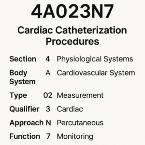
Measurement (J02H)
-
Definition: Determining the level of a physiological or physical function.
-
Cath Lab Application: Hemodynamic pressure measurements. This includes measuring pressures in the aorta, left ventricle (left heart cath), or right atrium, right ventricle, and pulmonary artery (right heart cath). Cardiac output measurements also fall under this root operation.
-
Example: A Swan-Ganz catheter is floated into the pulmonary artery to measure pulmonary artery wedge pressure. This is coded as Measurement of the Pulmonary Artery.
<a name=”extirpation”>Extirpation (J03C)</a>
-
Definition: Taking or cutting out solid matter, such as a blood clot or plaque, from a body part.
-
Cath Lab Application: Coronary Atherectomy (Rotational, Directional, Orbital). The catheter is physically removing plaque from the coronary artery.
-
Example: A Rotablator burr is used to abrade a calcified plaque in the Right Coronary Artery. This is coded as Extirpation of matter from the Right Coronary Artery.
Insertion (J02H)
-
Definition: Putting in a non-biological device that remains after the procedure.
-
Cath Lab Application: This is used for the initial placement of a vascular access closure device (e.g., Angio-Seal, Mynx, Perclose). It is not used for stents or pacemakers in this context.
-
Example: A percutaneous vascular closure device is deployed in the right common femoral artery at the end of the procedure. This is coded as Insertion of a Vascular Access Device into the Right Common Femoral Artery.
Supplement (J0HQ)
-
Definition: Putting in a biological substance or a device that physically reinforces and/or augments the function of a body part.
-
Cath Lab Application: Coronary Stent Placement. A stent is a device that “reinforces” the wall of the coronary artery.
-
Example: A drug-eluting stent is deployed in the Left Anterior Descending Artery. This is coded as Supplement of the Left Anterior Descending Artery with a Drug-eluting Intraluminal Device.
Dilation (J07H)
-
Definition: Expanding an orifice or the lumen of a tubular body part.
-
Cath Lab Application: Coronary Balloon Angioplasty (PTCA). The balloon is used to dilate the stenotic portion of the artery.
-
Example: A balloon catheter is inflated within a lesion in the Left Circumflex Artery. This is coded as Dilation of the Left Circumflex Artery.
Restriction (J08V)Definition: Partially closing an orifice or the lumen of a tubular body part.
-
Cath Lab Application: Coil Embolization of a vessel (e.g., for a coronary artery fistula or a vessel tear).
-
Example: Coils are placed in a small arteriovenous malformation to restrict blood flow. This is coded as Restriction of the specified vessel.
Occlusion (J09L)
-
Definition: Completely closing an orifice or the lumen of a tubular body part.
-
Cath Lab Application: Embolization with a device that causes a complete blockage (e.g., a vascular plug).
-
Example: An Amplatzer plug is deployed to occlude a patent ductus arteriosus (PDA). This is coded as Occlusion of the Aorta (using the qualifier for PDA).
Revision (J0W9)
-
Definition: Correcting a malfunctioning or displaced device.
-
Cath Lab Application: Balloon angioplasty of a previously placed stent (in-stent restenosis). The objective is to “revise” the stent that is no longer functioning properly.
-
Example: A balloon is inflated inside a stenotic drug-eluting stent in the LAD. This is coded as Revision of a Device in the Left Anterior Descending Artery.
Removal (J0WP)
-
Definition: Taking out a device from a body part.
-
Cath Lab Application: Removal of a catheter fragment or a misplaced device.
-
Example: A fractured pacemaker lead is percutaneously snared and removed from the right ventricle. This is coded as Removal of a Device from the Right Ventricle.
ICD-10-PCS Root Operations for Common Cardiac Cath Procedures
| Procedure | Root Operation | Definition & Rationale |
|---|---|---|
| Coronary Angiogram | Inspection | Visual examination of the lumen of the coronary arteries. |
| Pressure Measurement | Measurement | Determining the level of physiological pressure. |
| Balloon Angioplasty (PTCA) | Dilation | Expanding the lumen of the stenotic artery. |
| Stent Placement | Supplement | Putting in a device that reinforces the arterial wall. |
| Atherectomy | Extirpation | Taking out solid matter (plaque) from the artery. |
| Stent Angioplasty (for in-stent restenosis) | Revision | Correcting a malfunctioning device (the stenotic stent). |
| Vascular Closure Device | Insertion | Putting in a device that remains after the procedure. |
| Coil Embolization | Restriction | Partially closing the lumen of a vessel. |
6. Body Part: Precision in Anatomical Location
The fourth character specifies the exact body part. For coronary procedures, this requires precise documentation from the physician.
Key Body Part Values for Coronary Arteries (02):
-
2: Coronary Artery, single vessel (used when only one distinct coronary artery is treated, but the specific one isn’t documented).
-
3: Coronary Artery, two vessels (used when two distinct arteries are treated, but they aren’t specified).
-
4: Coronary Artery, three vessels (used when three distinct arteries are treated).
-
5: Coronary Artery, four or more vessels.
-
6: Left Main Coronary Artery
-
7: Left Anterior Descending Coronary Artery
-
8: Left Circumflex Coronary Artery
-
9: Right Coronary Artery
-
B: Obtuse Marginal Coronary Artery
-
C: Diagonal Coronary Artery
-
D: Ramus Intermedius Coronary Artery
Other Relevant Body Parts:
-
0: Right Atrium
-
1: Right Ventricle
-
2: Left Atrium
-
3: Left Ventricle
-
4: Pulmonary Artery, Right
-
5: Pulmonary Artery, Left
-
6: Pulmonary Artery, Main
-
7: Aorta
Crucial Note: The body part character is defined by the specific anatomical structure the procedure was performed on. If a stent is placed in the LAD, the body part is the LAD (7), not the “coronary artery, one” (2). Always code to the highest level of specificity documented.
7. Approach: The Pathway to the Procedure
The fifth character describes how the procedure site was reached.
-
Open (0): Cutting through the skin or mucous membrane and underlying tissues.
-
Percutaneous (3): Entry by puncture or minor incision of the skin, through which instrumentation is threaded. This is the most common approach in the cath lab.
-
Percutaneous Endoscopic (4): Percutaneous approach aided by endoscopic visualization.
-
Via Natural or Artificial Opening (7): Entry through a natural orifice (e.g., mouth, urethra).
-
Via Natural or Artificial Opening Endoscopic (8): Entry through a natural orifice with endoscopic aid.
For nearly all cardiac cath procedures, the approach will be Percutaneous (3).
8. Device: The Key to Interventional Coding
The sixth character is critical for differentiating between, for example, a simple angioplasty and a stent placement.
Key Device Values:
-
D: Intraluminal Device, Drug-eluting (e.g., Drug-Eluting Stent – DES)
-
J: Intraluminal Device (e.g., Bare-Metal Stent – BMS)
-
L: Intraluminal Device, Radioactive
-
0: Autologous Tissue Substitute (rarely used in cath lab)
-
3: Synthetic Substitute (e.g., for patch closure)
-
C: Extraluminal Device (e.g., a vascular clip)
-
Y: Other Device (used for vascular closure devices)
Important: The device character is only used if a device remains after the procedure is completed. A balloon catheter is a device, but it is removed, so no device character is used for a stand-alone Dilation (angioplasty). A stent remains, so the device character is required for a Supplement procedure.
9. Qualifier: The Finishing Detail
The seventh character provides additional information. This is often used to specify the diagnostic nature of a procedure or a specific anatomical qualifier.
-
X: Diagnostic
-
0: Aorta (used as a qualifier for procedures on structures like the PDA)
-
Z: No Qualifier
The Diagnostic (X) qualifier is used with root operations like Inspection and Measurement when the procedure is performed solely for diagnostic purposes and no therapeutic intervention is performed during the same session on that body part.
10. Putting It All Together: Coding Real-World Scenarios
Let’s apply our knowledge to full coding scenarios.
Scenario 1: Diagnostic Left Heart Catheterization with Coronary Angiogram
Procedure Note Summary: Via right femoral artery access, a pigtail catheter was advanced into the aortic root. An aortogram was performed. The catheter was then advanced into the left ventricle, and a left ventriculogram was performed, revealing normal systolic function. Pressures were measured. Next, a JL4 catheter was used to selectively engage the left main coronary artery. Coronary angiography of the left system was performed, revealing no significant disease. An JR4 catheter was used to engage the right coronary artery, and angiography was performed, revealing no significant disease.
Analysis:
-
Measurement of Aorta/Left Ventricle: The provider measured pressures. Root Operation = Measurement. Body Part = Aorta and Left Ventricle. Approach = Percutaneous. No device. Qualifier = Diagnostic.
-
Aorta: 02JA3ZZ (Measurement, Heart & Great Vessels, Aorta, Percutaneous, Diagnostic)
-
Left Ventricle: 02H63ZZ (Measurement, Heart & Great Vessels, Left Ventricle, Percutaneous, Diagnostic)
-
-
Inspection of Coronary Arteries: The provider visually examined the coronaries with contrast. Root Operation = Inspection. Body Parts = Left Main, LAD, Circumflex (coded as Coronary Artery, Left) and Right Coronary Artery. Approach = Percutaneous. No device. Qualifier = Diagnostic.
-
Left Coronary System: 02BJ3ZX (Inspection, Heart & Great Vessels, Coronary Artery, Left, Percutaneous, Diagnostic)
-
Right Coronary Artery: 02BJ3ZX (Inspection, Heart & Great Vessels, Coronary Artery, Right, Percutaneous, Diagnostic)
-
Final Codes: 02JA3ZZ, 02H63ZZ, 02BJ3ZX, 02BJ3ZX
Scenario 2: Percutaneous Coronary Intervention (PCI) with Stenting
Procedure Note Summary: *…A significant 90% lesion was found in the mid LAD. A guidewire was advanced across the lesion. A 3.0 x 20 mm drug-eluting stent was advanced to the lesion and deployed successfully. Post-dilation was performed with a 3.5 mm non-compliant balloon. Final angiography showed an excellent result with 0% residual stenosis and TIMI-3 flow.*
Analysis:
-
Stent Placement: The primary therapeutic procedure. Root Operation = Supplement (reinforcing the artery). Body Part = Left Anterior Descending Artery. Approach = Percutaneous. Device = Intraluminal Device, Drug-eluting. No qualifier.
-
Code: 027034D (Supplement, Heart & Great Vessels, LAD, Percutaneous, Drug-eluting Intraluminal Device)
-
-
Post-Dilation Angioplasty: The post-dilation is an integral part of the stent deployment process to optimize stent apposition. It is not coded separately.
-
Angiography: The angiograms performed before, during, and after the stent placement are inherent to the procedure and are not coded separately.
Final Code: 027034D
Scenario 3: Right Heart Catheterization with Biopsy
Procedure Note Summary: …Via right femoral venous access, a balloon-tipped catheter was advanced into the right atrium, right ventricle, and pulmonary artery. Pressures were measured. The catheter was then exchanged for a bioptome. Under fluoroscopic guidance, four samples of endomyocardial tissue were obtained from the right ventricular septum.
Analysis:
-
Measurement of Pressures: Root Operation = Measurement. Body Parts = Right Atrium, Right Ventricle, Pulmonary Artery. Approach = Percutaneous. No device. Qualifier = Diagnostic.
-
Right Atrium: 02H03ZZ
-
Right Ventricle: 02H13ZZ
-
Pulmonary Artery: 02H63ZZ
-
-
Endomyocardial Biopsy: This is the taking of tissue for pathology. The correct root operation is Excision (cutting out a portion of a body part). Body Part = Right Ventricle. Approach = Percutaneous. No device. Qualifier = Diagnostic.
-
Code: 02B13ZX (Excision, Heart & Great Vessels, Right Ventricle, Percutaneous, Diagnostic)
-
Final Codes: 02H03ZZ, 02H13ZZ, 02H63ZZ, 02B13ZX
Scenario 4: Coronary Atherectomy with Stent Placement
Procedure Note Summary: *…A severely calcified 99% lesion was noted in the proximal RCA. After wire placement, rotational atherectomy was performed using a 1.5 mm burr. Following atherectomy, a 4.0 x 28 mm drug-eluting stent was deployed across the lesion with an excellent result.*
Analysis:
-
Atherectomy: The removal of plaque. Root Operation = Extirpation. Body Part = Right Coronary Artery. Approach = Percutaneous. No device (the burr is removed). Qualifier = No Qualifier.
-
Code: 02C93ZZ (Extirpation, Heart & Great Vessels, RCA, Percutaneous)
-
-
Stent Placement: Root Operation = Supplement. Body Part = Right Coronary Artery. Approach = Percutaneous. Device = Intraluminal Device, Drug-eluting.
-
Code: 027034D (Note: This is the same code as in Scenario 2, but for a different body part, the RCA. The body part character is ‘9’ for RCA).
-
Final Codes: 02C93ZZ, 027034D
11. Coding for Imaging Guidance
Fluoroscopic guidance is an integral part of every cardiac catheterization procedure. According to ICD-10-PCS Official Guidelines, imaging guidance is included in the root procedure and is not coded separately. This is a key difference from CPT coding.
12. Common Pitfalls and How to Avoid Them
-
Confusing Root Operations: Don’t code a diagnostic angiogram (Inspection) as a catheter placement. The catheter is the tool, not the objective. The objective is to visually inspect.
-
Over-coding Angiography: Angiography performed to guide a therapeutic intervention is not coded separately. Only code it if it’s a standalone diagnostic procedure.
-
Incorrect Body Part Specificity: Do not default to “Coronary Artery, One” (2). Always code to the most specific artery named (e.g., LAD, RCA, Obtuse Marginal).
-
Missing the Device Character: Forgetting to add the device character for a stent placement (D or J) is a major error that changes the DRG and reimbursement.
-
Misapplying the Diagnostic Qualifier: Only use the X qualifier if the procedure was solely diagnostic. If any therapeutic intervention (angioplasty, stent, atherectomy) is performed on a vessel, the Inspection/Measurement of that vessel is no longer “diagnostic” for coding purposes.
13. The Importance of Clinical Documentation Integrity (CDI)
Accurate coding is impossible without precise documentation. Coders must work closely with physicians and CDI specialists to ensure the procedure report clearly states:
-
The specific arteries treated.
-
The exact procedures performed (e.g., “rotational atherectomy,” “stent placed,” “balloon angioplasty”).
-
The type of devices used (e.g., “3.5 x 28 mm drug-eluting stent”).
-
The approach (though typically percutaneous, it should be implied).
A query may be necessary if the documentation is unclear or conflicting.
14. Conclusion
Mastering ICD-10-PCS for cardiac catheterization requires a methodical understanding of heart anatomy, procedural objectives, and the PCS framework itself. By focusing on the root operation to define the procedure’s intent and using the other six characters to add layers of specificity for body part, approach, device, and qualifier, coders can achieve a high level of accuracy. This precision ensures proper reimbursement, supports quality patient data, and reflects the true complexity and value of the life-saving work performed in the cardiac catheterization laboratory. Continuous education and a strong partnership with clinical documentation are the keys to success in this challenging and rewarding field.
15. Frequently Asked Questions (FAQs)
Q1: How do I code a left heart cath that turns into a stent placement? Do I still code the diagnostic left heart cath?
A: No. When a diagnostic procedure leads to a therapeutic intervention on the same body part during the same session, the diagnostic procedure for that body part is not coded separately. You would only code the therapeutic procedure (e.g., the stent placement). However, if pressures were measured in the aorta and left ventricle, those Measurement codes (02JA3ZZ, 02H63ZZ) would still be coded, as they are performed on different body parts than the stent.
Q2: What is the difference between a “drug-eluting” and “bare-metal” stent in the device character?
A: A drug-eluting stent (DES) is coded with device character D (Intraluminal Device, Drug-eluting). A bare-metal stent (BMS) is coded with device character J (Intraluminal Device). This distinction is critical as it can impact reimbursement.
Q3: How do I code multiple stents placed in the same coronary artery?
A: If multiple stents are placed in the same coronary artery (e.g., two stents in the LAD), it is coded only once. The code represents the procedure performed on the body part. However, if stents are placed in different coronary arteries (e.g., one in the LAD and one in the RCA), you would code each one separately with the appropriate body part character.
Q4: Why is fluoroscopy not coded separately in ICD-10-PCS?
A: The ICD-10-PCS Official Guidelines state that the method of visualization (e.g., fluoroscopic guidance) is an integral part of a procedure and is not coded separately. This is a fundamental difference from CPT coding.
Q5: How do I code a thrombectomy (clot removal) in a coronary artery?
A: The removal of a blood clot (thrombus) is coded to the root operation Extirpation (taking out solid matter), just like atherectomy. The code would be 02C93ZZ for a percutaneous thrombectomy of the RCA, for example.


