In the high-stakes world of neurosurgery, few procedures are as simultaneously routine and critical as the placement of an External Ventricular Drain (EVD). This life-saving intervention, often performed under urgent or emergent conditions, serves as a direct conduit to the very core of the human central nervous system. For the neurosurgeon, it is a skill honed through years of training, a precise maneuver to relieve devastating pressure. For the patient, it is a lifeline. For the medical coder, however, it represents a complex puzzle—a sequence of alphanumeric characters that must perfectly encapsulate the intricate reality of the procedure. The accuracy of this code, ICD-10-PCS 00H03YZ, or its variants, is not merely an administrative task; it is a fundamental component of modern healthcare infrastructure, influencing patient care trajectories, hospital reimbursement, and the integrity of national health data.
This article is designed to be the definitive guide for medical coders, auditors, students, and healthcare information management professionals seeking to master the coding of EVD placement. We will embark on a detailed journey, beginning with the clinical “why,” moving through the surgical “how,” and culminating in the precise “how to code.” We will dissect the ICD-10-PCS system, not as a set of arbitrary rules, but as a logical language built to describe procedures with exacting specificity. By integrating clinical knowledge with coding expertise, we will transform a complex surgical report into an accurate and defensible code, ensuring that the vital work of the neurosurgical team is correctly represented in the patient’s permanent record.
ICD-10-PCS code for External Ventricular Drain (EVD) placement
2. Understanding the “Why”: The Clinical Imperative for External Ventricular Drainage
To code a procedure correctly, one must first understand its purpose. An EVD is not placed arbitrarily; it is a targeted solution to a potentially fatal problem, most commonly hydrocephalus.
The Physiology of Cerebrospinal Fluid (CSF)
Cerebrospinal Fluid is a clear, colorless liquid that bathes the brain and spinal cord, providing mechanical cushioning, chemical stability, and waste removal. It is produced primarily in the choroid plexus within the brain’s ventricles—a series of interconnected cavities. Under normal conditions, CSF circulates from the lateral ventricles to the third and fourth ventricles, then into the subarachnoid space where it is eventually absorbed into the bloodstream. This production, circulation, and absorption maintain a delicate balance and a stable intracranial pressure (ICP), typically between 5-15 mmHg in a healthy adult.
Pathologies Leading to Hydrocephalus and Increased ICP
Hydrocephalus, colloquially known as “water on the brain,” is a condition characterized by an excessive accumulation of CSF within the ventricles. This leads to ventricular enlargement and a dangerous increase in ICP. If left untreated, rising ICP can compress and damage delicate brain tissue, leading to herniation, neurological deficit, and death. The causes of hydrocephalus are broadly categorized as:
-
Obstructive (Non-communicating): A blockage within the ventricular system prevents CSF from flowing out. Common causes include:
-
Intraventricular Hemorrhage (IVH): Bleeding into the ventricles, common in premature infants or after trauma.
-
Brain Tumors: Masses that physically obstruct CSF pathways.
-
Aqueductal Stenosis: A congenital or acquired narrowing of the cerebral aqueduct.
-
Subarachnoid Hemorrhage (SAH): Blood clots in the basal cisterns can impede CSF absorption.
-
-
Communicating: The CSF pathways are open, but there is a failure of absorption at the arachnoid granulations. Causes include:
-
Meningitis: Inflammation from infection can scar the arachnoid granulations.
-
Normal Pressure Hydrocephalus (NPH): A condition typically in older adults with a triad of symptoms: gait disturbance, dementia, and urinary incontinence.
-
The Role of the EVD as a Lifesaving Intervention
The External Ventricular Drain is the primary tool for managing acute hydrocephalus and dangerously elevated ICP. It functions as a controlled “overflow valve.” By inserting a catheter directly into a ventricle (usually the frontal horn of the lateral ventricle) and tunneling it under the skin to an external collection system, the surgical team can:
-
Drain Excess CSF: Immediately lower intracranial pressure.
-
Monitor ICP Continuously: The EVD system is transduced, providing real-time ICP readings.
-
Administer Medications: Intrathecal drugs (e.g., antibiotics for meningitis) can be delivered directly into the CSF.
-
Sample CSF: Analyze CSF for infection, blood, or tumor markers.
The decision to place an EVD is often a race against time, making the procedure a cornerstone of neurocritical care.
3. Deconstructing the Procedure: A Step-by-Step Surgical Overview
A coder must be able to read an operative report and visualize the procedure. Here is a detailed breakdown of a typical EVD placement.
Preoperative Planning and Patient Positioning
The procedure begins with meticulous planning. The surgeon reviews preoperative imaging (CT or MRI) to identify the size and shift of the ventricles, the optimal trajectory for the catheter, and to avoid major blood vessels. The most common entry point is Kocher’s point, located approximately 11 cm posterior to the nasion and 3 cm lateral to the midline. The patient is positioned supine, with the head in a neutral position. The scalp is shaved, prepped, and draped in a sterile fashion.
The Surgical Technique: From Incision to Closure
-
Incision and Burr Hole: A small linear incision (typically 3-4 cm) is made in the scalp down to the skull. A self-retaining retractor is placed. A high-speed drill is used to create a single burr hole through the skull.
-
Dural Opening: The underlying dura mater (the tough outer membrane covering the brain) is sharply incised and coagulated to expose the pia-arachnoid surface of the brain.
-
Catheter Pass: The ventricular catheter, which is radio-opaque with depth markings, is then passed through the brain parenchyma along a predetermined trajectory aiming for the frontal horn of the lateral ventricle. The target is typically 5-6 cm deep from the cortical surface. A “pop” or give is often felt as the catheter enters the ventricle.
-
CSF Confirmation: The stylet within the catheter is removed. The spontaneous egress of CSF is the definitive confirmation of successful intraventricular placement.
-
Tunneling and Securing: The catheter is then tunneled several centimeters under the scalp away from the incision site to reduce the risk of infection. It is secured to the skin with a suture.
-
Connection and Closure: The proximal end of the catheter is connected to a closed, sterile drainage system. The scalp incision is closed in layers.
-
System Setup: The EVD system is leveled and zeroed at the foramen of Monro (an anatomical landmark approximating the external auditory meatus) to ensure accurate ICP monitoring. The drainage chamber is set at a specific height (e.g., 10 cm above the zero point) to control the pressure at which CSF will drain.
Postoperative Management and Monitoring
Postoperatively, the patient is managed in an intensive care unit (ICU). The nursing and medical staff meticulously monitor the ICP waveforms, the volume and character of the CSF output, and the patient’s neurological status. The EVD is weaned gradually once the underlying condition improves, and the catheter is removed at the bedside or in a controlled setting.
4. Navigating the ICD-10-PCS Universe: Foundational Principles
ICD-10-PCS (Procedure Coding System) is a completely different system from its diagnosis counterpart, ICD-10-CM. It is a procedural nomenclature designed for the precise description of medical and surgical interventions.
The Structure of an ICD-10-PCS Code: Seven Characters Explained
Every ICD-10-PCS code is seven characters long, and each character represents a specific aspect of the procedure. The characters are always alphanumeric (using numbers 0-9 and letters A-H, J-N, P-Z, excluding O and I). The structure is consistent:
-
Character 1: Section – The broadest category (e.g., Medical and Surgical, Obstetrics, Imaging).
-
Character 2: Body System – The general body system (e.g., Central Nervous System, Heart and Great Vessels).
-
Character 3: Root Operation – The objective of the procedure (e.g., Drainage, Resection, Insertion). This is the most critical conceptual element.
-
Character 4: Body Part – The specific part of the body system on which the procedure was performed.
-
Character 5: Approach – The technique used to reach the body part (e.g., Open, Percutaneous).
-
Character 6: Device – The device that remains after the procedure.
-
Character 7: Qualifier – An additional attribute that adds specificity.
The Importance of the Medical and Surgical Section (Section 0)
For EVD placement, we operate exclusively within the Medical and Surgical section, identified by the first character 0. This section contains the vast majority of inpatient procedures.
The Axial Approach to Code Building
ICD-10-PCS is built on an “axial” structure, meaning each character is an independent axis of classification. You build the code by making a selection from each column (character), moving from left to right.
5. The Heart of the Matter: Selecting the Correct Root Operation for EVD Placement
The root operation is the cornerstone of the code. Misidentifying it will result in a completely incorrect code. For EVD placement, the correct root operation is unequivocally Drainage.
Root Operation: Drainage – The Primary and Correct Choice
The official ICD-10-PCS definition of Drainage is: “Taking or letting out fluids and/or gases from a body part.”
Let’s analyze the EVD procedure against this definition:
-
Taking or letting out: The EVD allows CSF to flow out of the ventricle.
-
Fluids and/or gases: The fluid is cerebrospinal fluid (CSF).
-
From a body part: The body part is the cerebral ventricle.
The primary goal of the procedure is to remove (drain) excess CSF to lower ICP. The fact that a catheter is placed to achieve this is a means to an end. The root operation captures the objective, not the tool.
Why “Insertion” is Incorrect
The root operation Insertion is defined as: “Putting in a non-biological device that remains after the procedure.” While an EVD catheter is indeed a device that remains, the purpose of the procedure is not simply to “put in” a device. The purpose is to drain fluid. The device facilitates the drainage. If the objective were merely to place a device, “Insertion” would be correct, but that is not the clinical reality. Coding “Insertion” for an EVD misrepresents the procedure’s intent.
Why “Introduction” is Incorrect
The root operation Introduction is defined as: “Putting in or on a therapeutic, diagnostic, nutritional, physiological, or prophylactic substance except blood or blood products.” This root operation is for substances (e.g., injecting a medication, infusing IV fluids), not for placing mechanical devices like a drainage catheter.
Clinical Scenarios Solidifying the “Drainage” Decision
-
Scenario A: A patient with a ruptured aneurysm and SAH develops acute hydrocephalus. An EVD is placed. The objective is to drain bloody CSF and control ICP. Root Operation: Drainage.
-
Scenario B: A patient with meningitis and hydrocephalus has an EVD placed to drain CSF and monitor ICP. Root Operation: Drainage.
-
Scenario C: A patient has an EVD placed primarily for ICP monitoring after a severe TBI. Even though drainage may be secondary, the act of placing the catheter—which allows for drainage—is still classified under the objective of the procedure, which is to enable the taking of fluid and pressure. Root Operation: Drainage.
6. Character 4: Body Part – The Nuances of Ventricular System Anatomy
With the root operation established as Drainage (0), and the body system being Central Nervous System (H), we move to the body part.
0H9 – Ventricular, Cerebral – The Standard Code
In the Central Nervous System body system table, under Root Operation “Drainage,” the most specific body part value for a standard EVD is 0H9 – Ventricular, Cerebral. This value encompasses the entire cerebral ventricular system, including the lateral, third, and fourth ventricles. Unless the documentation specifies a different part of the system, 0H9 is the default and correct choice.
Distinguishing Lateral, Third, and Fourth Ventricles in Documentation
While “Ventricular, Cerebral” is the standard, a coder must be alert to more specific documentation. For example, if a surgeon’s note states, “The catheter was advanced into the third ventricle,” the coder might look for a more specific body part. However, in the standard PCS tables for the Central Nervous System, “Ventricular, Cerebral” (0H9) is the highest level of specificity provided for the ventricles in this context. The coder should not use a less specific “Brain” code. They must use the available specificity within the table.
Bilateral EVD Placement: A Complex Scenario
In rare, severe cases of intraventricular hemorrhage, a surgeon may place an EVD in each lateral ventricle. This presents a coding challenge, as ICD-10-PCS does not have a built-in “bilateral” indicator for this body part. According to the ICD-10-PCS Official Guidelines for Coding and Reporting (B3.10a), if the same root operation is performed on identical body parts (e.g., both lateral ventricles) via the same approach, and there is a separate body part value for each, the procedure is coded only once.
However, the body part value “0H9 – Ventricular, Cerebral” is a single value representing the entire system. Therefore, the placement of bilateral EVDs is still represented by a single code: 00H03YZ. The medical record must clearly document the bilateral placement, but the code itself does not change. This is a critical nuance.
7. Character 5: The Approach – Mapping the Surgical Pathway
The approach describes the technique used to reach the site of the procedure. For an EVD, the two most common approaches are Open and Percutaneous.
Open (0) Approach
The official definition of Open is: “Cutting through the skin or mucous membrane and any other body layers necessary to expose the site of the procedure.”
An EVD placement is coded as Open because the surgeon:
-
Makes an incision through the skin and subcutaneous tissue.
-
Retracts the tissues.
-
Drills a burr hole through the skull (cutting through bone).
-
Incises the dura mater.
This series of steps to “expose” the brain surface meets the definition of an Open approach. Many coders mistakenly think “Open” means a large craniotomy, but in PCS terminology, any procedure that cuts through layers to expose the site is “Open.”
Percutaneous (3) Approach
The official definition of Percutaneous is: “Entry, by puncture or minor incision, of instrumentation through the skin or mucous membrane and any other body layers necessary to reach the site of the procedure.”
While an EVD involves a skin incision, it is not typically considered a “minor incision” in this context because it involves drilling a burr hole, which is not a “puncture.” The Percutaneous approach is more appropriate for procedures like a lumbar puncture or a stereotactic biopsy using a twist-drill craniostomy, which is a smaller, less invasive entry than a formal burr hole.
Percutaneous Endoscopic (4) Approach
This is rarely used for standard EVD placement but may be employed in certain complex or elective scenarios. The documentation would explicitly mention the use of an endoscope.
Determining the Approach from Operative Reports
The coder must rely on the surgeon’s documentation. Key phrases that indicate an Open approach include: “scalpel incision,” “burr hole created,” “dura incised,” “galea closed.” The presence of a burr hole is the key differentiator from a true percutaneous puncture.
8. Character 6: Device – The “Y” Factor and Understanding the Drain
The device character identifies any device that remains after the procedure is completed.
Device Character: Y – Drainage Device
In the table for Drainage of the Central Nervous System, the device character is Y – Drainage Device. The EVD catheter is the quintessential example of a drainage device. It is a tube placed to allow for the continuous or intermittent drainage of fluid.
The Nature of an EVD as a Short-Term Drain
It is important to distinguish a temporary EVD from a permanent shunt. An EVD is an external, temporary system, while a shunt (e.g., Ventriculoperitoneal or VP shunt) is an internal, permanent (or long-term) system. Both, however, serve a drainage function and, when placed, are coded with the device value “Drainage Device.”
Differentiating from Shunt Systems
If a surgeon places a VP shunt, the root operation is still Drainage, but the device is a Shunt (character 6 = C). The key is in the operative report: an EVD drains to an external collection bag, while a shunt has an internal terminus (e.g., the peritoneal cavity). Do not confuse the device value for an EVD (Y) with that of a shunt (C).
9. Character 7: Qualifier – The Final Piece of the Puzzle
The qualifier provides additional information about the procedure. For the Drainage procedure on the Central Nervous System, the standard qualifier is Z – No Qualifier. There are no other qualifiers in this specific table that would typically apply to a standard EVD placement. Therefore, the final character is almost always Z.
10. Putting It All Together: Building the Complete ICD-10-PCS Code
Let’s assemble the code from our character selections:
-
Section: 0 – Medical and Surgical
-
Body System: H – Central Nervous System
-
Root Operation: 0 – Drainage
-
Body Part: 9 – Ventricular, Cerebral
-
Approach: 0 – Open
-
Device: Y – Drainage Device
-
Qualifier: Z – No Qualifier
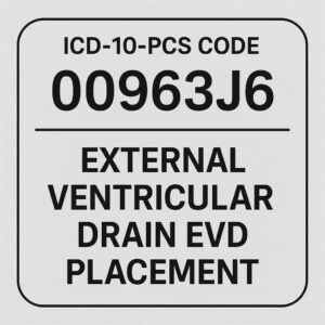
The complete, and most common, ICD-10-PCS code for an Open placement of an External Ventricular Drain is: 00H03YZ.
Case Study 1: Standard EVD for Subarachnoid Hemorrhage
-
Presentation: A 55-year-old female presents with a thunderclap headache. CT scan shows a diffuse subarachnoid hemorrhage. She becomes lethargic, and a repeat CT shows developing hydrocephalus.
-
Procedure: The neurosurgeon performs a right frontal EVD placement via a burr hole.
-
Operative Note Key Phrases: “A linear incision was made… a burr hole was drilled… the dura was opened… the ventricular catheter was passed and CSF return was noted… the catheter was tunneled and connected to a drainage system.”
-
ICD-10-PCS Code: 00H03YZ (Drainage of Ventricular, Cerebral, Open Approach, Drainage Device).
Case Study 2: Percutaneous EVD for Traumatic Brain Injury
-
Presentation: A 30-year-old male with a severe traumatic brain injury and intraventricular hemorrhage. The neurosurgeon decides to place an EVD at the bedside in the ICU using a twist-drill kit.
-
Procedure: A small stab incision is made in the scalp. A twist-drill is used to create a small cranial opening. The catheter is passed percutaneously.
-
Operative Note Key Phrases: “A twist-drill craniostomy was performed… the catheter was advanced percutaneously into the lateral ventricle…”
-
ICD-10-PCS Code: In this specific scenario, the use of a twist-drill may be classified by some as a Percutaneous (3) approach. The code would be 00H33YZ. Crucial Note: The coder must follow their facility’s coding clinic and internal guidance on classifying twist-drill EVDs, as this can be an area of interpretation. The presence of a “drill” often leans towards “Open,” but the minimal nature of a twist-drill may allow for “Percutaneous.”
Case Study 3: EVD Placement with Subsequent Conversion to Shunt
-
Presentation: An elderly patient with normal pressure hydrocephalus has an EVD placed for diagnostic continuous pressure monitoring and therapeutic drainage. After a positive response, the decision is made to convert to a permanent VP shunt.
-
Coding: These are two separate procedures.
-
Procedure 1 (Day 1): EVD Placement. Code: 00H03YZ.
-
Procedure 2 (Day 5): VP Shunt Placement. This would be coded as 00H10CZ (Drainage of Ventricular, Cerebral, Open Approach, Shunt). The EVD removal is inherent to the procedure and not separately coded.
-
11. Common Pitfalls and Coding Challenges: Avoiding Costly Errors
Accurate coding is as much about knowing what not to do as it is about knowing what to do.
-
Pitfall 1: Misinterpreting the Root Operation. The most common and serious error is coding this as “Insertion.” Always ask: “What was the goal? To put in a tube, or to drain fluid?” The answer is always to drain fluid.
-
Pitfall 2: Inadequate Documentation on Body Part and Approach. If the report does not specify the body part (e.g., “ventricular catheter placed”) or the approach (e.g., “burr hole” vs. “twist-drill”), the coder must query the physician for clarification.
-
Pitfall 3: Confusing an EVD with a VP Shunt. An EVD is external and temporary, coded with Device Y. A VP shunt is internal and permanent/long-term, coded with Device C.
-
Pitfall 4: Coding the Removal or Revision of an EVD. The removal of an EVD is not typically coded separately. It is considered part of the aftercare or is inherent to a subsequent procedure (like a shunt placement). If an EVD becomes dislodged and is replaced, the replacement is coded as a new procedure.
12. The Power of Precision: How Accurate EVD Coding Impacts Healthcare
The consequences of an incorrect code extend far beyond the medical record.
-
Reimbursement and DRG Assignment: Inpatient reimbursement is primarily driven by Diagnosis-Related Groups (DRGs). The procedures performed are a major factor in DRG assignment. Coding an EVD as “Insertion” instead of “Drainage” could place the case in an incorrect, and potentially lower-paying, DRG. For example, a DRG for “Craniotomy for Trauma” with a correctly coded EVD may reimburse at a higher rate than one without.
-
Data Analytics and Clinical Research: Health systems and public health agencies use coded data to track outcomes, complications, and the effectiveness of treatments. If EVD placements are miscoded, the data on the frequency, outcomes, and complications of this specific procedure become unreliable, hindering research and quality improvement initiatives.
-
Quality Metrics and Patient Safety Indicators: Accurate procedure coding is essential for calculating hospital quality measures, such as rates of hospital-acquired infections (e.g., ventriculitis). An incorrectly coded EVD would skew these vital statistics.
ICD-10-PCS Code Building for EVD Placement
| PCS Character | Component | Correct Value | Explanation & Clinical Correlation |
|---|---|---|---|
| 1 | Section | 0 | Medical and Surgical |
| 2 | Body System | H | Central Nervous System |
| 3 | Root Operation | 0 | Drainage: Taking or letting out fluids/gases. The goal of an EVD is to drain CSF. |
| 4 | Body Part | 9 | Ventricular, Cerebral: The catheter tip resides in the cerebral ventricle. |
| 5 | Approach | 0 | Open: Cutting through skin, creating a burr hole in the skull, and incising the dura to expose the brain. |
| 6 | Device | Y | Drainage Device: The EVD catheter is a tube that remains to facilitate drainage. |
| 7 | Qualifier | Z | No Qualifier: No additional specificity is required for this procedure. |
| FINAL CODE | 00H03YZ | Drainage of Ventricular, Cerebral, Open Approach, Drainage Device |
13. Conclusion: Mastering the Code for a Critical Procedure
The accurate ICD-10-PCS coding for External Ventricular Drain placement hinges on a deep understanding of its clinical purpose as a drainage procedure. By methodically building the code from its root operation, “Drainage,” through the specific body part, “Ventricular, Cerebral,” and the standard “Open” approach, the coder arrives at the definitive code 00H03YZ. This precision ensures clinical intent is captured, reimbursement is appropriate, and the healthcare data ecosystem remains robust and reliable, ultimately supporting the high-quality care that patients undergoing this critical neurosurgical intervention require.
14. Frequently Asked Questions (FAQs)
Q1: The surgeon documented “insertion of an EVD.” Should I code the root operation as Insertion?
A: No. You must code based on the objective of the procedure, not the verb used in the documentation. The objective is to drain CSF to treat hydrocephalus/monitor ICP. Therefore, the root operation is Drainage. The act of inserting the catheter is the method to achieve drainage.
Q2: How do I code the removal of an EVD?
A: According to the ICD-10-PCS Official Guidelines, the removal of a device without a replacement is not coded separately if it is part of the normal postoperative care. Therefore, the routine removal of an EVD is not assigned a code.
Q3: What if the EVD is placed in the operating room under stereotactic guidance?
A: The primary code for the EVD placement remains 00H03YZ (assuming an open approach). The stereotactic guidance is a separate function and is coded separately from the Imaging Section or the Adjuvant Sections of ICD-10-PCS (e.g., code X-ray guidance). You would assign two codes: one for the Drainage procedure and one for the guidance.
Q4: The patient had an EVD placed, which was later replaced a week later due to malfunction. How is this coded?
A: The replacement of the EVD is coded as a new procedure. You would assign the same code, 00H03YZ, for the new EVD placement. The fact that it is a replacement is captured in the sequence of procedures and the timing; there is no separate “replacement” qualifier in this context.
Q5: Is there a difference in coding an EVD placed at the bedside in the ICU versus one placed in the operating room?
A: The location does not determine the code; the technique (approach) does. An EVD placed at the bedside using a twist-drill kit may be coded as Percutaneous (00H33YZ), while one placed in the OR with a formal burr hole is coded as Open (00H03YZ). The coder must rely on the documented approach.
Date: November 23, 2025
Author: Neurosurgical Coding Specialist
Disclaimer: This article is intended for educational purposes and to illustrate professional coding principles. It is not a substitute for the official ICD-10-PCS guidelines, code books, or clinical documentation. Medical coders must use the current year’s official resources and consult with their clinical team for specific patient encounters. The author and publisher are not responsible for coding errors resulting from the use of this information.


