Imagine the human pulmonary circuit as a vast, intricate highway system, responsible for transporting deoxygenated blood to the lungs for revitalization. When a blockage, malformation, or rupture occurs on this critical pathway, the consequences can be catastrophic, leading to conditions like pulmonary embolism, arteriovenous malformations, or life-threatening hemorrhage. In the face of such diagnostic dilemmas, physicians turn to a powerful and definitive tool: pulmonary angiography. Long hailed as the “gold standard” for evaluating the pulmonary vasculature, this procedure provides a dynamic, real-time map of the blood vessels, allowing for both precise diagnosis and, increasingly, immediate therapeutic intervention.
For the medical coder, however, pulmonary angiography presents a labyrinth of complexity. Unlike its counterpart in the CPT® (Current Procedural Terminology) system, which often has a single, encompassing code for the angiographic procedure, the ICD-10-PCS (International Classification of Diseases, Tenth Revision, Procedure Coding System) demands a far more granular and nuanced approach. There is no single “ICD-10-PCS code for pulmonary angiography.” Instead, the coder must construct a code—or more often, a series of codes—that accurately reflects the intent and actions taken during the procedure. Was the procedure purely diagnostic? Was a blockage removed? Was a stent placed to keep a vessel open? Was a bleeding vessel sealed? Each of these questions leads to a different “Root Operation,” the very heart of the ICD-10-PCS code.
This article serves as a definitive guide to navigating this intricate terrain. We will embark on a detailed journey, beginning with the clinical fundamentals of the procedure itself, then delving deep into the architectural logic of ICD-10-PCS. We will deconstruct the code character by character, explore the most common and complex root operations associated with pulmonary angiography, and work through real-world coding scenarios. Our goal is to transform confusion into clarity, empowering coders, auditors, students, and healthcare professionals with the knowledge to accurately and confidently capture the full scope of this vital procedure, ensuring precise documentation, appropriate reimbursement, and robust data integrity.
ICD-10-PCS Code for Pulmonary Angiography
2. Understanding the Fundamentals: What is Pulmonary Angiography?
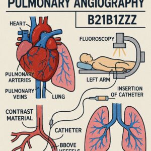
Before a single code can be assigned, a foundational understanding of the procedure is paramount. Pulmonary angiography is an invasive diagnostic and/or therapeutic procedure that involves the direct injection of radiopaque contrast material into the pulmonary arteries to visualize their structure and blood flow under X-ray guidance.
2.1. Clinical Indications: When is Pulmonary Angiography Necessary?
The procedure is typically reserved for situations where non-invasive tests like CT Pulmonary Angiography (CTPA) or V/Q scans are inconclusive, unavailable, or when a therapeutic intervention is anticipated. Key indications include:
-
Suspected Acute Pulmonary Embolism (PE): To confirm or rule out a blood clot in the pulmonary arteries, especially in high-risk patients with conflicting imaging results.
-
Chronic Thromboembolic Pulmonary Hypertension (CTEPH): To assess the extent and surgical accessibility of chronic clots.
-
Pulmonary Arteriovenous Malformations (PAVMs): To diagnose and often treat abnormal connections between arteries and veins.
-
Pulmonary Artery Stenosis or Aneurysm: To evaluate narrowing or abnormal dilation of the vessels.
-
Pre-operative Assessment: Prior to major lung resection surgery to map the vascular anatomy.
-
Hemoptysis (Coughing Blood) of Unknown Origin: To locate the source of bleeding.
-
Pulmonary Vasculitis: To assess inflammatory involvement of the vessels.
2.2. The Procedure: A Step-by-Step Walkthrough
A standard pulmonary angiography procedure unfolds in a series of deliberate steps:
-
Patient Preparation and Access: The patient is placed under sedation or general anesthesia. Access is typically gained via the femoral vein in the groin, though the jugular or brachial veins can also be used. The site is sterilized and locally anesthetized.
-
Venous Access and Sheath Placement: A needle is used to puncture the vein, followed by the insertion of a guidewire. Over this guidewire, an introducer sheath is placed, providing a stable port into the vascular system.
-
Catheter Navigation: A long, flexible catheter is then advanced under fluoroscopic (live X-ray) guidance. It travels through the venous system—the iliac vein, inferior vena cava, right atrium, and right ventricle—before entering the main pulmonary artery.
-
Pressure Measurement and Positioning: The physician measures pressures within the heart chambers and pulmonary artery. The catheter tip is then positioned in the specific branch of the pulmonary artery to be studied (e.g., right main, left main, or a lobar branch).
-
Contrast Injection and Image Acquisition: A mechanical injector rapidly delivers a bolus of iodinated contrast material through the catheter. Simultaneously, a rapid series of X-ray images (cineangiography) is captured, showing the contrast flowing through the arteries, capillaries, and veins.
-
Diagnostic Interpretation and Potential Intervention: The physician interprets the images in real-time. If a pathology is identified, the procedure may transition from diagnostic to therapeutic. The same vascular access can be used to perform interventions.
-
Catheter and Sheath Removal: At the conclusion of the procedure, the catheter and sheath are removed, and pressure is applied to the access site to achieve hemostasis.
This potential for a seamless shift from diagnosis to therapy is precisely what makes ICD-10-PCS coding for this procedure so complex.
3. The Architecture of ICD-10-PCS: A Primer for Procedural Coding
ICD-10-PCS is a procedural classification system, not a nomenclature like CPT®. Its primary purpose is to collect data on the types of procedures performed, for use in epidemiology, quality reporting, and reimbursement. Its structure is fundamentally different and is built on a logical, multi-axial framework.
3.1. The Seven-Character Alphanumeric System
Every ICD-10-PCS code is seven characters long, and each character represents a specific aspect of the procedure. The value for each character is selected from a predefined table.
-
Character 1: Section – The broadest category (e.g., Medical and Surgical, Obstetrics, Imaging).
-
Character 2: Body System – The general physiological system involved.
-
Character 3: Root Operation – The objective or intent of the procedure. This is the most critical character.
-
Character 4: Body Part – The specific anatomical site where the root operation was performed.
-
Character 5: Approach – The technique used to reach the site (e.g., open, percutaneous).
-
Character 6: Device – Any device that remains after the procedure is completed.
-
Character 7: Qualifier – Adds additional information to further specify the procedure.
3.2. The Importance of the Medical and Surgical Section (Section 0)
Pulmonary angiography, being an invasive procedure that involves catheter-based instrumentation, is almost exclusively coded in the Medical and Surgical Section (Section 0). The imaging component (the fluoroscopy and image acquisition) is considered an integral part of the procedure and is not coded separately in ICD-10-PCS, unlike in CPT® where supervision and interpretation codes exist.
4. Deconstructing the ICD-10-PCS Code for Pulmonary Angiography
Now, we will build the code from the ground up, character by character, focusing on the choices relevant to pulmonary angiography.
4.1. Character 1: Section – Medical and Surgical (0)
For all invasive, catheter-based pulmonary angiographic procedures, the section is always 0.
4.2. Character 2: Body System – Heart and Great Vessels (2)
The pulmonary arteries are classified under the “Heart and Great Vessels” body system. Therefore, the second character is 2.
4.3. Character 3: Root Operation – The Core of the Procedure
This is the most pivotal character and is entirely dependent on the objective of the procedure as documented in the operative report. The “angiography” itself is the imaging that confirms the success of the root operation or guides it; it is not the root operation. The following table outlines the common root operations for pulmonary angiography.
Common Root Operations in Pulmonary Angiography Procedures
| Root Operation | Code | Definition | Example in Pulmonary Angiography |
|---|---|---|---|
| Introduction | Y | Putting in or on a therapeutic, diagnostic, nutritional, physiological, or prophylactic substance except blood or blood products. | Injection of contrast material for a purely diagnostic angiogram. |
| Inspection | J | Visually and/or manually exploring a body part. | Not used. Angiographic visualization is not “Inspection” in ICD-10-PCS. |
| Occlusion | L | Completely closing an orifice or lumen of a tubular body part. | Placing coils or plugs to block off a Pulmonary AVM or a bleeding vessel. |
| Restriction | V | Partially closing an orifice or lumen of a tubular body part. | Placing a stent-graft that narrows but does not fully close the vessel lumen. |
| Extirpation | D | Taking or cutting out solid matter from a body part. | Mechanical thrombectomy; removing a clot from a pulmonary artery. |
| Fragmentation | F | Breaking solid matter in a body part into pieces. | Using a device to break up a large clot into smaller pieces. |
| Bypass | 1 | Altering the route of passage of the contents of a tubular body part. | Creating a shunt from the pulmonary artery to another vessel. |
| Dilation | 7 | Expanding an orifice or lumen of a tubular body part. | Balloon angioplasty of a stenotic pulmonary artery. |
Let’s explore the most common ones in detail.
4.3.10. Root Operation Y: Introduction
This is the root operation for a purely diagnostic pulmonary angiogram where no therapeutic intervention is performed. The definition is key: “Putting in… a diagnostic substance.” The contrast dye is the diagnostic substance. It is introduced into a vein (the access point) or an artery (the target) to make it visible. The objective is to obtain images. If the report states only “diagnostic pulmonary angiography” and describes the injection of contrast and acquisition of images, the root operation is Y: Introduction.
4.4. Character 4: Body Part/Region – Specifying the Anatomical Target
The body part character specifies where the root operation was performed. This can be tricky.
-
For Introduction (Y), the body part is the site where the substance is introduced. If the contrast is injected into a peripheral vein (e.g., femoral vein) and the catheter does not selectively enter a specific pulmonary branch, the body part is the Peripheral Vein.
-
If the catheter is advanced selectively into a specific pulmonary artery branch (e.g., the right pulmonary artery) and contrast is injected there, the body part would be that specific artery (e.g., Right Pulmonary Artery).
-
For therapeutic root operations like Occlusion (L) or Extirpation (D), the body part is the specific anatomical site where that action took place (e.g., Left Lower Lobe Pulmonary Artery).
The coder must carefully review the report to determine the precise site of action.
4.5. Character 5: Approach – The Path to the Site
The approach describes how the procedure reached the body part. In pulmonary angiography, this is almost always Percutaneous (3) or Percutaneous Endoscopic (4). The initial venous access is through the skin (percutaneous), and the entire procedure is performed through that access point.
-
Percutaneous (3): Entry through the skin via needle puncture, with the procedure performed using intraluminal instrumentation (the catheter).
-
Open (0): A surgical incision; not typical for standard angiography.
-
Via Natural or Artificial Opening (7): Not applicable.
4.6. Character 6: Device – Retained or Not?
This character identifies a device that remains in the body after the procedure is completed. For a diagnostic angiogram (Introduction), no device remains, so the character is Z: No Device.
However, if a therapeutic intervention occurs, a device may be used:
-
Occlusion Device (C): For coils, plugs, or vascular clips.
-
Intraluminal Device (D): For stents.
-
Autologous Tissue Substitute (7): For a graft, though rare in this context.
If a device is placed, Character 6 specifies the type. If no device remains, it is Z.
4.7. Character 7: Qualifier – Adding Specificity
The qualifier provides additional context. For many procedures in the Heart and Great Vessels body system, this character is Z: No Qualifier. However, it becomes crucial for specific root operations.
-
For Introduction (Y), the qualifier specifies the substance introduced. For contrast, it is X: Diagnostic Substance.
-
For Bypass (1), the qualifier specifies the destination of the bypass.
-
For Dilation (7) with a balloon, the qualifier is Z for no qualifier, but if a drug-eluting balloon is used, it would be specified here.
5. Coding Scenarios: From Operative Report to Accurate Code
Let’s apply this knowledge to realistic operative reports.
5.1. Scenario 1: Diagnostic Pulmonary Angiography via Femoral Artery
Operative Report Snippet: “After obtaining informed consent, the right femoral vein was accessed percutaneously using the Seldinger technique. A 6-French sheath was placed. A pigtail catheter was advanced into the main pulmonary artery. Contrast was injected, and multiple angiographic images were obtained of the right and left pulmonary arteries. The study was unremarkable. The catheter and sheath were removed. Hemostasis was achieved by manual compression.”
Coding Analysis:
-
Objective: To obtain diagnostic images. No therapy was performed.
-
Root Operation: Y – Introduction (of a diagnostic substance).
-
Body Part: The contrast was injected into the Main Pulmonary Artery.
-
Approach: 3 – Percutaneous.
-
Device: Z – No Device (the catheter was removed).
-
Qualifier: X – Diagnostic Substance.
Constructed ICD-10-PCS Code: B21 (Medical/Surgical, Heart & Great Vessels, Introduction) 6 (Main Pulmonary Artery) 3 (Percutaneous) Z (No Device) X (Diagnostic Substance).
Final Code: B2163ZX – Introduction of Diagnostic Substance into Main Pulmonary Artery, Percutaneous Approach.
5.2. Scenario 2: Pulmonary Angiography with Embolectomy and Stent Placement
Operative Report Snippet: “Diagnostic angiography revealed a large saddle embolus at the bifurcation of the main pulmonary artery. Decision was made to intervene. A mechanical thrombectomy device was advanced, and the bulk of the thrombus was removed. Post-thrombectomy, a significant residual stenosis was noted in the right pulmonary artery. A 10mm x 4cm self-expanding stent was deployed across the stenosis with an excellent angiographic result.”
Coding Analysis: This requires multiple codes.
-
Code 1: For the thrombus removal.
-
Root Operation: D – Extirpation (taking out solid matter).
-
Body Part: 5 – Pulmonary Artery, Right (the specific site of action).
-
Approach: 3 – Percutaneous.
-
Device: Z – No Device (the thrombectomy device was removed).
-
Qualifier: Z – No Qualifier.
-
Code: B2153ZZ – Extirpation of Matter from Right Pulmonary Artery, Percutaneous Approach.
-
-
Code 2: For the stent placement.
-
Root Operation: Dilation (7) is often debated, but the official ICD-10-PCS guideline for the body system “Heart and Great Vessels” directs that placing an intraluminal device (stent) constitutes the root operation Dilation. The stent is used to expand the lumen.
-
Body Part: 5 – Pulmonary Artery, Right.
-
Approach: 3 – Percutaneous.
-
Device: D – Intraluminal Device (the stent).
-
Qualifier: Z – No Qualifier.
-
Code: B2153DZ – Dilation of Right Pulmonary Artery with Intraluminal Device, Percutaneous Approach.
-
5.3. Scenario 3: Pulmonary Angiography with Coil Embolization for AVM
Operative Report Snippet: “Angiography confirmed a 1.5 cm AVM in the left lower lobe pulmonary artery. A microcatheter was superselectively advanced into the feeding vessel. Under fluoroscopic guidance, three platinum microcoils were deployed until angiography confirmed complete occlusion of the AVM nidus.”
Coding Analysis:
-
Objective: To block off the AVM.
-
Root Operation: L – Occlusion (completely closing the lumen).
-
Body Part: 8 – Pulmonary Artery, Left Lower Lobe.
-
Approach: 3 – Percutaneous.
-
Device: C – Occlusion Device (the coils).
-
Qualifier: Z – No Qualifier.
Constructed ICD-10-PCS Code: B2183CZ – Occlusion of Left Lower Lobe Pulmonary Artery with Occlusion Device, Percutaneous Approach.
6. Navigating Common Pitfalls and Challenges
6.1. Diagnostic vs. Therapeutic Intent
The single biggest pitfall is misinterpreting the intent. If the physician’s plan is “pulmonary angiography,” but they find and treat a problem, the diagnostic angiogram is not coded separately as “Introduction.” The therapeutic procedure(s) take precedence. The angiography is the imaging used to perform the root operation.
6.2. Differentiating Root Operations: Occlusion vs. Restriction
-
Occlusion (L): The lumen is completely closed. Think coils, plugs, vessel ligation. The goal is to stop all flow.
-
Restriction (V): The lumen is narrowed but not fully closed. This is less common but could apply to certain stent-grafts or bands that reduce flow without stopping it entirely.
6.3. Coding the “Catheterization” Itself
The placement of the venous sheath and the navigation of the catheter through the venous system to the pulmonary artery are considered inherent components of the procedure. These steps are not coded separately in ICD-10-PCS.
7. The Role of Imaging Guidance in Coding
In ICD-10-PCS, the fluoroscopic guidance used to navigate the catheter, position devices, and perform the angiography is considered an integral part of the procedure. There is no separate code for it in the Medical and Surgical section. This is a fundamental difference from CPT® coding, where a separate “Supervision and Interpretation” code (e.g., 75746) is reported alongside the procedural code.
8. ICD-10-PCS and CPT® Coordination: A Necessary Distinction
It is critical for coders to understand that ICD-10-PCS and CPT® serve different purposes and have different structures. They are used together on claims but represent different things.
-
CPT® (Professional Fee): Describes the “what” from the physician’s work perspective. A single CPT® code like 36013 (Selective catheter placement, venous) and 75746 (Angiography, pulmonary, radiological supervision and interpretation) might be used.
-
ICD-10-PCS (Hospital/Technical Fee): Describes the “what” from the facility’s resource use perspective. It details the specific therapeutic actions taken (e.g., Extirpation, Occlusion, Dilation).
A coder must be fluent in both systems, recognizing that a single operative report will generate codes in both classifications that complement but do not duplicate each other.
9. Conclusion: Summarizing the Content of the Article in Three Lines
Accurate ICD-10-PCS coding for pulmonary angiography hinges on identifying the procedure’s therapeutic objective, not the imaging component. The root operation is the cornerstone of the code, determined by the physician’s documented intent, whether it be Introduction for diagnosis, Extirpation for clot removal, or Occlusion for embolization. Mastering this granular, intent-driven approach is essential for capturing the full clinical picture, ensuring data integrity, and supporting appropriate reimbursement in today’s complex healthcare environment.
10. Frequently Asked Questions (FAQs)
Q1: What is the ICD-10-PCS code for a simple, diagnostic pulmonary angiogram?
A: There is no single code; it depends on the approach and injection site. A common code is B2163ZX (Introduction of Diagnostic Substance into Main Pulmonary Artery, Percutaneous Approach).
Q2: Why isn’t there a specific root operation for “Angiography”?
A: ICD-10-PCS classifies procedures by their objective. The objective of angiography is either to introduce a diagnostic substance (Root Operation Y) or to obtain images to guide a therapeutic root operation (which is then the primary procedure coded).
Q3: How do I code a pulmonary angiogram that starts as diagnostic but turns into a therapeutic embolization?
A: You code only the therapeutic procedure(s). The diagnostic angiogram is not coded separately. You would assign a code for the embolization (e.g., Occlusion with an occlusion device).
Q4: Do I need to code the venous access (e.g., femoral vein puncture) separately?
A: No. The vascular access is considered the initial approach for the procedure and is inherent to the root operation code. It is not coded separately in ICD-10-PCS.
Q5: What is the difference between coding a stent placement as Dilation versus a different root operation?
A: Per the ICD-10-PCS Official Guidelines for Coding and Reporting, in the “Heart and Great Vessels” body system, the placement of an intraluminal device (stent) is coded to the root operation Dilation, as the objective is to expand the lumen of the vessel.
Date: November 17, 2025
Disclaimer: This article is intended for educational and informational purposes only. It is not a substitute for professional medical coding advice, official coding guidelines, or the current, complete ICD-10-PCS code set. Coders must always consult the most recent official resources and facility-specific policies for accurate code assignment.


