Uterine fibroids, known in medical terms as leiomyomas or myomas, are far more than an alphanumeric entry in a patient’s chart or a line item on an insurance claim. They are a complex, common, and often life-altering health concern for millions of individuals with a uterus worldwide. For some, they are a silent, asymptomatic discovery during a routine exam. For others, they are the source of debilitating pain, profound anemia, urinary dysfunction, and heartbreaking infertility. The journey from initial symptoms to final diagnosis and treatment is paved with clinical expertise, advanced technology, and, crucially, a language that translates this journey into actionable data: medical coding.
The International Classification of Diseases, Tenth Revision, Clinical Modification (ICD-10-CM) is that universal language. For healthcare providers, medical coders, billers, and administrators, understanding the nuanced world of ICD-10 codes for uterine fibroids is not merely an administrative task—it is a critical component of patient care, revenue cycle management, and population health data analysis. An accurately chosen code tells a precise story. It communicates the patient’s specific condition, justifies the medical necessity of procedures performed, ensures appropriate reimbursement, and contributes to valuable research that shapes future treatments.
This article serves as a definitive guide. We will delve deep into the medical reality of uterine fibroids, exploring their biology, symptoms, and the array of modern treatment options. Then, we will master the art and science of ICD-10 coding for this condition, moving beyond the basics to a level of detail that ensures accuracy, compliance, and clarity. Whether you are a patient seeking to understand your diagnosis, a medical student, a seasoned clinician, or a coding professional, this comprehensive resource is designed to provide the depth of knowledge you need.
icd-10 code for uterine fibroids
2. What Are Uterine Fibroids? Demystifying the Benign Tumor
At its core, a uterine fibroid is a benign (non-cancerous) tumor that develops from the smooth muscle tissue (myometrium) of the uterus. The term “tumor” can be alarming, but it simply denotes an abnormal mass or growth of cells. In the case of fibroids, these cells multiply at a higher rate than surrounding tissue, forming a firm, rubbery lump distinct from the normal uterine wall.
Key Characteristics:
-
Prevalence: They are exceedingly common. It is estimated that 70% to 80% of women will develop at least one fibroid by the age of 50, though many will never know it due to a lack of symptoms.
-
Hormonal Dependence: Fibroids are hormonally responsive, particularly to estrogen and progesterone. They typically grow during the reproductive years when hormone levels are highest and often stabilize or shrink after menopause.
-
Size Variability: Fibroids exhibit a remarkable range in size. They can be as minuscule as a seed, undetectable by the human eye, or grow to the size of a grapefruit, melon, or even larger, dramatically distorting the shape and size of the uterus. A uterus enlarged by multiple large fibroids can reach the size of a pregnancy well into the second trimester.
-
Multiplicity: It is common for an individual to have multiple fibroids (a condition sometimes called uterine fibromatosis) of different sizes and in different locations simultaneously.
Understanding that fibroids are benign, common, and hormonally driven is the first step in demystifying the condition and reducing the anxiety that often accompanies a diagnosis.
3. The Clinical Spectrum: Types and Locations of Uterine Fibroids
The clinical impact of a fibroid—its symptoms and the treatment it necessitates—is profoundly influenced by its location within the uterus. The ICD-10-CM coding system reflects this importance by requiring coders to specify the fibroid’s type. The primary classification is based on location:
1. Intramural Fibroids: These are the most common type. They develop and remain embedded within the muscular wall of the uterus. As they grow, they can stretch and distort the uterine wall from within. They may cause symptoms like heavy menstrual bleeding, a feeling of fullness or pressure in the lower abdomen, and pelvic pain.
2. Subserosal Fibroids: These fibroids project outward from the outer wall of the uterus (the serosa) into the pelvic cavity. Their primary impact is often on surrounding structures rather than on menstrual bleeding. A large subserosal fibroid can press on the bladder, causing frequent urination; on the rectum, leading to constipation or backache; or on nerves, resulting in pain.
3. Submucosal Fibroids: These are the least common but often the most symptomatic type regarding menstrual changes. They develop just underneath the inner lining of the uterus (the endometrium) and bulge into the uterine cavity. Even a small submucosal fibroid can disrupt the endometrial lining, leading to exceptionally heavy and prolonged menstrual bleeding (menorrhagia), severe cramping, and can act as a natural intrauterine device (IUD), interfering with implantation and causing infertility or pregnancy loss.
4. Pedunculated Fibroids: This is not a primary location-based category but a sub-type. Both subserosal and submucosal fibroids can become pedunculated, meaning they grow on a stalk-like structure (a peduncle). A pedunculated submucosal fibroid may dangle into the uterine cavity, while a pedunculated subserosal fibroid extends into the pelvic space. The peduncle can sometimes twist, cutting off the blood supply to the fibroid and causing a sudden, sharp, severe pain—a surgical emergency known as infarction.
Infographic Description: A detailed cross-section diagram of the female pelvis showing a uterus with the different types of fibroids labeled: Submucosal (protruding into the endometrial cavity), Intramural (within the myometrial wall), Subserosal (on the outer surface), and Pedunculated (on a stalk, both inside and outside).
4. Unveiling the Mystery: Causes and Risk Factors
The exact cause of uterine fibroids remains unknown, but research has pinpointed several key factors that influence their development and growth.
-
Genetic Alterations: Many fibroids contain altered genes that differ from those in normal uterine muscle cells. There is often a family history, suggesting a genetic predisposition.
-
Hormones: As established, estrogen and progesterone promote fibroid growth. They contain a higher density of estrogen and progesterone receptors than normal uterine muscle.
-
Growth Factors: Substances that help the body maintain tissues, such as insulin-like growth factor, may affect fibroid growth.
-
Extracellular Matrix (ECM): This is the material that makes cells stick together. Fibroids have an increased ECM, making them fibrous and dense.
Known Risk Factors:
-
Age: Risk increases during the 30s and 40s, declining after menopause.
-
Race: Black women are disproportionately affected. They are 2-3 times more likely to develop fibroids, to develop them at a younger age, to have more and larger fibroids, and to experience more severe symptoms. The reasons for this disparity are complex and likely involve genetic, socioeconomic, and environmental factors.
-
Family History: Having a mother or sister with fibroids increases your risk.
-
Early Menarche: Beginning menstruation at an early age increases risk.
-
Weight: Being overweight or obese increases the risk, as fat tissue produces additional estrogen.
-
Diet: A diet high in red meat and ham appears to increase risk, while a diet rich in green vegetables and fruit may be protective.
-
Nulliparity: Women who have never given birth may have a higher risk.
5. A Silent Presence or a Loud Disruption? Recognizing the Symptoms
The symptomatology of uterine fibroids exists on a vast spectrum. An estimated 50% of women with fibroids are entirely asymptomatic.
For the other 50%, symptoms can be significant and include:
-
Heavy Menstrual Bleeding (Menorrhagia): The most common symptom. This includes periods lasting more than 7 days, soaking through sanitary products in an hour, passing large blood clots, and anemia resulting from blood loss.
-
Pelvic Pressure and Pain: A constant feeling of fullness, pressure, or heaviness in the lower abdomen, often described as a “bearing-down” sensation. Dull aching or sharp pain is also common.
-
Urinary Symptoms: Frequency (needing to urinate often), urgency (a sudden, strong need to urinate), or retention (inability to empty the bladder) caused by a fibroid pressing on the bladder.
-
Bowel Symptoms: Constipation or difficulty with bowel movements due to rectal pressure.
-
Backache or Leg Pains: Resulting from pressure on nerves in the pelvis and back.
-
Pain During Intercourse (Dyspareunia): Often associated with the location and size of the fibroids.
-
Reproductive Challenges: Submucosal fibroids can interfere with implantation of a fertilized egg, contributing to infertility. They can also increase the risk of pregnancy complications like placental abruption, fetal growth restriction, and preterm birth.
-
Increase in Abdominal Girth: Large fibroids can cause a noticeable enlargement of the abdomen, often mistaken for weight gain or pregnancy.
6. From Consultation to Diagnosis: The Diagnostic Journey
Diagnosis typically begins with a visit to a gynecologist prompted by symptoms or a finding during a routine pelvic exam.
-
Pelvic Exam: A physician may feel an irregularly shaped, enlarged, or firm uterus, suggesting the presence of fibroids.
-
Ultrasound (Sonography): The primary and most common imaging tool. A transabdominal or transvaginal ultrasound uses sound waves to create a picture of the uterus, confirming the presence, number, size, and location of fibroids.
-
Lab Tests: A complete blood count (CBC) may be ordered to check for anemia due to chronic blood loss. Other tests may rule out bleeding disorders or thyroid problems.
-
Magnetic Resonance Imaging (MRI): This provides highly detailed images and can precisely map the size and location of fibroids, differentiate them from other masses, and is invaluable for planning surgical interventions, particularly uterine artery embolization (UAE) or MRI-guided focused ultrasound.
-
Hysterosonography (Saline Infusion Sonography): Saline is injected into the uterine cavity to expand it, providing a clearer ultrasound image of submucosal fibroids and the endometrial lining.
-
Hysterosalpingography (HSG): A dye is injected into the uterus and fallopian tubes, and X-rays are taken. It is primarily used to evaluate the fallopian tubes but can also reveal submucosal fibroids.
-
Hysteroscopy: A thin, lighted telescope (hysteroscope) is inserted through the cervix into the uterus. This allows the physician to visually inspect the interior of the uterus and identify submucosal fibroids.
7. Navigating Treatment Options: From Watchful Waiting to Surgery
Treatment is highly individualized, based on symptom severity, fibroid size and location, age, and desire for future fertility.
1. Watchful Waiting (Expectant Management): For asymptomatic or minimally symptomatic women, no treatment is necessary. Regular monitoring to ensure no significant growth occurs is often the recommended course.
2. Medications: Aim to manage symptoms, not eliminate fibroids.
-
NSAIDs: Ibuprofen or naproxen can help relieve menstrual cramping.
-
Tranexamic Acid: An anti-fibrinolytic drug taken during menstruation to reduce heavy bleeding.
-
Hormonal Therapies:
-
Gonadotropin-releasing hormone (GnRH) agonists: (e.g., Leuprolide) induce a temporary, medical menopause, shrinking fibroids by drastically lowering estrogen levels. Used preoperatively to reduce fibroid size and control bleeding or in perimenopausal women. Side effects can be significant (hot flashes, bone density loss).
-
Progestin-Releasing Intrauterine Device (IUD): Can dramatically reduce heavy menstrual bleeding but does not shrink fibroids themselves.
-
Oral Contraceptives: Can help regulate bleeding cycles and reduce menstrual flow but do not shrink fibroids.
-
3. Minimally Invasive Procedures:
-
Uterine Artery Embolization (UAE): An interventional radiologist injects tiny particles into the arteries supplying the uterus, blocking blood flow to the fibroids and causing them to shrink. It preserves the uterus but is not typically recommended for women desiring future pregnancy.
-
MRI-Guided Focused Ultrasound Surgery (FUS): Uses high-intensity, focused ultrasound waves to heat and destroy (ablate) fibroid tissue through the abdominal wall, guided by real-time MRI.
-
Myomectomy: The surgical removal of fibroids while preserving the uterus. It is the preferred treatment for women who wish to maintain fertility. It can be performed via:
-
Hysteroscopy: For submucosal fibroids.
-
Laparoscopy: Minimally invasive surgery with small incisions.
-
Laparotomy: Open abdominal surgery for numerous or very large fibroids.
-
-
Endometrial Ablation: Destroys the lining of the uterus to reduce heavy bleeding. It is not a treatment for the fibroids themselves and is not an option for women wanting future pregnancy.
4. Surgical Intervention:
-
Hysterectomy: The surgical removal of the uterus. It is the only definitive cure for uterine fibroids and eliminates any possibility of recurrence. It is a major surgery considered when other treatments have failed or are not desired, and childbearing is complete. It can be performed laparoscopically, vaginally, or via laparotomy.
8. The Cornerstone of Medical Documentation: Mastering ICD-10 Coding for Uterine Fibroids
This section is the technical heart of the article, providing the detailed coding knowledge essential for accurate medical records and billing.
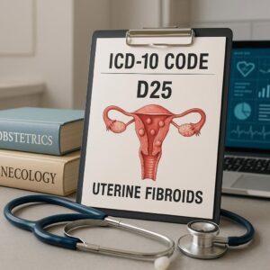
The ICD-10-CM codes for leiomyoma of the uterus are found in Chapter 14: Diseases of the Genitourinary System (N00-N99), specifically under category N80- Endometriosis and then the block D25- Leiomyoma of uterus.
The coding structure requires specificity. The foundational code is D25.9 – Leiomyoma of uterus, unspecified. However, using this code should be a last resort when the medical documentation provides no further detail. Coders must always review the patient’s record—specifically the physician’s notes, operative reports, and imaging results—to assign the most specific code possible.
The ICD-10-CM codes for uterine fibroids are as follows:
| ICD-10-CM Code | Code Description | Clinical Scenario & Documentation Requirements |
|---|---|---|
| D25.0 | Submucous leiomyoma of uterus | The fibroid is located underneath the endometrial lining and bulges into the uterine cavity. Documentation must specify “submucosal” or “submucous.” |
| D25.1 | Intramural leiomyoma of uterus | The fibroid is embedded within the myometrial (muscular) wall of the uterus. Documentation must specify “intramural.” |
| D25.2 | Subserosal leiomyoma of uterus | The fibroid is located on the outer surface of the uterus, projecting into the pelvic cavity. Documentation must specify “subserosal.” |
| D25.9 | Leiomyoma of uterus, unspecified | Use only if the medical record does not specify the type (e.g., only states “uterine fibroids” or “leiomyoma”). |
| D26. | Other benign neoplasms of uterus | This is for other, rarer benign uterine growths. D26.0 is for the cervix uteri, D26.1 for the corpus uteri (body), and D26.7 for other specified parts. |
| E10.9 / E11.9, etc. | Diabetes mellitus | Important Note: This is an example of a co-morbidity that must be coded separately if present. Fibroids themselves are not caused by diabetes, but if a patient has both, both conditions must be coded. |
Coding for Complications: The 4th and 5th Characters
The true power of ICD-10-CM lies in its ability to capture complexity. For categories D25.0, D25.1, and D25.2, a 5th character is required to indicate whether the fibroid is causing specific complications.
-
D25.X0 – … without mention of degeneration. This is the most common scenario. The fibroid is present but there is no mention of it undergoing any degenerative changes (e.g., hyaline, cystic, red degeneration).
-
D25.X1 – … with degeneration. This is used when the physician documents that the fibroid has undergone a type of degenerative change. This often occurs when a fibroid outgrows its blood supply, causing tissue death. This can be a source of acute pain.
Example 1: A patient’s MRI report states: “A 5 cm intramural fibroid in the fundus with signs of cystic degeneration.” The correct code would be D25.11 – Intramural leiomyoma of uterus with degeneration.
Example 2: An operative report from a hysterectomy describes “a large subserosal fibroid on the posterior wall.” There is no mention of degeneration. The correct code is D25.20 – Subserosal leiomyoma of uterus without mention of degeneration.
Coding for Multiple Types and Associated Conditions
A patient often has more than one type of fibroid. The ICD-10-CM Official Guidelines for Coding and Reporting state: “Code all documented conditions that coexist.” Therefore, if a patient’s record documents both an intramural and a submucosal fibroid, both codes should be assigned, sequencing first the one that is most responsible for the encounter.
Furthermore, conditions directly caused by the fibroids must be coded separately.
-
Heavy Menstrual Bleeding: Code N92.0 – Excessive and frequent menstruation with regular cycle (or other codes from category N92) in addition to the fibroid code.
-
Iron Deficiency Anemia: If the heavy bleeding has resulted in anemia, code D50.9 – Iron deficiency anemia, unspecified (or a more specific anemia code) as a secondary diagnosis.
-
Pelvic Pain: Code R10.2 – Pelvic and perineal pain.
-
Infertility: If attributed to the fibroid, code N97.0 – Female infertility associated with anovulation or another code from N97.
Coding Scenario: Putting It All Together
Patient Presentation: A 42-year-old female presents to her gynecologist with complaints of increasingly heavy menstrual periods, passing large clots, and new onset pelvic pressure. She is also feeling unusually tired. An ultrasound and subsequent MRI reveal a 4cm submucosal fibroid and two smaller intramural fibroids. Her CBC shows a hemoglobin of 9.8 g/dL (low).
Physician’s Final Diagnoses:
-
Symptomatic uterine fibroids, specifically a submucosal fibroid and intramural fibroids.
-
Menorrhagia.
-
Iron deficiency anemia secondary to menorrhagia.
Correct ICD-10-CM Code Assignment:
-
D25.00 – Submucous leiomyoma of uterus without mention of degeneration (This is sequenced first as it is the submucosal fibroid likely causing the primary symptom of menorrhagia).
-
D25.10 – Intramural leiomyoma of uterus without mention of degeneration (The intramural fibroids are also documented and coded).
-
N92.0 – Excessive and frequent menstruation with regular cycle (The menorrhagia).
-
D50.9 – Iron deficiency anemia, unspecified (The anemia resulting from the blood loss).
This precise coding paints a complete clinical picture for anyone reviewing the claim or the patient’s record, justifying the medical necessity of any treatment proposed, such as a hysteroscopic myomectomy.
9. The Impact on Quality of Life and Future Health
The impact of symptomatic fibroids extends far beyond physical discomfort. The chronic pain, unpredictable and heavy bleeding, and fear of embarrassing accidents can lead to social isolation, anxiety, and depression. The fatigue from anemia can cripple professional and personal productivity. Concerns about fertility can cause significant emotional distress. The economic burden is also substantial, including costs of sanitary products, missed days of work, and medical expenses. Recognizing this holistic impact is crucial for providing empathetic, comprehensive care.
10. Conclusion: Empowering Patients and Perfecting Practice
Uterine fibroids represent a complex intersection of biology, symptomatology, and personalized medicine. Accurate ICD-10 coding is the critical link that translates this clinical complexity into data, ensuring precise communication, appropriate reimbursement, and valuable contributions to health outcomes research. For patients, understanding this condition is the first step toward empowered advocacy; for healthcare professionals, mastering its documentation is a fundamental component of excellence in patient care and practice management.
11. Frequently Asked Questions (FAQs)
Q1: Can uterine fibroids turn into cancer?
A: Leiomyosarcoma, a cancerous tumor of the uterine muscle, is extremely rare and is not believed to arise from pre-existing benign fibroids. The risk of a presumed fibroid being cancerous is estimated to be less than 1 in 1,000. However, rapid growth, particularly after menopause, can be a red flag that warrants further investigation.
Q2: I’ve been diagnosed with fibroids but want to have children. What should I do?
A: Many women with fibroids have successful pregnancies. However, submucosal fibroids, in particular, can interfere with implantation. It is essential to discuss your goals with a gynecologist or a fertility specialist. A myomectomy is often a highly effective option for preserving fertility while treating problematic fibroids.
Q3: What is the difference between ICD-10 code D25.9 and the more specific codes?
A: D25.9 is a nonspecific code meaning “unspecified.” It should only be used when the medical documentation (doctor’s notes, ultrasound report) does not describe the location of the fibroid(s). Using specific codes (D25.0, D25.1, D25.2) is always preferred as it provides a clearer clinical picture and is often required for accurate billing.
Q4: Are there any new treatments on the horizon for uterine fibroids?
A: Yes, research is ongoing. New oral medications known as selective progesterone receptor modulators (SPRMs), like ulipristal acetate, have shown promise in shrinking fibroids and controlling bleeding, though their long-term use requires more study. Additionally, newer minimally invasive techniques are constantly being refined.
12. Additional Resources
-
The American College of Obstetricians and Gynecologists (ACOG): Provides detailed patient education materials and clinical practice guidelines for providers. https://www.acog.org
-
The Society of Interventional Radiology (SIR): Offers information on Uterine Artery Emolization (UAE). https://www.sirweb.org
-
The Centers for Disease Control and Prevention (CDC) – Women’s Health: Features information on uterine fibroids and other health topics. https://www.cdc.gov/women
-
The ICD-10-CM Official Guidelines for Coding and Reporting: The definitive source for coding rules and conventions. Published annually by the Centers for Medicare & Medicaid Services (CMS) and the National Center for Health Statistics (NCHS).
Date: September 17, 2025
Author: The Medical Coding Specialist Team
Disclaimer: *The information provided in this article is for educational and informational purposes only and does not constitute medical advice. It is not a substitute for professional medical advice, diagnosis, or treatment. Always seek the advice of your physician or other qualified health provider with any questions you may have regarding a medical condition. Never disregard professional medical advice or delay in seeking it because of something you have read in this article. The codes and guidelines referenced are for illustrative purposes and are subject to change. Always consult the most current, official ICD-10-CM coding manuals and guidelines for accurate coding.*


