Walking into a dental office can sometimes feel like stepping into a world with its own language. Between the sounds of the equipment and the terms your dentist uses, it is easy to feel a bit lost. Then, you get a treatment plan or an insurance statement, and you see a string of codes that look more like a secret cipher than a bill.
One of the most common codes you will encounter is the one for a full mouth series of X-rays. If you have ever looked at a dental claim and wondered what “D0210” means, you are not alone.
This guide is designed to be your friendly companion through the world of dental imaging codes. We will break down exactly what the dental code for full mouth x ray entails, why it is so important for your oral health, how insurance typically handles it, and what you can expect during the procedure. Whether you are a new patient, a seasoned dental veteran, or just someone who likes to understand their healthcare bills, this article will give you the clarity you need.
Dental Code for Full Mouth X Ray
What Exactly is a Full Mouth X Ray?
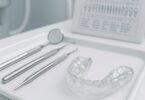
Before we dive into the codes themselves, it helps to understand what you are actually getting when your dentist recommends this procedure. A full mouth x ray, often abbreviated as FMX, is exactly what it sounds like: a comprehensive set of images that captures every tooth in your mouth, from root to crown.
Think of it as creating a detailed map of your oral landscape. While a dentist can see the surfaces of your teeth during a visual exam, they cannot see what is happening beneath the gums, inside the bone, or between the teeth. This is where the FMX becomes invaluable.
The Two Main Types of Images in an FMX
A traditional full mouth series is actually a combination of two types of X-rays:
-
Periapical Images: These X-rays focus on capturing the entire tooth, from the biting surface down to the very tip of the root and the surrounding bone structure. The term “periapical” literally means “around the apex” or the root tip. These are crucial for diagnosing issues like abscesses, deep decay, bone loss, and cysts that originate at the root.
-
Bitewing Images: You have likely had bitewing X-rays before. These involve biting down on a small tab or holder while the X-ray machine captures the crowns of your upper and lower teeth simultaneously. Bitewings are the gold standard for detecting cavities between teeth (interproximal caries) and checking the height of the bone that supports your teeth.
A standard full mouth series usually includes 14 to 20 individual images. The exact number can vary slightly depending on the size of your mouth, the number of teeth you have, and the specific protocol of your dental office. For an adult with a full set of teeth, the combination is typically 4 bitewings and 14 periapicals, totaling 18 images.
The Modern Alternative: Panoramic X-rays
It is important to distinguish a full mouth series from a panoramic X-ray. A panoramic X-ray (code D0330) is a single, broad image that shows the entire mouth in one shot. It is excellent for seeing impacted wisdom teeth, jaw disorders, and overall bone structure, but it does not offer the fine detail needed to diagnose small cavities between teeth or subtle root fractures.
While some patients ask if a panoramic can replace a full mouth series, they serve different purposes. Your dentist might use a panoramic for an initial assessment but still require an FMX for a comprehensive diagnosis, especially if you are a new patient.
The Official Code: D0210
Let us get to the heart of the matter. The current dental terminology (CDT) code used for a full mouth x ray is D0210.
The American Dental Association (ADA) maintains the Current Dental Terminology (CDT) code set. These codes are updated periodically to reflect changes in dental practice and technology. Under this system, D0210 is defined as “Intraoral – complete series of radiographic images.”
This code is specifically designated for a set of images that includes both periapical and bitewing projections. It is the standard code used for a comprehensive radiographic examination of the entire dentition.
When is D0210 Used?
Dental offices use this code primarily for new patients or for established patients who have not had a full set of X-rays in a significant period of time. The standard of care often dictates that a full mouth series is taken every three to five years.
However, there are other scenarios where D0210 might be used:
-
As a baseline: For a new patient, an FMX provides the dentist with a baseline of your oral health. This allows them to compare future X-rays to see if any changes have occurred.
-
Before extensive treatment: If you are planning for major work like crowns, bridges, implants, or orthodontics, a full mouth series gives the dentist the detailed information needed to plan the treatment safely and effectively.
-
For diagnosing complex issues: If you are experiencing unexplained pain, swelling, or other symptoms, a full set of images can help pinpoint the source of the problem.
Other Related Codes: What They Mean
To avoid confusion, it is helpful to understand other radiographic codes you might see on your treatment plan. This ensures you know exactly what your dentist is recommending and what you are being billed for.
| CDT Code | Description | What It Includes |
|---|---|---|
| D0210 | Full Mouth X Ray (FMX) | A complete series of periapical and bitewing images. Usually 14-20 images. |
| D0220 | Intraoral – Periapical, first image | A single X-ray focused on one or two specific teeth, capturing the entire root. |
| D0230 | Intraoral – Periapical, each additional image | Used when multiple single periapical images are taken (e.g., to follow up on a specific area). |
| D0272 | Bitewings – two images | A set of two bitewing X-rays, often taken for recall exams when a patient has no history of decay. |
| D0274 | Bitewings – four images | A standard set of four bitewings, often taken annually to check for cavities between teeth. |
| D0330 | Panoramic film | A single, extraoral image showing the entire mouth, jaws, and sinuses. |
| D0350 | Oral/facial image capture | This is for digital or photographic images, often used for documentation or cosmetic planning. |
Important Note: If you see codes D0220 and D0230 listed several times in a row on a treatment plan, it may add up to the equivalent of a full mouth series. However, offices typically use the single code D0210 for a full series, which is often more straightforward for insurance processing.
Why Your Dentist Recommends a Full Mouth X Ray
It is natural to wonder if all these X-rays are truly necessary. After all, they do take time, and there is a small amount of radiation exposure involved. However, the diagnostic value of an FMX is immense. It allows your dentist to see problems long before they become visible to the naked eye or cause you pain.
Detecting Hidden Problems
Many dental issues are silent. They do not cause symptoms until they have progressed significantly. A full mouth series can reveal:
-
Hidden Decay: Cavities between teeth are a classic example. The biting surfaces of your teeth may look perfect, but a bitewing X-ray can reveal decay that is eating away at the enamel from the side.
-
Recurrent Decay: If you have existing fillings or crowns, X-rays can show if decay is developing underneath these restorations.
-
Periodontal (Gum) Disease: The periapical images show the bone level around each tooth. Bone loss is a key indicator of periodontal disease. Without X-rays, it is impossible to measure this accurately below the gumline.
-
Infections and Abscesses: A dark spot at the tip of a root on a periapical image often indicates an abscess, a serious infection that can spread and lead to tooth loss.
-
Cysts and Tumors: While rare, abnormal growths in the jawbone can be detected on X-rays, allowing for early intervention.
-
Impacted Teeth: The images can reveal teeth, such as wisdom teeth, that are stuck in the bone and unable to erupt properly.
Establishing a Baseline for Future Care
One of the most important functions of a full mouth series is that it serves as a benchmark. Years from now, your dentist can take new X-rays and overlay them with your current ones to see if there have been any changes. This ability to compare images over time is crucial for monitoring the progression of conditions like gum disease or the health of existing dental work.
Insurance Coverage and the D0210 Code
Navigating dental insurance can be one of the most frustrating parts of dental care. Understanding how your plan handles the D0210 code can help you avoid unexpected bills.
Frequency Limitations
Almost all dental insurance plans have frequency limitations. This means they will only pay for a specific procedure a certain number of times within a given period. For a full mouth series (D0210), the most common frequency limit is once every 36 months (3 years) or once every 60 months (5 years) .
If your dentist recommends a new FMX before this time limit has expired, your insurance will likely deny the claim, and the cost may fall to you. It is always a good idea to confirm your specific plan’s frequency limitations with your insurance carrier before scheduling the appointment.
New Patient vs. Established Patient
Many plans make an exception for new patients. If you are joining a new dental practice, your insurance may approve a full mouth series even if you had one at your previous dentist less than three years ago. The reasoning is that the new dentist needs their own set of baseline images to establish care. However, this is not a universal rule, so it is best to verify.
Percentage of Coverage (Co-insurance)
Dental insurance plans often categorize procedures into three tiers: preventive, basic, and major. Full mouth X-rays are typically classified as a basic service.
-
Preventive: (Cleanings, exams, bitewings) Often covered at 100%.
-
Basic: (Fillings, extractions, full mouth X-rays) Often covered at 70-80%.
-
Major: (Crowns, bridges, dentures) Often covered at 50%.
This means that if your plan covers basic services at 80%, you will be responsible for the remaining 20% after your deductible is met.
The Role of the Deductible
Most dental insurance plans have an annual deductible, which is a set amount you must pay out-of-pocket before the insurance company starts paying. The deductible usually applies to basic and major services. So, if you are having an FMX early in the year, you might be responsible for the full cost of the X-rays until you meet your deductible for the year.
The Cost of a Full Mouth X Ray Without Insurance
Understanding the cash price for a full mouth series is important, whether you are uninsured or considering paying out-of-pocket to avoid insurance limitations. The cost can vary significantly based on geographic location, the type of dental practice (private practice vs. corporate chain), and whether the images are taken with traditional film or advanced digital sensors.
On average, the cost for a full mouth series (D0210) in the United States typically ranges from $100 to $300.
Here is a rough breakdown of factors that influence the price:
-
Digital vs. Film: Digital X-rays are becoming the standard. While the cost to the patient is often similar, digital sensors reduce radiation exposure significantly and allow for instant viewing. Some offices may charge slightly less for digital because it eliminates the cost of film and chemicals.
-
Geographic Location: As with most services, prices tend to be higher in major metropolitan areas and lower in rural communities.
-
Type of Practice: A high-end cosmetic or specialty practice may have higher overhead costs, which can be reflected in the price of their X-rays. A community health center or a dental school often offers these services at a reduced rate.
Dental Schools and Community Clinics
If cost is a significant barrier, dental schools are an excellent resource. At a dental school, the work is performed by students under the close supervision of experienced faculty. The cost for a full mouth series at a dental school can be 50-70% less than at a private practice. The trade-off is that appointments are often longer due to the teaching environment.
Federally Qualified Health Centers (FQHCs) also often have dental departments that offer sliding scale fees based on income, making this essential diagnostic tool accessible to more people.
The Procedure: What to Expect
If your dentist has recommended a full mouth series, knowing what to expect can ease any anxiety. The process is straightforward and, with modern technology, faster than ever.
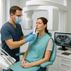
Step 1: Preparation
You will be seated in the dental chair. A lead apron with a thyroid collar will be placed over your chest and lap. This protective gear shields your body from any stray radiation. The thyroid collar is especially important for protecting the thyroid gland, and it is standard practice, particularly for women who are or may be pregnant.
Step 2: Positioning the Sensor
In a digital office, a small, flat electronic sensor will be used. For traditional film, a small piece of film in a plastic holder is used. The dental assistant or dentist will place this sensor inside your mouth.
You will be asked to hold the sensor in place by gently biting down on a holder or a cotton roll. This can feel a little awkward, but it only lasts for a few seconds per image.
Step 3: Capturing the Images
The X-ray machine will be positioned outside your cheek. The dental professional will step out of the room or behind a protective barrier and press a button to take the image. You will hear a brief beep or buzz, and the image will appear on a computer screen almost instantly if it is digital.
The process is repeated for each angle. For a full series, this will be repeated 14 to 20 times. Your dental team will work systematically around your mouth, starting in one area and moving to the next.
Step 4: Review
Once all the images are taken, the dentist will review them. Often, they will use a large monitor to show you what they see. They may point out areas of interest, such as an old filling, a developing cavity, or the health of the bone around your teeth. This is a great time to ask questions.
A Note on Gag Reflex: Some people worry about gagging during X-rays. If you have a sensitive gag reflex, let your dental team know beforehand. They have techniques to help, such as using smaller sensors, positioning you differently in the chair (leaning slightly forward), or using a topical anesthetic spray on the soft palate to desensitize it temporarily.
Radiation Safety and the FMX
One of the most common concerns patients have is about radiation exposure. It is a valid concern, and it is important to have accurate information.
How Much Radiation is in a Full Mouth Series?
The amount of radiation from a dental X-ray is extremely low, especially with modern digital technology. To put it in perspective, a full mouth series of digital X-rays exposes a patient to about the same amount of radiation as a short airplane flight (roughly 0.005 millisieverts). This is also a fraction of the background radiation we are naturally exposed to from the sun, soil, and air over the course of a year.
The ALARA Principle
Dentists follow the ALARA principle, which stands for “As Low As Reasonably Achievable.” This means they are committed to using the minimum amount of radiation necessary to obtain a diagnostic image. The use of lead aprons, thyroid collars, high-speed film, and digital sensors are all part of this commitment.
Pregnancy and X-rays
If you are pregnant or suspect you might be, it is crucial to inform your dentist. While the risk from dental X-rays is considered extremely low, many dentists will postpone non-emergency X-rays until after the first trimester or until after delivery as a precaution. In the case of a dental emergency, X-rays can be taken with double lead shielding to ensure the safety of the fetus.
Insurance Reimbursement and Claim Forms
For those who handle their own insurance claims or want to understand the process, knowing how the D0210 code appears on a claim form is helpful. The standardized ADA claim form has specific fields for the procedure code.
When an office submits a claim for D0210, they are attesting that a complete series of images was taken and that it was medically necessary. If an insurance company denies a claim for a full mouth series, common reasons include:
-
Frequency limitation exceeded: The patient had an FMX within the plan’s specified time frame (e.g., 3 years).
-
Lack of necessity: The insurance company may argue that a panoramic or a set of bitewings would have sufficed, especially if the patient is not new to the practice.
-
Missing documentation: Sometimes, an insurance company will ask for the actual X-rays to be submitted for review to verify that the procedure was indeed a complete series and not just a few images.
If you receive a denial, your dental office’s billing coordinator can often help you file an appeal with the insurance company, providing the necessary documentation to prove the service was required.
The Future of Dental Imaging
The world of dental radiography is not static. Technology continues to evolve, offering new ways to diagnose and treat patients. While the D0210 code remains the standard for a full mouth series, how those images are captured is changing.
Cone Beam Computed Tomography (CBCT)
CBCT (code D0367 or similar, depending on the area) is a three-dimensional imaging technology. It rotates around the patient’s head, capturing hundreds of images that are reconstructed into a 3D model of the teeth, jaws, and nerves. This technology is invaluable for complex procedures like implant placement, root canal therapy, and orthodontics. It does not replace an FMX but is used in conjunction with it or as a more advanced alternative for specific treatment planning.
AI-Assisted Diagnosis
Artificial intelligence is beginning to play a role in dental radiography. AI algorithms can analyze X-ray images and highlight areas of potential concern, such as cavities, bone loss, or even signs of osteoporosis. This acts as a second set of eyes for the dentist, helping to ensure that nothing is missed. It enhances the diagnostic power of the FMX, making it an even more valuable tool.
Making an Informed Decision
Ultimately, the decision to get a full mouth x ray is a collaborative one between you and your dentist. It is based on your medical history, your oral health status, and your risk factors for disease.
Here are some questions you can ask your dentist to help you feel more confident in the recommendation:
-
“What specific information are you hoping to gain from this full mouth series?”
-
“How will these X-rays change or improve the treatment plan you are recommending for me?”
-
“When was my last full mouth series, and is this timing appropriate based on my insurance coverage and my oral health?”
-
“Are there any alternative imaging options that might be suitable in my case?”
A good dentist will be happy to answer these questions. They understand that an informed patient is an empowered patient, and that transparency builds trust.
Common Misconceptions
There are a few persistent myths about full mouth X-rays that are worth addressing directly.
Myth: “My dentist just takes X-rays to make more money.”
Reality: While no one can deny that X-rays are a revenue stream for a practice, they are also a non-negotiable part of modern diagnostic dentistry. The cost of not taking X-rays can be far higher, both financially and in terms of your health. Treating a small cavity found early on an X-ray is much less expensive and invasive than treating a large cavity, an abscess, or a tooth that requires extraction and replacement years later.
Myth: “I don’t need X-rays because my teeth feel fine.”
Reality: This is perhaps the most dangerous misconception. The problems X-rays detect—cavities between teeth, root infections, bone loss—are often completely painless in their early stages. By the time a problem causes pain, it has typically progressed to a point where the treatment is more complex and costly. X-rays allow for proactive, rather than reactive, care.
Myth: “A full mouth series exposes me to dangerous levels of radiation.”
Reality: As discussed earlier, the radiation exposure from a full mouth series is minimal, especially with digital sensors. It is well within safe limits established by the National Council on Radiation Protection and Measurements (NCRP). The diagnostic benefits of a full mouth series far outweigh the minuscule risk.
Tips for Working with Your Dental Office
A smooth experience with your dental X-rays often comes down to good communication. Here are a few practical tips:
-
Be transparent about your insurance: When you call to schedule your appointment, let the front desk know if you are concerned about insurance coverage for X-rays. They can often verify your benefits beforehand and give you an estimate of your out-of-pocket cost.
-
Keep a record of your X-rays: If you change dentists, you have the right to have your X-rays transferred. This can save you time and money, as your new dentist may not need to take a new full mouth series if they have recent, diagnostic-quality images from your previous office. Some offices charge a small fee for copying and transferring records.
-
Ask about payment plans: If you do not have insurance or if your insurance denies coverage for a necessary FMX, ask the office if they offer in-house payment plans or if they work with third-party financing options like CareCredit.
-
Don’t be afraid to speak up: If you are anxious about the X-ray process, or if you have a small mouth or a strong gag reflex, let the team know. They are professionals who deal with these situations every day and have strategies to help you.
Conclusion
The dental code for a full mouth x ray, D0210, represents much more than a billing entry. It is a foundational tool for comprehensive dental care, providing a detailed and critical view of your oral health that is invisible to the naked eye. From detecting hidden cavities and infections to establishing a baseline for future comparisons, this series of images plays an indispensable role in helping your dentist keep your smile healthy.
Understanding what D0210 entails, how insurance typically covers it, and what to expect during the procedure empowers you to make informed decisions about your health. While navigating dental codes and insurance policies can be complex, having this knowledge turns you from a passive patient into an active, confident participant in your own dental care. Remember, a conversation with your dentist is the best way to understand why an FMX is recommended for you and how it fits into your long-term health journey.
Frequently Asked Questions (FAQ)
1. Is D0210 the only code for a full mouth x ray?
Yes, D0210 is the standard Current Dental Terminology (CDT) code used for a complete intraoral series of radiographic images (periapicals and bitewings). There is no alternative code for the same service.
2. How often can I get a full mouth x ray covered by insurance?
Most dental insurance plans cover a full mouth series once every 3 to 5 years. However, many plans make an exception for new patients to allow a new dentist to establish a baseline. You should check with your specific insurance carrier to confirm your plan’s frequency limitation.
3. What is the difference between a panoramic x ray (D0330) and a full mouth series (D0210)?
A panoramic x ray is a single, broad image that shows the entire mouth, jaws, and sinuses. It is excellent for seeing impacted teeth and bone structure but lacks the fine detail to detect small cavities between teeth. A full mouth series uses multiple detailed images to capture each tooth individually, including the roots and the surrounding bone, making it more effective for diagnosing decay and gum disease.
4. Are digital X-rays more expensive than film X-rays?
The cost to the patient is often similar. However, digital X-rays are preferred because they use significantly less radiation, provide instant images that can be enhanced and enlarged for better diagnosis, and are more environmentally friendly. The procedure code (D0210) is the same regardless of whether the images are captured digitally or on film.
5. What should I do if my insurance denies my claim for D0210?
First, do not panic. Contact your dental office’s billing coordinator. They can help you understand the reason for the denial (often a frequency limitation). If the service was necessary, they can help you file an appeal with your insurance company, providing the clinical notes and images to justify the medical necessity.
6. Is it safe to get a full mouth x ray while pregnant?
It is crucial to inform your dentist if you are pregnant or suspect you may be. As a precaution, most dentists will postpone routine, non-emergency X-rays until after the first trimester or until after delivery. In the case of a dental emergency, X-rays can be taken using double lead shielding to ensure the safety of the fetus.
7. Why does my dentist want new X-rays if I just had them at my previous dentist?
Your new dentist needs their own baseline images to establish a standard of care. While they can request your X-rays from your previous dentist, the quality may be poor, or the images may be outdated. If you have recent X-rays, ask your previous office to transfer them to your new dentist to potentially avoid the need for a new full mouth series.
Additional Resource
For the most accurate and up-to-date information on dental procedure codes and terminology, the best resource is the American Dental Association (ADA). They publish the official Current Dental Terminology (CDT) code book.
-
Link: American Dental Association – CDT Codes (This link directs you to the ADA’s official page for coding resources.)