In the intricate world of medical coding, few procedures seem as deceptively simple as a thoracentesis. To the uninitiated, it is a single needle puncture to drain fluid from the chest. For the seasoned medical coder, biller, and healthcare administrator, it represents a complex intersection of clinical medicine, precise documentation, and nuanced regulatory rules. The difference between accurate and inaccurate coding for this procedure can mean the difference between appropriate reimbursement and a costly denial, between a clean audit and a compliance nightmare.
This article is designed to be the definitive guide for coders, billers, physicians, and practice managers seeking to master the coding and billing of thoracentesis. We will move beyond a simple code lookup and delve into the anatomy of the procedure, the intricacies of the CPT® code set published by the American Medical Association (AMA), the critical link to ICD-10-CM diagnoses, and the labyrinth of payer policies. We will dissect common pitfalls, explore real-world case studies, and provide the tools necessary to build clean, defensible, and reimbursable claims for one of the most common bedside procedures in medicine. By understanding the “why” behind the “what,” you will transform your approach from reactive code assignment to proactive revenue cycle management.
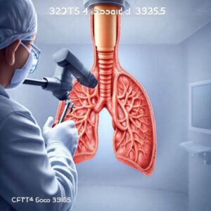
CPT Code 32554 and 32555
2. Understanding the Procedure: What is a Thoracentesis?
A thoracentesis (also known as a pleural tap) is a percutaneous invasive procedure performed to remove fluid or, less commonly, air from the pleural space—the potential space between the visceral pleura (lining the lungs) and the parietal pleura (lining the chest wall). Under normal conditions, this space contains a small amount of lubricating fluid. However, a multitude of pathological processes can cause an abnormal accumulation of fluid, known as a pleural effusion, which can compress the lung and lead to significant respiratory distress, hypoxia, and other complications.
Clinical Indications: Why is it Performed?
A thoracentesis is performed for two primary purposes:
-
Diagnostic: To obtain a sample of pleural fluid for laboratory analysis to determine the cause of the effusion. Tests include cell count and differential, Gram stain and culture, pH, glucose, protein, lactate dehydrogenase (LDH), cytology, and sometimes specific markers for tuberculosis or fungi.
-
Therapeutic: To relieve symptoms of respiratory compromise, such as shortness of breath (dyspnea), chest pain, and cough, by removing a large volume of fluid. Large, symptomatic effusions are often drained for patient comfort even if the cause is already known.
Common conditions leading to a pleural effusion requiring thoracentesis include:
-
Transudative Effusions: Caused by an imbalance of hydrostatic and oncotic pressures.
-
Congestive Heart Failure (CHF)
-
Cirrhosis (Hepatic hydrothorax)
-
Nephrotic syndrome
-
Pulmonary embolism
-
-
Exudative Effusions: Caused by inflammation or increased capillary permeability from disease of the pleura or lung.
-
Malignancy (e.g., lung cancer, breast cancer, mesothelioma, lymphoma)
-
Infection (Parapneumonic effusion, empyema, tuberculosis)
-
Inflammatory diseases (Lupus, rheumatoid arthritis)
-
Trauma (Hemothorax)
-
Chylothorax (leakage of lymphatic fluid)
-
Contraindications and Risks
While generally safe, thoracentesis is not without risks. Absolute contraindications are few but include an uncooperative patient and an uncorrected bleeding diathesis. Relative contraindications require careful risk-benefit analysis and include:
-
Very small volume of fluid
-
Mechanical ventilation (increases risk of complications)
-
Known bullous emphysema in the puncture area
-
Anticoagulant medication use
Potential complications, though uncommon, can be serious:
-
Pneumothorax (collapsed lung): The most common complication, occurring in 5-30% of procedures, though only a small percentage require a chest tube.
-
Hemothorax (bleeding into the pleural space): Rare, but can be life-threatening.
-
Infection (empyema): Very rare if proper sterile technique is used.
-
Pulmonary edema: Can occur if a very large volume of fluid (>1.5L) is removed too rapidly due to re-expansion of the lung.
-
Spleen or liver puncture: If the needle is placed too low.
-
Vasovagal reaction.
-
Cough or chest pain.
The Procedural Steps: A Walkthrough
Understanding the procedure is paramount for accurate coding. A typical thoracentesis follows these steps:
-
Informed Consent: The physician explains the procedure, benefits, risks, and alternatives, and obtains written consent.
-
Positioning: The patient is positioned sitting upright, leaning forward over a bedside table, to allow the fluid to gravitate to the dependent parts of the pleural space.
-
Site Selection and Preparation: The physician identifies the appropriate puncture site, often by percussing the chest for dullness and confirming with ultrasound. The skin is then widely cleaned with an antiseptic solution and draped sterilely.
-
Local Anesthesia: The skin, subcutaneous tissue, muscle, and down to the parietal pleura are infiltrated with a local anesthetic (e.g., lidocaine).
-
Puncture and Aspiration: A needle or, more commonly, a catheter-over-needle device is advanced through the anesthetized track into the pleural space. The needle is withdrawn, and the catheter remains. The catheter is connected to a three-way stopcock and syringe or to vacuum bottles for drainage.
-
Fluid Management: For diagnostic taps, 30-100 mL of fluid is aspirated into syringes and sent to the lab. For therapeutic taps, up to 1.5 liters may be drained slowly to prevent re-expansion pulmonary edema.
-
Catheter Removal and Post-Procedure Care: The catheter is swiftly removed, pressure is applied, and a bandage is placed. A post-procedure chest X-ray is almost always obtained to rule out an iatrogenic pneumothorax.
3. The Cornerstone of Coding: CPT Codes 32554 and 32555
The American Medical Association’s CPT® (Current Procedural Terminology) code set provides the standardized language for describing medical, surgical, and diagnostic services. For thoracentesis, the codes are found in the “Surgery / Respiratory System” section of the codebook.
CPT 32554: Thoracentesis, needle or catheter, aspiration of the pleural space; without imaging guidance
This code is used when the physician performs the thoracentesis without the use of any real-time imaging to guide the needle/catheter placement. The physician relies solely on physical exam findings (e.g., percussion, palpation) and their anatomical knowledge. This approach is becoming less common due to the demonstrated benefits of ultrasound guidance in improving safety and success rates.
CPT 32555: Thoracentesis, needle or catheter, aspiration of the pleural space; with imaging guidance
This code is used when the physician utilizes real-time imaging to visualize the needle/catheter entering the pleural space. This is the current standard of care for most settings. It is crucial to note that code 32555 includes the imaging guidance. The code descriptor is a “with” code, meaning the imaging guidance is an inherent part of the procedure and should not be reported separately by the same physician.
The Critical Role of Imaging Guidance
The use of imaging, particularly bedside ultrasound, has revolutionized thoracentesis. It allows the physician to:
-
Confirm the presence of an adequate fluid volume.
-
Identify the optimal and safest puncture site, avoiding organs, blood vessels, and consolidated lung.
-
Visualize the needle in real-time as it enters the fluid collection.
-
Significantly reduce the rate of complications, most notably pneumothorax.
Because of these proven benefits, many hospital policies and payer medical necessity guidelines now mandate the use of image guidance for thoracentesis. Coding 32554 without strong, documented justification (e.g., emergent situation where equipment was unavailable) may lead to denials based on “not medically necessary” or “not the standard of care.”
4. A Deep Dive into Imaging Guidance Codes (76000, 76942, 77002)
While the physician performing the thoracentesis cannot separately report the imaging guidance when using 32555, there are specific circumstances where these codes come into play. Understanding them is critical for correct billing in different scenarios.
Fluoroscopic Guidance (76000)
-
CPT 76000: Fluoroscopy (separate procedure), up to 1 hour physician time, other than 71023 or 71034 (e.g., cardiac fluoroscopy)
-
Usage: This is typically used when the thoracentesis is performed in an interventional radiology (IR) suite under continuous fluoroscopic guidance. The radiologist would report the appropriate thoracentesis code (32554 or 32555, though it’s almost always 32555 in this setting) and cannot also report 76000, as the guidance is bundled. However, if a portable C-arm is brought to the bedside in the ICU and used for guidance, the same rule applies when performed by a single physician.
Ultrasonic Guidance (76942)
-
CPT 76942: Ultrasonic guidance for needle placement (e.g., biopsy, aspiration, injection, localization device), imaging supervision and interpretation
-
Usage: This is the most common guidance modality for thoracenteses performed at the bedside by pulmonologists, intensivists, or hospitalists. Crucially, the physician performing the thoracentesis reports only 32555. They cannot separately report 76942. The work of performing and interpreting the ultrasound is included in the valuation of 32555.
-
Exception: If one physician performs and interprets the ultrasound to mark the site and then a different physician performs the actual thoracentesis puncture, the first physician could report 76942 with modifier 26 (Professional Component) if they document a separate, distinct report. This scenario is very uncommon.
-
CT Guidance (77012)
-
CPT 77012: Computed tomography guidance for needle placement (e.g., biopsy, aspiration, injection, localization device), radiological supervision and interpretation
-
Usage: CT guidance is rarely used for a simple thoracentesis due to its cost, lack of portability, and radiation exposure. It is reserved for very complex cases, such as localized or loculated effusions that are not accessible by ultrasound. As with the other guidance codes, if the same physician uses CT to guide their own thoracentesis, only 32555 is reported. The CT guidance is bundled.
Bundling and Modifier Use
The National Correct Coding Initiative (NCCI) edits, maintained by the Centers for Medicare & Medicaid Services (CMS), explicitly bundle the imaging guidance codes (76000, 76942, 77012) into CPT 32555. This means that for the same patient on the same date of service, if the same physician provides both services, the guidance code is not separately reimbursable. A modifier (like 59) should not be used to override this edit, as it is a correct bundling based on the CPT code definition.
5. The Diagnostic Puzzle: Coding the Reason with ICD-10-CM
The CPT code describes what was done. The ICD-10-CM (International Classification of Diseases, 10th Revision, Clinical Modification) code describes why it was done. This link is the cornerstone of medical necessity, which is the fundamental principle of healthcare reimbursement. Payers will only pay for services that are deemed medically necessary to diagnose or treat a patient’s condition.
Common ICD-10-CM Codes for Thoracentesis
The primary diagnosis code must be the condition that necessitated the fluid removal. Here is a table of common, highly specific ICD-10-CM codes.
Common ICD-10-CM Codes for Thoracentesis
| ICD-10-CM Code | Code Description | Clinical Scenario |
|---|---|---|
| J91.0 | Malignant pleural effusion | Effusion due to any underlying cancer (e.g., lung, breast). |
| J90 | Pleural effusion, not elsewhere classified | Use only if the effusion is unspecified or the cause is not yet known (e.g., for a diagnostic tap). |
| J81.0 | Acute pulmonary edema due to heart failure | Use if the effusion is a direct result of acute CHF. |
| J18.1 | Lobar pneumonia, unspecified organism | For a parapneumonic effusion. |
| J86.9 | Pyothorax without fistula | For an empyema (infected pleural space). |
| R06.02 | Shortness of breath | For a therapeutic tap; often used as a secondary diagnosis. |
| J94.0 | Chylous effusion | For a chylothorax. |
| J94.2 | Hemothorax | For blood in the pleural space due to trauma or other cause. |
| J84.84 | Pleural plaque with presence of asbestos | For effusions related to asbestos exposure. |
| I26.99 | Other pulmonary embolism without acute cor pulmonale | PE can cause pleural effusions. |
Linking Medical Necessity: The Key to Clean Claims
The connection must be clear in the medical record. The patient’s history and physical (H&P) should document the symptoms (e.g., dyspnea, hypoxia) and the physical/radiographic findings (e.g., “large right pleural effusion on CXR”). The procedure note should restate the indication. The coder must ensure the ICD-10-CM code matches the physician’s documented reason.
-
Example of a Good Claim: ICD-10-CM: J91.0 (Malignant pleural effusion) -> CPT: 32555 (Thoracentesis with US guidance). The medical record documents a known lung cancer patient with worsening shortness of breath and a large effusion on imaging.
-
Example of a Denied Claim: ICD-10-CM: R05.9 (Cough) -> CPT: 32555. A cough alone is not sufficient medical necessity for an invasive procedure. The underlying cause of the cough (e.g., the effusion) must be coded.
6. Modifiers: The Fine-Tuning Tools of Medical Coding
Modifiers are two-character suffixes (alphabetic or alphanumeric) added to a CPT or HCPCS code to provide additional information about the service performed. They can affect reimbursement and are essential for accurate claim submission.
Modifier 26: Professional Component
-
Usage: Used to indicate that only the professional component of a service was provided. This is the physician’s work of performing, supervising, and interpreting the service. It is used when the physician performs a service in a facility (e.g., hospital, ASC) that owns the equipment.
-
Application to Thoracentesis: A physician would not use modifier 26 on 32554 or 32555. These are global procedure codes that include both the professional and technical components when performed in an office. In a facility setting, the physician still reports the full code (e.g., 32555). The facility will bill separately for its resources (room, equipment, nursing staff) using the same code with a different modifier or on a separate institutional claim (UB-04).
Modifier TC: Technical Component
-
Usage: Used to indicate that only the technical component of a service was provided. This includes the equipment, supplies, and technician costs.
-
Application to Thoracentesis: A physician in a private office would not use modifier TC. If they own the ultrasound machine and perform the procedure in their office, they bill the global service (32555). The facility (hospital) is the entity that bills the technical component for a hospital-based service. Physicians do not typically bill with modifier TC.
Modifier 59: Distinct Procedural Service
-
Usage: Used to indicate that a procedure or service was distinct or independent from other services performed on the same day. It is most commonly used to bypass NCCI edits when two procedures that are usually bundled are performed on separate organs, separate lesions, or at separate patient encounters.
-
Application to Thoracentesis: This could be applied if a physician performs a thoracentesis and another separately identifiable procedure on the same day. For example:
-
A patient has a large right-sided effusion and a small left-sided effusion. The physician performs a thoracentesis on each side. The second thoracentesis (e.g., 32555-59 on the left) could be modified with 59 to indicate it was a distinct procedure at a separate site.
-
A thoracentesis (32555) is performed and, during the same session, a closed pleural biopsy (CPT 32400) is performed. Modifier 59 might be appended to 32400 to indicate it was a distinct service. Note: Always check the NCCI edits first; some combinations may be allowed without a modifier.
-
Other Relevant Modifiers (e.g., LT, RT, XE, XS)
-
LT (Left side) and RT (Right side): Anatomical modifiers are crucial if a procedure is performed on a paired organ. While thoracentesis is inherently a unilateral procedure, using LT or RT provides clarity, especially if multiple procedures are done.
-
Example: 32555-RT (Thoracentesis with guidance, right side).
-
-
X{EPSU} Modifiers: These are more specific replacements for modifier 59, introduced to reduce misuse.
-
XE (Separate Encounter): Service performed during a separate encounter.
-
XS (Separate Structure): Service performed on a separate organ/structure. This would be appropriate for a thoracentesis on the right and left sides (e.g., 32555-RT, 32555-LT-XS).
-
XP (Separate Practitioner): Service performed by a different practitioner.
-
XU (Unusual Non-Overlapping Service): Service does not overlap usual components of the main service.
-
7. The Billing Ecosystem: Place of Service and Reimbursement
Reimbursement for medical services is not a fixed number; it varies based on where the service is performed and who is billing for it.
Facility vs. Non-Facility Pricing
The Medicare Physician Fee Schedule (MPFS) has two columns for each CPT code:
-
Non-Facility Price: This is the total allowed amount when the service is performed in a place like a physician’s office. It includes both the professional work and the practice expense (overhead, equipment, supplies).
-
Facility Price: This is the allowed amount for the professional component only when the service is performed in a facility (e.g., hospital inpatient, hospital outpatient, ASC). The facility bills separately for its costs (the technical component) on a UB-04 form.
CPT 32555 will have a lower reimbursement rate when billed by a physician for a hospital inpatient because the physician’s claim only includes their professional work. The same code billed for a procedure done in a freestanding physician’s office has a higher reimbursement because it covers all costs.
Impact of Place of Service (POS) Codes
The Place of Service code on the physician’s claim (CMS-1500 form) tells the payer where the service was rendered and triggers the correct payment rate.
-
POS 21: Inpatient Hospital
-
POS 22: Outpatient Hospital (e.g., ER, hospital-based clinic)
-
POS 19: Off Campus-Outpatient Hospital
-
POS 11: Office
Using the wrong POS code is a common billing error that leads to incorrect payment and potential audit flags.
Understanding the RVUs (Relative Value Units)
Medicare and many other payers calculate payment using RVUs, which quantify the resources required to provide a service. There are three components:
-
Work RVU (wRVU): Measures the physician’s time, skill, effort, and stress.
-
Practice Expense RVU (peRVU): Measures overhead (e.g., rent, staff, equipment).
-
Malpractice RVU (mRVU): Measures the cost of professional liability insurance.
These RVUs are multiplied by a geographic practice cost index (GPCI) and a conversion factor (CF) to determine the dollar amount. For example, CPT 32555 has higher wRVUs and peRVUs than 32554, reflecting the increased work and resources required for image-guided procedures.
8. Navigating Payer-Specific Policies and Common Denials
Even with perfect CPT and ICD-10-CM coding, claims can be denied if they violate a specific payer’s medical policy.
Medicare (NCDs and LCDs)
Medicare rules are foundational. They publish:
-
National Coverage Determinations (NCDs): Country-wide policies.
-
Local Coverage Determinations (LCDs) and Local Billing Articles: Policies set by the Medicare Administrative Contractor (MAC) for your region (e.g., Novitas, Palmetto GBA, WPS). You must check your MAC’s website for their thoracentesis LCD. These documents explicitly state what they consider medically necessary, what documentation is required, and any coding restrictions.
Major Commercial Payers (UHC, Aetna, Cigna, etc.)
Each commercial payer has its own clinical payment and policy manuals. They often mirror Medicare but can have subtle differences. For instance, a payer may have stricter rules about the medical necessity of image guidance or may bundle the post-procedure chest X-ray into the thoracentesis code.
Top Denial Reasons and How to Appeal
-
Lack of Medical Necessity (LMN): The most common denial. Appeal with: Copies of the H&P, imaging report showing the effusion, and procedure note clearly stating the indication.
-
Bundled Service: Payer states the guidance code or another service is included. Appeal with: CPT guidelines and the NCCI edit manual showing that 32555 includes the guidance when performed by the same physician. If modifiers were used incorrectly, refile the claim.
-
Invalid or Incomplete ICD-10-CM Code: The diagnosis code doesn’t support the procedure. Appeal with: Clinical documentation that validates a more specific code.
-
Missing Information: e.g., no post-procedure chest X-ray report in the chart to prove it was done. Appeal with: The missing document.
A strong appeal is factual, references specific policies, and includes all relevant clinical documentation.
9. Documentation: The Foundation of a Defensible Claim
“If it wasn’t documented, it wasn’t done.” This old adage is the law in medical coding and billing. The medical record must paint a complete and coherent picture.
The Procedure Note Essentials
A robust procedure note should include:
-
Indication: The medical reason for the procedure (e.g., “Large right pleural effusion causing dyspnea, for diagnostic fluid analysis and therapeutic relief”).
-
Informed Consent: A note that risks, benefits, and alternatives were discussed.
-
Time Out: Verification of correct patient, procedure, and site.
-
Site: The specific puncture site (e.g., “8th intercostal space, posterior axillary line”).
-
Anesthetic: Type and amount used.
-
Image Guidance: Crucially, if ultrasound was used, it must be documented. “Ultrasound was used to identify a safe pocket of fluid and guide needle insertion in real-time.”
-
Description of Procedure: Step-by-step account.
-
Findings: Amount, color, and character of fluid removed (e.g., “900 mL of serosanguinous fluid”).
-
Specimens: What was sent to the lab.
-
Complications: “None” or a description of any issues.
-
Post-Procedure Condition: “Patient tolerated procedure well.”
-
Post-Procedure Plan: “CXR ordered to rule out pneumothorax.”
The Radiology Report (if applicable)
If a post-procedure chest X-ray was performed, the radiology report must be in the chart. Its findings (e.g., “No pneumothorax. Interval decrease in right pleural effusion.”) complete the procedural record.
Medical Necessity in the History & Physical
The H&P should establish the story before the procedure: the patient’s symptoms, physical exam findings (e.g., “dullness to percussion, decreased breath sounds at right base”), and imaging results that led to the decision to perform the thoracentesis.
10. Case Studies: Applying Knowledge to Real-World Scenarios
Case Study 1: The Hospital Inpatient
-
Scenario: A 68-year-old male is admitted with acute decompensated heart failure. A CXR shows a large right pleural effusion. The pulmonologist is consulted. At the bedside, she uses a portable ultrasound machine to identify a site, administers local anesthesia, and drains 1.2L of clear yellow fluid using a catheter kit. The patient’s breathing improves. A post-procedure CXR shows no pneumothorax.
-
Coding:
-
Physician (CMS-1500): CPT: 32555 (Includes the ultrasound guidance performed by the same physician). POS: 21 (Inpatient Hospital). ICD-10-CM: J81.0 (Acute pulmonary edema due to heart failure) and R06.02 (Shortness of breath).
-
Hospital (UB-04): The hospital will also bill 32555 on its institutional claim to be reimbursed for the supplies (catheter kit, tray), room use, and nursing support.
-
Case Study 2: The Interventional Radiology Suite
-
Scenario: An oncology patient with a known malignant pleural effusion presents for therapeutic thoracentesis. Due to prior surgery making anatomy difficult, the procedure is scheduled in the IR suite. An interventional radiologist performs the thoracentesis under continuous fluoroscopic guidance.
-
Coding:
-
Radiologist (CMS-1500): CPT: 32555. Even though fluoroscopy was used, it is included in 32555. Do not report 76000. POS: 22 (Outpatient Hospital) or 21 (Inpatient). ICD-10-CM: J91.0 (Malignant pleural effusion).
-
Hospital (UB-04): The hospital bills 32555 for the technical component, including the extensive use of the IR suite, fluoroscopy equipment, and highly specialized staff.
-
Case Study 3: Bilateral Thoracentesis
-
Scenario: A patient with lupus presents with symptomatic bilateral pleural effusions. The physician uses ultrasound guidance to perform a thoracentesis on the right side, removing 800mL. He then re-preps and drapes and performs a separate thoracentesis on the left side, removing 600mL.
-
Coding:
-
Physician (CMS-1500):
-
First Procedure: 32555-RT
-
Second Procedure: 32555-LT-XS (Modifier XS indicates a separate structure/organ). Some payers may still prefer modifier 59, so checking policy is key.
-
-
ICD-10-CM: M32.13 (Pleuropulmonary involvement in systemic lupus erythematosus) and R06.02 (Shortness of breath).
-
11. Conclusion: Mastering the Art and Science of Procedural Coding
Accurate coding for thoracentesis hinges on a deep understanding of the clinical procedure itself. The critical choice between 32554 and 32555 is dictated solely by the documented use of real-time image guidance. Coders must forge a strong link between the ICD-10-CM code representing medical necessity and the CPT code describing the service rendered. Navigating the complexities of bundled imaging guidance, appropriate modifier application, and payer-specific policies is essential for ensuring compliant and optimal reimbursement while maintaining a defensible audit trail.
12. Frequently Asked Questions (FAQs)
Q1: Can I bill both 32555 and 76942 if I use ultrasound guidance?
A: No. CPT code 32555 is defined as “with imaging guidance.” The work of performing and interpreting the ultrasound is included in the value of this code. Reporting 76942 separately for the same physician on the same patient for the same procedure is considered unbundling and will lead to a denial.
Q2: The physician used ultrasound just to mark the spot but not during the needle insertion. Can I use 32554?
A: This is a gray area and highly dependent on payer policy. Strictly interpreting the CPT description, 32555 requires imaging guidance for the “aspiration.” If the needle was not visualized entering the space in real-time, some argue 32554 may be appropriate. However, most payers and experts would consider the use of ultrasound for site selection an integral part of a modern, safe procedure and would expect 32555. The safest approach is to document thoroughly and follow your specific payer’s guidance. When in doubt, use 32555.
Q3: How do I code for the chest X-ray done after the thoracentesis?
A: The chest X-ray (e.g., CPT 71045 or 71046) is a separate and distinct radiological service. It is not included in the thoracentesis code and should be reported separately by the physician who interprets it, with the appropriate diagnosis code (e.g., Z09.8 for follow-up after other surgery, or the condition itself). The technical component is billed by the facility.
Q4: A resident performed the thoracentesis with the attending physician supervising. How is this billed?
A: In a teaching hospital setting, the attending physician must be physically present during the critical portions of the procedure to bill for the service. They must document their presence and participation. The service is billed under the attending physician’s National Provider Identifier (NPI) with no special modifier, as the “teaching physician” rules are a documentation requirement, not a coding modifier requirement.
13. Additional Resources
-
The American Medical Association (AMA): For the official CPT® codebook and guidelines. www.ama-assn.org
-
Centers for Medicare & Medicaid Services (CMS):
-
Medicare Physician Fee Schedule Lookup Tool: https://www.cms.gov/medicaremedicare-fee-service-paymentphysicianfeesched
-
National Correct Coding Initiative (NCCI) Edits: https://www.cms.gov/medicare/coding-billing/national-correct-coding-initiative-ncci-edits
-
-
Your Medicare Administrative Contractor (MAC): Find your local MAC’s website for LCDs and billing articles. A list is available on the CMS website.
-
American Academy of Professional Coders (AAPC): For continuing education, certifications, and networking. www.aapc.com
-
American Health Information Management Association (AHIMA): For resources on clinical documentation integrity. www.ahima.org
-
Peer-Reviewed Medical Journals: Journals like Chest or The New England Journal of Medicine often publish clinical guidelines that inform medical necessity policies.
Date: September 1, 2025
Author: Medical Coding & Billing Insights Team
Disclaimer: The information contained in this article is for educational and informational purposes only and is not intended as medical, legal, or financial advice. While every effort has been made to ensure accuracy, CPT® codes are owned by the American Medical Association (AMA), and users must have a license from the AMA to utilize them. Always consult the most current, official AMA CPT® codebook, payer-specific guidelines, and relevant government regulations for definitive coding and billing guidance.


