The human heart is a marvel of biological engineering, a relentless pump that beats over 100,000 times a day, circulating life-sustaining blood throughout the body. For centuries, physicians could only listen to its sounds and feel its rhythms from the outside, diagnosing its ailments through inference and experience. Then, in the mid-20th century, a revolution occurred: echocardiography. This technology, using harmless sound waves, pulled back the curtain, allowing clinicians to see the heart’s structures, watch its valves open and close, and measure its strength in real time.
Today, echocardiography is the most widely used cardiac imaging modality in the world. It is non-invasive, painless, radiation-free, and provides a wealth of diagnostic information. But behind every clear image of a beating heart lies a complex framework of codes, rules, and regulations that ensures this vital technology is appropriately documented, billed, and reimbursed. This framework is built on the Current Procedural Terminology (CPT®) code set.
For sonographers, cardiologists, practice managers, and medical coders, understanding echocardiography CPT codes is not merely an administrative task—it is a critical component of providing quality patient care. Accurate coding ensures that healthcare providers are fairly compensated for their expertise, technology, and time, which in turn supports the sustainability of cardiac care services. Inaccurate coding, however, can lead to claim denials, audits, fines, and even allegations of fraud.
This exhaustive guide is designed to be your definitive resource for navigating the intricate world of echocardiography CPT codes. We will move beyond simple code definitions to explore the clinical scenarios, documentation requirements, and nuanced rules that govern proper code selection. Whether you are a seasoned coder looking for a refresher or a new sonographer seeking to understand the business side of your craft, this article will provide the depth and clarity you need.
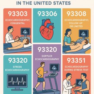
CPT Codes for Echocardiography in the US
2. The Language of Reimbursement: Understanding CPT® and Its Importance
Before diving into specific echo codes, one must understand the language they are written in. The Current Procedural Terminology (CPT®) is a uniform coding system developed and maintained by the American Medical Association (AMA). Its primary purpose is to provide a standardized language for describing medical, surgical, and diagnostic services, thereby streamlining communication between physicians, coders, patients, and payers like Medicare and private insurance companies.
CPT codes are five-digit numeric codes that represent specific procedures or services. They are used by providers to report the services they perform on claims submitted to payers. Payers, in turn, use these codes to determine the amount of reimbursement the provider will receive based on a fee schedule or contract.
Why Accurate Echo Coding is Critical:
-
Financial Integrity: Correct coding ensures appropriate reimbursement for the complex work and expensive technology involved in echocardiography. Undercoding leaves money on the table, while overcoding can be construed as fraud.
-
Compliance: The healthcare industry is heavily regulated. Adhering to CPT guidelines, National Coverage Determinations (NCDs), and Local Coverage Determinations (LCDs) is mandatory to avoid audits, recoupments, and penalties from entities like the Office of Inspector General (OIG) and the Centers for Medicare & Medicaid Services (CMS).
-
Data Accuracy: CPT codes are used for healthcare analytics, research, and public health reporting. Accurate coding ensures that data on the prevalence and treatment of cardiac conditions is reliable.
-
Clinical Relevance: The process of coding requires a thorough review of the medical record. This process can itself be a quality check, ensuring that the documented findings justify the medical necessity of the procedure performed.
CPT codes are updated annually by the AMA. It is absolutely imperative that coding professionals use the current year’s codebook and stay informed of changes, as using outdated codes is a common source of claim errors.
3. The Foundational Echo: A Deep Dive into the Complete Transthoracic Echocardiogram (CPT 93306)
The transthoracic echocardiogram (TTE) is the cornerstone of cardiac ultrasound. It is performed by placing a transducer on the patient’s chest wall (thorax) and using ultrasound to obtain images of the heart. CPT code 93306 is used to report a complete TTE.
Description: *Echocardiography, transthoracic, real-time with image documentation (2D), with or without M-mode recording, complete, with spectral Doppler echocardiography, and with color flow Doppler echocardiography.*
The key word in this description is “complete.” A service reported with 93306 must include all of the following components:
-
2D Imaging: Real-time two-dimensional imaging of cardiac structures. This is the primary modality for assessing heart anatomy.
-
M-mode Recording: This older technique provides a one-dimensional “ice-pick” view of cardiac structures, useful for making precise measurements of chamber sizes and wall thicknesses over time. While its use has declined with advanced 2D imaging, it is still a required element of a “complete” study.
-
Spectral Doppler Echocardiography: This includes both Pulse Wave (PW) Doppler and Continuous Wave (CW) Doppler.
-
PW Doppler: Used to assess blood flow velocities at specific, localized points (e.g., mitral valve inflow, pulmonary vein flow).
-
CW Doppler: Used to accurately measure high-velocity blood flows (e.g., across a stenotic aortic valve or a regurgitant mitral valve).
-
-
Color Flow Doppler Echocardiography: This technology superimposes a color-coded map of blood flow velocities on the 2D image. It is indispensable for visualizing and assessing valvular regurgitation (leaking) and stenosis (narrowing), as well as intracardiac shunts.
Documentation Requirements: The final report must reflect that all elements of a complete study were performed and interpreted. The report should include:
-
Measurements of cardiac chambers (left atrium, left ventricle in diastole and systole, right ventricle, aortic root).
-
Assessment of ventricular function (qualitative and quantitative, e.g., ejection fraction).
-
Evaluation of all four heart valves (aortic, mitral, tricuspid, pulmonary) for structure and function, including Doppler-derived gradients and velocities.
-
Evaluation of the pericardium.
-
A summary of findings and an interpretation.
If any of these components are missing, the study may not be reported as a complete TTE. Code 93306 is typically reported once per session, regardless of how long the study takes or how many images are acquired.
4. The Limited Study: Purpose and Nuances of CPT 93308
There are clinical scenarios where a full, complete echocardiogram is not medically necessary. The physician may only need to answer a specific, focused question. This is where the limited echocardiogram comes into play, reported with CPT code 93308.
Description: *Echocardiography, transthoracic, real-time with image documentation (2D), with or without M-mode recording, limited, follow-up or repeat study.*
The AMA CPT guidelines explicitly state that a limited study is performed when less than a complete exam is performed. Common examples include:
-
A follow-up study to re-check a previously identified ejection fraction after starting new heart failure medication.
-
Assessing for a pericardial effusion (fluid around the heart) after a procedure.
-
Re-evaluating a single valve known to have mild disease.
-
Guiding pericardiocentesis (draining fluid from the pericardium).
-
A quick intraoperative assessment.
Critical Distinction: The difference between 93306 and 93308 is not determined by time, difficulty, or image quality. It is determined by the scope of the examination. If the sonographer performs a full protocol but the cardiologist only reports on a limited finding, the complete code 93306 must still be used. The code reflects the work performed, not the complexity of the findings.
Using 93308 inappropriately to report a less-than-complete study that was actually performed (e.g., because the patient was difficult to image) is incorrect and constitutes down-coding. If a complete study is attempted and performed to the best of the sonographer’s ability, even if image quality is suboptimal, 93306 is typically still appropriate.
5. Seeing the Heart from Within: Transesophageal Echocardiography (TEE) Codes
A transthoracic echo has limitations. Air in the lungs, body habitus, and bandages can obscure the ultrasound waves. When a clearer, more detailed image is required, a transesophageal echocardiogram (TEE) is performed. This involves passing a specialized transducer on a flexible endoscope down the patient’s esophagus, which sits directly behind the heart, providing exceptionally clear images.
TEE coding is more complex due to the involvement of both a probe placement component and an imaging component. The codes are often used in combination.
TEE for Diagnostic Purposes
-
93312: *Echocardiography, transesophageal, real-time with image documentation (2D) (with or without M-mode recording); including probe placement, image acquisition, interpretation and report.*
-
This is a “monolithic” code that includes everything: placing the TEE probe, acquiring the images, and interpreting the study. It is used for a diagnostic TEE performed on its own.
-
-
93313: … placement of probe only.
-
93314: … image acquisition, interpretation and report only.
-
These codes are used when the services are performed by different providers. For example, an anesthesiologist may place the probe (93313) for intraoperative monitoring, and a cardiologist later acquires the diagnostic images and interprets them (93314).
-
-
93315: … including probe placement, image acquisition, interpretation and report, with 3D rendering of the mitral valve and surrounding structures, when performed.
-
This is an add-on code used in conjunction with 93312. It describes the additional work of acquiring and rendering a 3D dataset of the mitral valve complex, which is crucial for planning mitral valve repairs.
-
TEE for Guidance of Non-Coronary Interventions
TEE is frequently used to guide structural heart procedures (e.g., mitral valve clip, left atrial appendage closure, ASD/PFO closure). A separate set of codes exists for this specific purpose.
-
93317: *Echocardiography, transesophageal (TEE) for guidance of a transcatheter intracardiac or great vessel(s) structural intervention(s) (eg, TAVR, transcatheter mitral valve repair, paravalvular regurgitation repair, left atrial appendage occlusion/closure, ventricular septal defect closure) (peri-and intra-procedural), real-time with image documentation (2D) (with or without M-mode recording) and 3D, when performed, including probe placement, image acquisition, interpretation, and report;*
-
93318: … placement of probe only.
-
93321: *… for guidance of transcatheter endocardial stent(s) transplantation(s) (eg, mitral valve, tricuspid valve, pulmonary valve, left atrial appendage, right ventricular outflow tract) (peri-and intra-procedural), real-time with image documentation (2D) (with or without M-mode recording) and 3D, when performed, including probe placement, image acquisition, interpretation, and report;*
-
Crucial Note: Codes 93317-93321 include all echocardiographic guidance during the entire procedure. They are typically reported only once per procedure, not per hour. Code 93325 is an add-on code used with 93321.
-
TEE for Congenital Heart Anomalies
-
93355: *Echocardiography, transesophageal (TEE) for monitoring purposes, including probe placement, real-time 2-dimensional image acquisition and interpretation leading to ongoing (continuous) assessment of (dynamically changing) cardiac pumping function and to therapeutic measures on an immediate time basis.*
-
This is used for continuous monitoring during surgery, typically by an anesthesiologist.
-
-
93356: Echocardiography, transesophageal (TEE) for congenital cardiac anomalies; including probe placement, image acquisition, interpretation and report.
-
This is a specific code for diagnosing complex congenital heart defects via TEE.
-
Summary of Key TEE CPT Codes
| CPT Code | Description | Key Use Case |
|---|---|---|
| 93312 | Diagnostic TEE, complete service | Stand-alone diagnostic TEE |
| 93313 | TEE probe placement only | Often by anesthesiologist for monitoring |
| 93314 | TEE imaging & interpretation only | By cardiologist after probe is placed |
| 93315 | Add-on for 3D mitral valve rendering | Used with 93312 for mitral valve assessment |
| 93317 | TEE guidance for transcatheter intervention | Guidance for procedures like MitraClip, TAVR |
| 93355 | TEE for monitoring | Continuous intraoperative monitoring |
| 93356 | TEE for congenital anomalies | Diagnosing complex congenital heart d |
6. Stress Testing the Heart: Echo Codes for Ischemia and Function
Stress echocardiography assesses how the heart functions under physical or pharmacological stress. It is primarily used to diagnose coronary artery disease (CAD). The heart is imaged at rest and then immediately after stress. New or worsening wall motion abnormalities induced by stress indicate reduced blood flow (ischemia) from a blocked coronary artery.
The codes for stress echocardiography are structured as “base” codes plus “add-on” codes.
-
93350: *Echocardiography, transthoracic, real-time with image documentation (2D) (with or without M-mode recording), during rest and cardiovascular stress test using treadmill, bicycle exercise and/or pharmacologically induced stress, with interpretation and report;*
-
This is the base code. It includes the complete echocardiographic imaging at rest and at peak stress, as well as the interpretation and report.
-
-
93351: … including performance of cardiovascular stress test
-
This is an add-on code used with 93350. It describes the work of the physician or qualified healthcare professional who directly supervises and performs the stress portion of the test (e.g., administering the drug, monitoring the ECG and blood pressure). This code is reported in addition to 93350.
-
Coding Scenario: A patient undergoes a dobutamine stress echocardiogram. The cardiologist supervises the stress portion (93351) and also performs and interprets the echocardiographic images (93350). Both codes are reported. If one physician performs the stress and a different physician interprets the echo images, they would each report their respective services (93351 and 93350).
7. The Unseen Forces: Doppler and Color Flow Mapping Demystified
It is vital to understand that Spectral Doppler (PW and CW) and Color Flow Doppler are not separately reportable services when performed as part of a complete echocardiogram (93306, 93312, etc.). Their use is an inherent and required part of those complete procedures.
The AMA CPT guidelines are explicit: “Doppler echocardiography is included in 93306, 93307, 93308, 93312, 93313, 93314, 93315, 93317, 93318, 93320, 93321, 93325, 93350, 93351, 93355, and 93356, when performed.”
Attempting to report codes like 93320 (Doppler echocardiography, pulsed wave or continuous wave) or 93321 (Doppler echocardiography, color flow mapping) separately with a complete echo code is considered “unbundling” and will result in a denial. These codes are largely historical and are only used in extremely rare circumstances where Doppler is performed as a standalone, targeted study without 2D imaging—a scenario that is almost non-existent in modern practice.
8. 3D Echocardiography: Capturing the Heart in a New Dimension
Three-dimensional echocardiography has transformed the visualization of complex cardiac anatomy, particularly for valves and congenital heart disease. However, there is no specific CPT code for 3D echocardiography itself.
The work of acquiring, rendering, and interpreting 3D images is considered an inherent part of the primary echocardiographic procedure when it is performed. If a 3D dataset is acquired during a complete TTE (93306), no additional code is reported. The technical work and professional interpretation of the 3D images are bundled into 93306.
The exception, as mentioned earlier, is 93315, which is an add-on code specifically for the 3D rendering of the mitral valve and surrounding structures during a transesophageal echocardiogram (93312). This code recognizes the additional work and expertise required for this specific application in planning mitral valve surgery.
It is also important to note the radiology codes for 3D rendering:
-
76376: 3D rendering with interpretation and reporting of computed tomography, magnetic resonance imaging, ultrasound, or other tomographic modality; not requiring image postprocessing on an independent workstation
-
76377: … requiring image postprocessing on an independent workstation
These codes are for the post-processing and interpretation of a 3D dataset that is performed separately from the primary interpretation. Their use in echocardiography is highly controversial and generally not supported by CPT guidelines or payer policies (including Medicare). Most payers consider 3D rendering an integral part of the echo interpretation, and reporting 76376/76377 with an echo code would be considered unbundling. These codes are primarily used in radiology for CT and MRI.
9. Contrast Echocardiography: Enhancing the Image (93352)
In some patients, the TTE images are suboptimal due to body habitus, lung disease, or other factors. Ultrasound contrast agents are microbubble solutions that are injected intravenously to opacify the left ventricular chamber and dramatically improve the delineation of the endocardial border (the inner heart wall). This allows for a much more accurate assessment of wall motion and ejection fraction.
CPT code 93352 is used to report this service:
-
Description: Use of contrast agent during transthoracic echocardiography, including all necessary injections/administrations and imaging (List separately in addition to code for echocardiographic imaging).
Key Points for 93352:
-
It is an add-on code. It must always be reported in addition to the primary echocardiography code (93306 or 93308).
-
It includes the cost of the contrast agent and the work of administering it.
-
Its use is subject to medical necessity. Documentation should note the reason for contrast use (e.g., “technically difficult study,” “inadequate endocardial border delineation”).
-
It is not reported for the use of saline bubble studies (agitated saline) used to detect intracardiac shunts. The saline bubble study is considered an inherent part of the complete echocardiographic examination.
10. Beyond the Scan: The Critical Role of Documentation and Interpretation
The echocardiogram is not complete when the sonographer saves the last clip. The procedure encompasses both the technical component (image acquisition) and the professional component (interpretation and report). The final report is the legal document that encapsulates the entire service and is the coder’s primary source of information.
A robust echo report must include:
-
Indication: The medical reason for the test (e.g., “dyspnea on exertion,” “evaluation for endocarditis”).
-
Description of Procedures: A brief note on how the study was performed (e.g., “complete 2D, Doppler, and color flow TTE performed”).
-
Quantitative Data: Measurements of chambers, valves, and Doppler velocities.
-
Findings: A narrative description of the anatomy and function of each major cardiac structure (ventricles, atria, valves, aorta, pericardium).
-
Impressions/Conclusion: A succinct summary of the abnormal and significant normal findings, answering the clinical question posed by the indication.
-
Comparison: If applicable, comparison to prior studies to show changes over time.
The physician’s signature and date on the report attest that they have personally interpreted the images and authored the report. Without a signed report, the service cannot be billed.
11. Navigating the Maze: Bundling, Modifiers, and Payer-Specific Rules
Coding is not just about picking the right number. It involves applying rules about how codes can be used together.
-
Bundling (NCCI Edits): The National Correct Coding Initiative (NCCI) edits are rules created by CMS to prevent improper payment when certain services are reported together. For example, a complete TTE (93306) is bundled with a stress echo (93350) if performed on the same day. The CCI edits dictate which code is column 1 (the comprehensive service) and which is column 2 (the component service). In this case, 93350 is column 2 and is not separately payable with 93306 unless a modifier is used appropriately.
-
Modifiers: Modifiers are two-character suffixes added to a CPT code to indicate that a service was altered in some way without changing the definition of the code itself.
-
Modifier -26: Professional Component. Used when the physician only performs the interpretation (e.g., interpreting an echo performed in a hospital outpatient department).
-
Modifier -TC: Technical Component. Used when the practice only owns the equipment and performs the scan, but the interpretation is done by an external physician (rare in echo).
-
Modifier -59: Distinct Procedural Service. Used to indicate that a procedure or service was distinct or independent from other services performed on the same day. This might be used to override an NCCI edit if, for example, a limited follow-up echo (93308) is performed for a new, unrelated reason on the same day as a complete stress echo (93350).
-
Modifier -XE: Separate Encounter. Used to indicate that a service was performed during a separate encounter on the same day.
-
-
LCDs/NCDs: Perhaps the most important rules are Local and National Coverage Determinations. These are policies issued by Medicare Administrative Contractors (MACs) and CMS that define the specific clinical circumstances under which a service is considered “reasonable and necessary.” They list covered and non-covered diagnoses. For example, an LCD may state that a routine screening echocardiogram without symptoms is not covered. Coders must be familiar with the LCDs issued by their regional MAC.
12. The Future of Echo Coding: AI, Automation, and Evolving Guidelines
The field of echocardiography is not static, and neither is its coding. Several trends will shape its future:
-
Artificial Intelligence (AI): AI algorithms are already being developed to automate measurements, enhance image quality, and even suggest diagnoses. This could change the workflow of sonographers and cardiologists, potentially impacting how work is valued and coded in the future.
-
Automated Coding Engines: Natural Language Processing (NLP) is being used to read physician reports and automatically suggest CPT and ICD-10 codes. While these tools can improve efficiency, human oversight remains critical to ensure accuracy and context understanding.
-
Value-Based Care: The shift from fee-for-service to value-based reimbursement may eventually change how imaging is paid for, moving away from per-procedure payments toward bundled payments or capitation. Accurate coding and documentation will remain essential for demonstrating the value of care provided.
-
Evolving Guidelines: The CPT code set will continue to evolve. Recent years have seen the introduction of specific codes for TEE guidance of interventions. Future updates may include more specific codes for strain imaging, 3D quantification, or other advanced techniques as they become standard of care.
Staying current through continuing education, attending webinars, and participating in professional organizations like the American Academy of Professional Coders (AAPC) or the American Health Information Management Association (AHIMA) is essential for anyone involved in the echo coding process.
13. Conclusion
Mastering echocardiography CPT coding requires a blend of clinical knowledge, meticulous attention to detail, and a firm understanding of constantly evolving regulations. The journey from patient scan to successful reimbursement is built upon accurate code selection, thorough documentation, and strict adherence to payer-specific guidelines. By viewing coding not as a mere administrative task but as an integral part of quality patient care, healthcare providers can ensure the financial stability necessary to continue delivering this vital window into the human heart.
14. Frequently Asked Questions (FAQs)
Q1: Can I report a complete TTE (93306) and a limited TTE (93308) on the same day for the same patient?
A: Generally, no. These codes are mutually exclusive. If a complete study is performed, report 93306. If a truly limited, focused study is performed, report 93308. Reporting both would require two separate and distinct medical reasons, supported by documentation, and would likely require a modifier (-59, -XE) to override NCCI edits, but this scenario is extremely rare and heavily scrutinized.
Q2: If a TEE is performed for monitoring during a surgery (e.g., 93355 by an anesthesiologist), can a cardiologist also report a diagnostic TEE code (93312) on the same day?
A: This is a complex situation. If the cardiologist is called in to perform a separate, comprehensive diagnostic TEE to answer a new clinical question that arose during surgery, and they generate a full formal report, it might be separately reportable with a modifier (-59, -XE) indicating a distinct service. However, if the cardiologist is simply re-interpreting the monitoring images, it is not separately billable. The documentation must clearly support two separate and distinct procedures.
Q3: How do I code for an echocardiogram that was attempted but yielded technically difficult images?
A: If the sonographer and physician perform a complete study to the best of their ability using the standard protocol, you should report the complete code (93306). The difficulty is factored into the work. You should not report a limited code (93308) simply because the images are suboptimal. If contrast was used to overcome the limitations, you would report 93352 in addition to 93306.
Q4: Are there specific ICD-10-CM diagnoses required for medical necessity?
A: Yes. Medicare and private payers publish lists of covered diagnoses in their LCDs. Common covered diagnoses for echocardiography include heart failure (I50.-), valvular heart disease (I34.-, I35.-, I36.-, I37.-), cardiomyopathy (I42.-), and atrial fibrillation (I48.-). A diagnosis of “routine exam” or “screening” without symptoms is typically not covered.
Q5: Who can perform and interpret an echocardiogram?
A: The technical component must be performed by a qualified sonographer or physician. The professional component (interpretation and report) must be performed by a physician (typically a cardiologist) who is trained and competent in echocardiography. The interpreting physician is legally responsible for the entire content of the report.
15. Additional Resources
-
American Medical Association (AMA): The official source for the CPT® codebook and guidelines. https://www.ama-assn.org/
-
Centers for Medicare & Medicaid Services (CMS): Source for NCCI edits, NCDs, and general Medicare policy. https://www.cms.gov/
-
Your Local Medicare Administrative Contractor (MAC): Find your MAC’s website to access your specific Local Coverage Determinations (LCDs) for echocardiography. (e.g., Novitas Solutions, First Coast Service Options, etc.)
-
American Society of Echocardiography (ASE): Provides clinical guidelines, educational resources, and advocacy for the field. https://www.asecho.org/
-
American Academy of Professional Coders (AAPC): Offers certifications (e.g., CPC, CIC), training, and networking for medical coders. https://www.aapc.com/
-
American College of Cardiology (ACC): Provides clinical guidelines and resources relevant to cardiovascular coding. https://www.acc.org
Date: September 4, 2025
Author: The Medical Coding Specialist
Disclaimer: This article is intended for informational and educational purposes only. It is not a substitute for professional medical coding, billing, or legal advice. CPT® codes are proprietary to the American Medical Association (AMA). Medical coders must use the current, official CPT® codebook and payer-specific guidelines for accurate coding and reimbursement. Always consult with a certified professional coder or your payer for specific guidance.


