Imagine the incessant, nagging sensation of tightness across your limbs, a feeling that your skin is a canvas stretched too far. Now, add to that a relentless itch—a pruritus that seems to ignite with every change of temperature, every piece of clothing that grazes your skin, and every wash. This is the daily reality for millions of individuals suffering from Xerosis Cutis, a dermatological condition whose name, derived from the Greek ‘xero’ (dry) and ‘osis’ (condition), barely captures the profound discomfort it inflicts. Far from being a mere cosmetic nuisance or a simple winter ailment, Xerosis Cutis is a complex, multifactorial disorder of the skin barrier that can significantly impair quality of life, disrupt sleep, and serve as a precursor to more severe inflammatory skin diseases. This article delves deep into the world of Xerosis Cutis, moving beyond superficial advice to explore its scientific underpinnings, its formal classification in the medical world under the ICD-10 code L85.3, and the comprehensive, evidence-based strategies for its effective management and long-term prevention. We will journey through the intricate architecture of the skin, unravel the biochemical pathways that lead to its desiccation, and equip you with the knowledge to not just treat, but truly understand and conquer chronic dry skin.
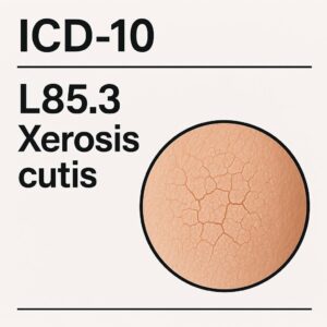
ICD-10 L85.3 Xerosis Cutis
2. Decoding the Diagnosis: The ICD-10 Code L85.3 for Xerosis Cutis
In the realm of modern medicine, precision is paramount. The International Classification of Diseases, 10th Revision (ICD-10) is the global standard for diagnosing, treating, and billing for health conditions. For Xerosis Cutis, the assigned code is L85.3. Understanding this code is crucial for both healthcare providers and patients navigating the healthcare system.
The code L85.3 is structured hierarchically:
-
L: Represents the chapter for “Diseases of the skin and subcutaneous tissue.”
-
L80-L99: Covers “Other disorders of the skin and subcutaneous tissue.”
-
L85: Specifically denotes “Other epidermal thickening,” a category that includes various forms of acquired ichthyosis and xerosis.
-
L85.3: Precisely identifies “Xerosis cutis,” excluding other conditions like vulgar ichthyosis (Q80.-) or dermatitis.
The use of this specific code ensures accurate communication between clinicians, facilitates epidemiological studies to understand the prevalence of the condition, and is essential for insurance and reimbursement purposes. When a dermatologist diagnoses you with severe dry skin that meets the clinical criteria, L85.3 is the identifier that formalizes that diagnosis within the medical record.
3. The Skin’s Barrier: A Primer on Hydration and Homeostasis
To comprehend Xerosis Cutis, one must first appreciate the masterpiece of biological engineering that is the skin, particularly its outermost layer, the stratum corneum. Often mistakenly thought of as a mere collection of dead cells, the stratum corneum is a dynamic, brick-and-mortar-like structure that is the primary defender against transepidermal water loss (TEWL).
The “Bricks and Mortar” Model:
-
The Bricks (Corneocytes): These are anucleate, keratin-filled cells that are the end-product of keratinocyte differentiation. They are flattened and densely packed, providing mechanical strength.
-
The Mortar (Lipid Matrix): The spaces between the corneocytes are filled with a meticulously organized lipid matrix composed primarily of ceramides, cholesterol, and free fatty acids. This lipid mortar is the critical component that forms a waterproof barrier, preventing the escape of internal water and the entry of external irritants and pathogens.
A second crucial component for skin hydration is the Natural Moisturizing Factor (NMF). Residing within the corneocytes, NMF is a complex mixture of low-molecular-weight, water-soluble compounds, including amino acids, pyrrolidone carboxylic acid, lactate, urea, and salts. These compounds are hygroscopic, meaning they attract and bind water from the atmosphere and the underlying dermis, thereby hydrating the corneocytes and keeping them plump and flexible.
When the stratum corneum is intact and healthy, with a robust lipid matrix and ample NMF, the skin appears smooth, supple, and resilient. Xerosis Cutis is, fundamentally, the clinical manifestation of a failure in this sophisticated system.
4. The Pathophysiology of Dryness: How Xerosis Cutis Takes Hold
Xerosis Cutis is not a single disease but a final common pathway resulting from the disruption of one or more components of the skin’s hydrating machinery. The pathophysiology can be broken down into several interconnected events:
-
Depletion of Natural Moisturizing Factor (NMF): The production of NMF is influenced by the skin’s hydration status and the external environment. In low-humidity conditions or with excessive washing, the process of NMF synthesis can be downregulated. Furthermore, NMF components are easily washed away by harsh surfactants, leading to a direct depletion. Without adequate NMF, corneocytes lose their ability to hold water, becoming brittle and inflexible.
-
Disruption of the Stratum Corneum Lipid Matrix: This is arguably the central event in Xerosis. The synthesis or organization of intercellular lipids (ceramides, cholesterol, fatty acids) can be impaired by several factors:
-
Aging: Enzymatic activity responsible for lipid synthesis declines with age.
-
Solvent Exposure: Frequent use of alcohol-based sanitizers or occupational exposure to solvents can dissolve these essential lipids.
-
Detergents: Harsh soaps can strip away lipids and damage the lipid bilayers.
A disrupted lipid matrix is like a wall with crumbling mortar; it becomes highly permeable, leading to a dramatic increase in Transepidermal Water Loss (TEWL).
-
-
Desquamation Impairment: The shedding of corneocytes in a controlled, invisible manner (desquamation) is an active enzymatic process. It requires proteases that degrade the corneodesmosomes (the protein “glue” holding corneocytes together). This process is optimal at a specific hydration level. In xerotic skin, reduced hydration inactivates these enzymes, leading to corneocyte retention. This results in the visible flaking and scaling characteristic of the condition.
-
The Itch-Scratch Cycle: As the skin becomes dry, tight, and scaly, it often triggers pruritus (itching). The act of scratching, while providing momentary relief, causes direct physical damage to the already compromised stratum corneum. This further breaches the skin barrier, promotes inflammation, and stimulates nerve fibers, creating a vicious and self-perpetuating cycle of itch and scratch that can lead to lichenification (thickening of the skin) and even secondary infections.
5. Clinical Presentation: Recognizing the Signs and Symptoms
The presentation of Xerosis Cutis can range from mild, asymptomatic dryness to a severe, debilitating condition. It most commonly affects the extremities, particularly the shins, arms, and thighs, as these areas have fewer sebaceous glands.
Common Signs (What you see):
-
Dryness: A general lack of the skin’s natural shine and suppleness.
-
Scaling: Fine, white, branny scales or, in more severe cases, larger, plate-like scales resembling fish skin (acquired ichthyosis).
-
Xerotic Eczema: Areas of redness, inflammation, and scaling that can crack and fissure.
-
Lichenification: Thickened, leathery skin with accentuated skin markings, resulting from chronic scratching or rubbing.
-
Fissures and Cracks: Deep, painful cracks in the skin, especially over joints or on the heels, which can bleed and pose a risk for infection.
Common Symptoms (What you feel):
-
Tightness: A sensation of the skin being stretched, particularly after bathing.
-
Pruritus (Itching): The hallmark symptom, which can be intermittent or constant, and is often worse at night.
-
Stinging or Burning: Especially upon application of certain products or when the skin is exposed to wind.
-
Pain: Associated with deep fissures and cracks.
6. Etiology and Risk Factors: Who Gets Xerosis Cutis and Why?
Xerosis Cutis is an etiologically diverse condition. Its development is typically the result of an interplay between intrinsic (internal) and extrinsic (external) factors.
Intrinsic Factors:
-
Advanced Age (“Senile Xerosis”): This is the most common cause. Aging skin undergoes a natural decline in sebum production, lipid synthesis, NMF content, and the skin’s ability to retain water.
-
Genetic Predisposition: Individuals with a personal or family history of atopic dermatitis are genetically prone to a dysfunctional skin barrier.
-
Endocrine Diseases: Hypothyroidism and diabetes mellitus are strongly associated with dry skin.
-
Chronic Renal Disease: Uremia in kidney failure is a potent cause of severe, often intractable, pruritus and xerosis.
-
Malnutrition: Deficiencies in essential fatty acids, vitamins (especially A, C, and D), and zinc can impair skin barrier function.
-
Certain Medications: Diuretics, retinoids, and some cholesterol-lowering drugs can have dry skin as a side effect.
Extrinsic Factors:
-
Environmental:
-
Low Humidity: Both winter (cold, dry air outdoors and heated indoor air) and desert climates are major triggers.
-
Cold Temperature: Constricts blood vessels, reducing the delivery of nutrients and moisture to the skin.
-
Excessive Sun Exposure: UV radiation damages skin cells and degrades collagen and lipids.
-
-
Lifestyle and Behavioral:
-
Frequent Hot Baths/Showers: Hot water strips the skin of its natural oils far more effectively than warm water.
-
Use of Harsh Soaps and Detergents: Alkaline soaps and surfactants disrupt the skin’s acidic pH and dissolve the lipid barrier.
-
Over-Exfoliation: Physical or chemical exfoliants can damage the stratum corneum if used too aggressively.
-
Occupational Exposures: Healthcare workers (frequent hand washing), hairdressers (chemicals, water), and mechanics (solvents) are at high risk.
-
7. Differential Diagnosis: Conditions That Mimic Xerosis
While Xerosis Cutis is common, it is essential to distinguish it from other dermatological conditions that may present with similar dryness and scaling but require different treatments. A dermatologist will consider:
-
Atopic Dermatitis: Often begins with xerosis but is characterized by intense itch, eczematous plaques in flexural areas, and a chronic relapsing course.
-
Psoriasis: Presents with well-demarcated, erythematous plaques with silvery scales, often on the elbows, knees, scalp, and lower back.
-
Ichthyosis Vulgaris: A genetic disorder causing persistent, adherent, fish-like scaling, often present from childhood and frequently associated with hyperlinear palms.
-
Allergic or Irritant Contact Dermatitis: Caused by an external agent; the distribution often provides a clue (e.g., on hands if due to gloves, or under a watch strap).
-
Hypothyroidism: Can cause generalized xerosis, along with other systemic symptoms like fatigue, weight gain, and cold intolerance.
-
Cutaneous T-Cell Lymphoma (Mycosis Fungoides): In its early stages, it can present as generalized, non-specific xerosis and eczema that is refractory to standard treatments.
8. A Tiered Approach to Management and Treatment
Effective management of Xerosis Cutis requires a proactive, multi-pronged strategy tailored to the severity of the condition.
8.1. First-Line Therapy: Emollients, Humectants, and Occlusives
The cornerstone of treatment is the regular and correct use of moisturizers. Not all moisturizers are created equal; they work through three primary mechanisms, and the best products often contain a combination of these ingredients.
The Triad of Effective Moisturization
| Component Type | How It Works | Key Examples | Ideal Use Case |
|---|---|---|---|
| Humectant | Attracts water from the dermis and the environment into the stratum corneum. | Glycerin, Hyaluronic Acid, Urea, Lactic Acid, Alpha-Hydroxy Acids (AHAs), Propylene Glycol | All cases of xerosis, especially when atmospheric humidity is above 60%. Best used under an occlusive. |
| Emollient | Sinks into the spaces between corneocytes, smoothing and filling the cracks, providing plasticity. | Squalane, Jojoba Oil, Caprylic/Capric Triglyceride, Diisopropyl Dimer Dilinoleate, Shea Butter | For rough, scaly, flaky skin to immediately improve texture and feel. |
| Occlusive | Forms a hydrophobic, inert film on the skin’s surface, physically blocking TEWL. | Petrolatum (most effective), Lanolin, Mineral Oil, Dimethicone, Beeswax | For severe xerosis, used after a humectant to “seal in” moisture. Essential in low-humidity environments. |
Practical Application:
-
Frequency: Apply at least twice daily, and immediately after bathing when the skin is still damp to lock in maximum moisture.
-
Choice of Product: For mild xerosis, a lotion may suffice. For moderate to severe cases, thicker creams or ointments are significantly more effective.
8.2. Advanced Topical Agents
For xerosis that does not respond adequately to basic moisturizers, several advanced agents can be incorporated:
-
Urea (5-40%): A dual-action molecule. At lower concentrations (</=10%), it is a potent humectant. At higher concentrations (>10%), it also acts as a gentle keratolytic, breaking down the accumulated scales and aiding in desquamation.
-
Lactic Acid and Other AHAs: Excellent humectants and keratolytics. They help to shed the retained corneocytes, smoothing the skin’s surface.
-
Ceramide-Dominant Formulations: These “barrier repair” creams are designed to directly replenish the lipids that are deficient in the xerotic stratum corneum, actively helping to rebuild the skin’s natural defense system.
-
Topical Steroids: For xerotic eczema or severe inflammation triggered by the itch-scratch cycle, a short course of a low- to mid-potency topical corticosteroid (e.g., hydrocortisone 1%, triamcinolone 0.1%) may be necessary to break the cycle of inflammation and itching.
8.3. Systemic and Procedural Interventions
In recalcitrant or severe cases, particularly when associated with systemic disease, further interventions may be considered:
-
Systemic Antipruritics: Oral antihistamines (e.g., cetirizine, fexofenadine) can help control itching, especially sedating ones like hydroxyzine or doxepin at night.
-
Phototherapy: Narrowband UVB phototherapy has been shown to be highly effective for severe, generalized xerosis and pruritus, particularly in the elderly and in patients with renal disease. It is thought to work by modulating immune responses in the skin and inducing vitamin D synthesis.
9. Special Populations: Xerosis in the Elderly, Children, and Those with Comorbidities
-
The Elderly (Asteatotic Eczema): Management must be aggressive and consistent. “Soak and smear” techniques—soaking in a warm (not hot) bath for 20 minutes, patting dry, and immediately applying a thick occlusive ointment—can be highly effective. Simplifying their skincare regimen is key to compliance.
-
Children: Xerosis is often the first sign of atopic dermatitis in children. Use of gentle, fragrance-free cleansers and thick, bland emollients is paramount. Caution is advised with keratolytics like high-concentration urea or lactic acid on young skin.
-
Diabetic Patients: Xerosis, especially on the feet, can lead to fissures that serve as a portal for infection, a serious complication in diabetes. Meticulous foot care and daily moisturizing (avoiding between the toes) are crucial.
10. The Psychosocial Impact: Quality of Life and Dry Skin
The impact of Xerosis Cutis extends far beyond the physical. Chronic pruritus can lead to:
-
Sleep Deprivation: Nocturnal itching is a major cause of sleep disruption, leading to daytime fatigue and irritability.
-
Anxiety and Embarrassment: Visible scaling and flaking can cause self-consciousness and social anxiety.
-
Impaired Concentration: The constant need to scratch can interfere with work, school, and daily activities.
Acknowledging and addressing this burden is a vital part of comprehensive patient care.
11. Prevention: Building a Proactive Skincare Regimen
An ounce of prevention is worth a pound of cure, especially for Xerosis Cutis.
-
Bathing Hygiene: Limit showers to 5-10 minutes using lukewarm water. Use a mild, syndet (synthetic detergent) cleanser with a pH close to that of skin (pH 5.5).
-
Pat Dry, Don’t Rub: After bathing, gently pat the skin with a towel, leaving it slightly damp.
-
Immediate Moisturization: Apply your moisturizer within 3 minutes of patting dry to trap the water in the skin.
-
Humidify: Use a humidifier, especially in bedrooms during winter months, to maintain ambient humidity above 40%.
-
Wear Soft, Breathable Fabrics: Choose cotton and silk over wool and synthetic fibers that can be irritating.
-
Protect Your Hands: Wear gloves when washing dishes, cleaning, or during exposure to cold weather.
12. Conclusion: Key Takeaways
Xerosis Cutis, classified under ICD-10 code L85.3, is a pervasive and often underestimated dermatological condition rooted in the dysfunction of the skin’s barrier. Its successful management hinges on a deep understanding of its pathophysiology, which involves the depletion of NMF, disruption of the lipid matrix, and impaired desquamation. A tiered therapeutic approach—beginning with a consistent regimen of combination moisturizers containing humectants, emollients, and occlusives, and escalating to advanced topicals or phototherapy when needed—is essential for control. Ultimately, a proactive, preventative skincare routine tailored to individual risk factors offers the best strategy for maintaining a healthy, hydrated, and comfortable skin barrier for life.
13. Frequently Asked Questions (FAQs)
Q1: Is Xerosis Cutis contagious?
A: No, Xerosis Cutis is not contagious. It is a disorder of the skin’s barrier function and cannot be spread from person to person.
Q2: What is the difference between Xerosis Cutis and Eczema?
A: Xerosis is a symptom of dry skin, while eczema (atopic dermatitis) is a specific inflammatory skin disease. Xerosis is often a feature of eczema, but not everyone with xerosis has eczema. Eczema typically involves significant redness, inflammation, and a specific distribution pattern.
Q3: Can diet affect my Xerosis?
A: Yes. Adequate water intake is crucial. A diet rich in omega-3 and omega-6 fatty acids (found in fish, flaxseeds, and walnuts) can help support the skin’s lipid barrier. Deficiencies in vitamins and zinc can also worsen dry skin.
Q4: I use moisturizer every day, but my skin is still very dry. What am I doing wrong?
A: Consider the type and timing of your moisturizer. You may need a thicker cream or ointment instead of a lotion. Ensure you are applying it immediately after bathing onto damp skin. Also, evaluate your bathing habits—using hot water and harsh soaps can negate the benefits of moisturizing.
Q5: When should I see a doctor for my dry skin?
A: You should consult a dermatologist or your primary care physician if:
-
Your dry skin is severe and doesn’t improve with consistent use of over-the-counter moisturizers.
-
The itching is so intense that it interferes with your sleep or daily life.
-
Your skin is red, inflamed, cracked, bleeding, or shows signs of infection (pus, yellow crusting).
-
You have large areas of scaling or peeling.
14. Additional Resources and References
-
American Academy of Dermatology (AAD): www.aad.org – Provides patient-friendly information on dry skin and eczema.
-
National Eczema Association: nationaleczema.org – Offers resources and support for individuals with eczema and related dry skin conditions.
-
World Health Organization (WHO): ICD-10 Online Browser – To view the official classification of L85.3.
Date: November 04, 2025
Author: The Digital Dermatology Team
Disclaimer: The information contained in this article is for educational and informational purposes only and is not intended as medical advice. It is not a substitute for professional medical advice, diagnosis, or treatment. Always seek the advice of your physician or other qualified health provider with any questions you may have regarding a medical condition. Never disregard professional medical advice or delay in seeking it because of something you have read in this article.


