The clavicle, or collarbone, is a slender, S-shaped bone that serves as a vital mechanical strut, connecting the arm to the body. It is one of the most commonly fractured bones in the human body, accounting for up to 5% of all adult fractures and a significant portion of orthopedic injuries. For centuries, the vast majority of these fractures were treated non-operatively with simple slings and braces, accepting a degree of deformity for the sake of healing. However, the modern era of orthopedics has ushered in a paradigm shift. With a deeper understanding of functional anatomy and patient outcomes, surgeons now more frequently turn to a precise and effective surgical solution: Open Reduction and Internal Fixation, universally identified in medical billing by the CPT code 23515.
This article serves as the definitive guide to CPT code 23515. We will embark on a detailed journey from the cellular anatomy of the clavicle to the intricate details of surgical technique, from the stark realities of postoperative recovery to the complex world of medical coding and reimbursement. Whether you are a medical student, a healthcare coder, a physical therapist, or a patient contemplating this procedure, this comprehensive resource is designed to provide you with an exclusive, in-depth, and human-centric understanding of what ORIF of the clavicle truly entails.
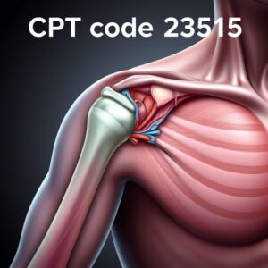
CPT Code 23515
2. Anatomy of the Clavicle: The Body’s Strut
To appreciate the fracture and its repair, one must first understand the anatomy. The clavicle is a tubular bone with a double curve, resembling a shallow ‘S’. It is the first bone in the body to begin ossification (around the 5th week of gestation) and one of the last to finish, completing ossification in the early 20s.
-
Joints: It articulates medially with the sternum (forming the sternoclavicular joint) and laterally with the acromion process of the scapula (forming the acromioclavicular joint). These joints are crucial for the wide range of motion of the shoulder.
-
Landmarks:
-
Medial (Sternal) End: Bulky and triangular, it provides attachment for the sternocleidomastoid muscle.
-
Lateral (Acromial) End: Flattened, it forms the AC joint.
-
Shaft: Divided into the medial two-thirds (which is cylindrical and convex anteriorly) and the lateral third (flattened and concave anteriorly). The transition between these two sections is a biomechanical weak point, predisposing it to fracture.
-
-
Muscle Attachments: A multitude of muscles and ligaments stabilize the clavicle, including the deltoid, trapezius, pectoralis major, and sternocleidomastoid. These muscular forces often cause significant displacement when a fracture occurs.
-
Neurovascular Relations: This is of critical surgical importance. The brachial plexus (a network of nerves controlling the arm) and the subclavian artery and vein pass directly behind the clavicle, protected only by a thin layer of tissue. A displaced fracture or errant surgical instrumentation can jeopardize these vital structures.
Diagram: Anatomy of the Clavicle and Surrounding Structures
(Note: This is a textual description of what a diagram would show)
[A diagram would show the S-shaped clavicle bone. Labels would indicate:
-
Sternal End: Connecting to the sternum.
-
Acromial End: Connecting to the acromion of the scapula.
-
Conoid Tubercle: Attachment point for the conoid ligament.
-
Subclavian Groove: On the inferior surface, where the subclavius muscle attaches.
-
Proximity to Brachial Plexus: Nerves shown running posterior and inferior to the clavicle.
-
Proximity to Subclavian Vessels: Artery and vein shown coursing behind the clavicle.]
3. The Nature of Clavicle Fractures: Causes, Types, and Classification
Mechanisms of Injury
The most common mechanism of injury is a direct fall onto the shoulder, such as during a bicycle accident, football tackle, or skiing fall. A less common mechanism is a fall onto an outstretched hand (FOOSH), which transmits force along the arm to the clavicle. Direct trauma to the bone itself is also possible.
Classification Systems
Fractures are classified to describe their location, pattern, and displacement, which guides treatment decisions.
-
Allman Classification (1967): The classic system, dividing fractures into three groups based on location:
-
Group I: Fractures of the middle third (shaft). Most common (80%).
-
Group II: Fractures of the lateral (distal) third. Account for 10-15%.
-
Group III: Fractures of the medial third. Rare (5%).
-
-
Robinson Classification (1998): A more modern and detailed system that further subdivides fractures based on displacement, comminution (bone shattered into multiple pieces), and articular involvement. This system is more predictive of outcome and is widely used in research and clinical practice.
Common Clavicle Fracture Classification Systems
| System | Group | Location | Description | Prevalence |
|---|---|---|---|---|
| Allman | I | Middle Third | Fracture of the clavicular shaft. | ~80% |
| II | Lateral (Distal) Third | Fracture near the AC joint. | ~10-15% | |
| III | Medial Third | Fracture near the sternum. | ~5% | |
| Robinson | 2A1 | Middle Third, Undisplaced | Minimal or no displacement. | Varies |
| 2B1 | Middle Third, Displaced | Fracture segments are shifted apart. | Varies | |
| 2B2 | Middle Third, Comminuted | Bone is broken into multiple pieces. | Varies | |
| 3A1/3B1 | Lateral Third | Further subdivided by ligament integrity. | Varies |
4. Clinical Presentation and Diagnosis
A patient with an acute clavicle fracture typically presents with a clear history of trauma followed by:
-
Pain: Immediate, severe, and localized to the fracture site.
-
Swelling and Bruising: Over the fracture area.
-
Deformity: Often a visible “bump” or sagging of the shoulder.
-
Guarding: The patient will typically hold the arm close to the body and support the elbow with the other hand to minimize motion.
-
Crepitus: A grating sensation felt if the arm is moved (though this should not be intentionally elicited).
Diagnosis is primarily clinical but is confirmed with imaging. An X-ray is the first-line investigation, typically including Antero-Posterior (AP), cephalic tilt (15-45° angled view), and sometimes a stress view. In complex cases, especially involving the joints, a CT scan may be ordered for superior bony detail and preoperative planning.
5. Treatment Options: Non-Operative vs. Operative Management
The choice between surgery and non-surgery is a nuanced decision made by the orthopedic surgeon in consultation with the patient.
Non-Operative Treatment: For decades, this was the standard. It involves immobilization in a sling or figure-of-eight brace for 4-6 weeks to manage pain and allow the bone to heal naturally. It is typically recommended for:
-
Non-displaced or minimally displaced fractures.
-
Fractures in pediatric patients (who have remarkable healing potential).
-
Low-demand elderly patients.
Indications for Surgery (ORIF with CPT 23515): Research has shown that non-operative management of significantly displaced fractures can lead to higher rates of nonunion (failure to heal), malunion (healing in a shortened or deformed position), and residual shoulder weakness. Surgery is therefore strongly considered for:
-
Significant Displacement: Fracture fragments shortened by >2 cm or displaced >100% (shaft width).
-
Comminution: Multiple fracture fragments.
-
Open Fractures: Where the bone pierces the skin (a surgical emergency).
-
Associated Neurovascular Injury: Fracture fragments compressing or injuring nerves/blood vessels.
-
Impending Skin Compromise: A sharp bone fragment threatening to poke through the skin.
-
“Floating Shoulder”: A combined injury involving both the clavicle and the scapular neck.
-
Symptomatic Nonunion: A fracture that has failed to heal after several months.
-
Polytrauma Patients: To improve mobility and pain control.
-
Active, High-Demand Patients: Athletes or laborers who require a rapid and predictable return to full function.
6. Deep Dive into CPT Code 23515
CPT (Current Procedural Terminology) codes are a uniform language for describing medical, surgical, and diagnostic services. CPT code 23515 is specifically defined as: “Open treatment of clavicular fracture, with internal fixation”.
-
“Open treatment” means the surgeon makes an incision to directly visualize the fracture site. This is in contrast to “closed treatment” (e.g., setting the bone without an incision, coded as 23500).
-
“With internal fixation” means the surgeon applies hardware (e.g., plates, screws, pins) to stabilize the fracture fragments from inside the body.
This code is a “global” service code. It bundles the surgical approach, fracture reduction, application of internal fixation, and wound closure into a single code. It does not include the diagnosis (which is represented by an ICD-10-CM code, like S42.031A for a displaced midshaft fracture) or the hardware used.
Contrasting Codes:
-
23500: Closed treatment of clavicular fracture; without manipulation. (e.g., placing a sling).
-
23505: Closed treatment with manipulation. (e.g., manually reducing the fracture before placing a sling).
-
23520: Open treatment of clavicular fracture, with internal fixation; with iliac or other autograft (includes graft harvest). This is used only if a bone graft is necessary, such as in a nonunion case.
7. The Surgical Procedure: A Step-by-Step Walkthrough
The performance of CPT 23515 is a meticulous process.
-
Preoperative Planning: The surgeon reviews all imaging to understand the fracture pattern and selects the appropriate implant, typically a pre-contoured anatomic clavicle plate.
-
Anesthesia: The procedure is performed under general anesthesia, often supplemented with a regional nerve block (e.g., interscalene block) for excellent postoperative pain control.
-
Positioning: The patient is placed in a beach-chair position (semi-seated), which provides excellent access to the clavicle.
-
Surgical Approach: A linear incision is made parallel to and just inferior to the clavicle. Layers of skin, subcutaneous tissue, and the platysma muscle are carefully dissected. The surgeon must meticulously protect the underlying supraclavicular nerves to prevent permanent numbness over the shoulder.
-
Fracture Reduction: The fracture fragments are carefully exposed, and any hematoma or interposed tissue is removed. Using specialized instruments, the surgeon manually re-aligns the bone fragments to their anatomic position. This “reduction” is confirmed visually and with intraoperative fluoroscopy (a live X-ray).
-
Internal Fixation: The pre-contoured plate is placed on the superior or anterior-inferior surface of the clavicle. It is held in place while the surgeon drills pilot holes and inserts screws through the plate into the stable bone fragments on either side of the fracture, compressing the fragments together. This creates a rigid, stable construct.
-
Closure: The wound is irrigated with saline. The muscular and fascial layers are closed with absorbable sutures, the subcutaneous layer is approximated, and the skin is closed with sutures, staples, or surgical glue. A sterile dressing is applied.
8. The Role of the Surgical Team and Technology
A successful ORIF is a symphony of coordinated effort.
-
The Orthopedic Surgeon leads the procedure, making critical decisions on approach, reduction, and fixation.
-
The Anesthesiologist manages the patient’s vital signs and pain levels throughout.
-
Scrub Nurses/Technologists anticipate the surgeon’s needs, managing instruments and implants.
-
Circulating Nurses manage the overall OR environment and documentation.
-
Intraoperative Fluoroscopy is indispensable, providing real-time feedback on fracture reduction and hardware placement, ensuring accuracy and safety.
9. Postoperative Recovery and Rehabilitation
Recovery is a phased process:
-
Phase 1 (Weeks 0-6): The arm is immobilized in a sling. The focus is on pain control, wound healing, and gentle pendulum exercises to prevent shoulder stiffness.
-
Phase 2 (Weeks 6-12): X-rays confirm healing. Physical therapy intensifies to restore full range of motion and begin gentle strengthening.
-
Phase 3 (Months 3-6+): Progressive strengthening and a gradual return to all activities, including contact sports, once cleared by the surgeon. Full healing and remodeling of the bone can take up to a year.
Hardware (plates and screws) is often left in place permanently unless it becomes symptomatic (e.g., painful, prominent, or irritating overlying tissue), in which case a second surgery for hardware removal may be performed after the fracture is fully healed.
10. Potential Risks and Complications
As with any surgery, risks exist:
-
Infection: Treated with antibiotics and sometimes irrigation and debridement.
-
Hardware Irritation: The most common complaint, due to the plate lying just under the skin.
-
Nonunion: Failure of the bone to heal (risk <5% with surgery).
-
Malunion: Healing in a deformed position.
-
Neurovascular Injury: Damage to nerves or blood vessels (rare with careful technique).
-
Anesthetic complications.
-
Clavicular Osteolysis: Bone resorption under the plate.
11. Coding and Billing for 23515: A Practical Guide
Accurate coding is essential for appropriate reimbursement.
-
Documentation is Key: The operative report must clearly document “open treatment,” “fracture reduction,” and “internal fixation” with a plate and screws. It should describe the approach and the specific hardware used.
-
Modifiers: The laterality modifier is crucial. Use -LT (Left side) or -RT (Right side). If a procedure is repeated on the same day by the same surgeon, modifier -76 may be needed.
-
Bundling: Code 23515 includes the exposure, reduction, fixation, and closure. Do not separately code for the incision or “application of plate.” However, if a bone graft is harvested and applied, code 23520 is used instead.
-
NCCI Edits: Coders must check the National Correct Coding Initiative edits to ensure no bundled components are incorrectly billed separately.
12. The Patient Perspective
Understanding the patient journey is vital. Pre-operatively, patients often experience significant pain and anxiety about the deformity and functional loss. The decision for surgery is often driven by the desire for a predictable outcome and a faster return to function. Postoperatively, the first two weeks are the most challenging, with pain and dependency on others. The gradual return of function through therapy is a powerful motivator. The most common long-term patient-reported outcome is satisfaction with function, though some may have numbness around the incision or awareness of the hardware.
13. Conclusion: Summarizing the Journey of Clavicle Fracture Repair
CPT code 23515 represents a sophisticated surgical solution for a common yet debilitating injury. It moves beyond simple stabilization to the precise restoration of anatomy and function. The procedure’s success hinges on meticulous surgical technique, comprehensive postoperative rehabilitation, and accurate clinical documentation. For patients with displaced clavicle fractures, ORIF offers a proven pathway to reduced complications, reliable healing, and a timely return to an active life, solidifying its critical role in modern orthopedic trauma care.
14. Frequently Asked Questions (FAQs)
Q1: How long does the surgery take?
A: The procedure itself typically takes between 60 and 90 minutes, though preoperative preparation and anesthesia can make the total time in the operating room longer.
Q2: Will I have a visible scar?
A: Yes, there will be a linear scar along the line of your clavicle. Surgeons place incisions along natural skin lines to minimize scarring. The scar will be red and raised initially but will fade significantly over 12-18 months.
Q3: When can I drive after surgery?
A: Most patients cannot drive safely while wearing a sling and while taking opioid pain medication. Driving is usually resumed once you are out of the sling, have adequate pain control without narcotics, and have regained enough reaction time and mobility (often around 6-8 weeks). Always follow your surgeon’s specific advice.
Q4: Is it normal to feel the plate under my skin?
A: Yes, especially in thinner individuals, it is very common to be able to feel the outline of the plate. This is not a sign of a problem unless it becomes painful or causes skin irritation.
Q5: Why might the hardware need to be removed?
A: Hardware is not routinely removed. Removal is only considered if it becomes symptomatic—causing persistent pain, skin irritation, or limiting movement—usually after the fracture has fully healed, which takes at least a year. This requires a second, smaller surgery.
15. Additional Resources
-
American Academy of Orthopaedic Surgeons (AAOS): OrthoInfo website provides excellent patient-friendly information on clavicle fractures and treatment options.
-
American Medical Association (AMA): Publisher of the CPT code set. For accurate coding, the CPT Professional Edition code book is the authoritative source.
-
National Institutes of Health (NIH) – PubMed: A database of medical literature for those seeking advanced clinical studies on clavicle fracture outcomes.
-
Your Orthopedic Surgeon and Physical Therapist: Always your best and most personalized resource for information pertaining to your specific case and recovery.
Disclaimer
The information contained in this article is for educational and informational purposes only and does not constitute medical advice, diagnosis, or treatment recommendations. Always seek the advice of your physician, orthopedic surgeon, or other qualified health provider with any questions you may have regarding a medical condition or surgical procedure. The content regarding CPT coding is based on guidelines from the American Medical Association (AMA) and is subject to change. It is intended for educational use only. Medical coding is complex and dependent on specific clinical circumstances. Always consult the most current AMA CPT Professional Edition code book and payer-specific policies for accurate, reimbursable coding. The authors and publishers are not responsible for any errors, omissions, or for any outcomes related to the use of this information.