
You sit in the dental chair. The procedure ends. The tooth is gone. Now you stare into the little mirror the dentist hands you. You see a dark hole where your tooth used to be. Your gums look strange. Swollen. Maybe a bit bloody. And immediately, the questions flood your mind. Is this normal? How long will this look so weird? What exactly happens to gums after tooth extraction?
Take a deep breath. Every single person who has ever had a tooth pulled has felt this exact anxiety. The mouth heals differently than any other part of your body. The gum tissue transforms in specific, predictable stages. Understanding these stages removes the fear. It lets you recognize normal healing versus warning signs. This guide walks you through every detail. No sugar-coating. No scary exaggerations. Just the honest, biological truth about gum healing after extraction.
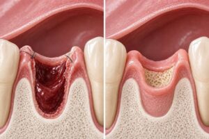
What Happens to Gums After Tooth Extraction
The Moment the Tooth Leaves the Socket
The seconds immediately after extraction set the stage for everything that follows. Your body launches a remarkable cascade of healing processes before you even leave the dental chair.
The Empty Socket: What You Actually See
When the dentist removes the tooth, they do not leave behind a simple hole. The socket actually contains the remnants of the periodontal ligament—tiny fibers that once connected the tooth root to the jawbone. These fibers tear during extraction. The bony socket walls remain intact, though potentially slightly expanded if the tooth required rocking to remove.
Your gum tissue around the extraction site immediately begins to collapse inward slightly. The elasticity of gum tissue means it does not stay perfectly rigid. Instead, it starts to drape toward the center of the empty socket. This initial movement marks the very beginning of wound closure.
First Blood Clot Formation: Nature’s Perfect Bandage
Within seconds to minutes, blood fills the socket. This is not just random bleeding. This blood contains platelets, fibrin, and countless healing factors. The blood coagulates into a gel-like plug that dentists call the “blood clot.” This clot serves as the single most critical element in the entire healing process.
The clot does several jobs simultaneously. It seals the underlying bone and nerve endings from air, food particles, and bacteria. It provides the scaffold upon which new tissue will grow. It releases growth factors that call healing cells to the area. Without a stable, healthy clot, everything goes wrong—a condition called dry socket that we will discuss in detail later.
Immediate Physical Sensations in the Gums
Your gums will feel strange right away. The local anesthetic typically keeps you numb for two to four hours after the procedure. During this time, you feel pressure more than pain. The gum tissue feels “full” or “puffy.” Some patients describe it as feeling like something is stuck between their teeth, even though the tooth is gone.
As the anesthetic fades, the gum tissue begins to throb gently. This throbbing matches your heartbeat. It signals the increased blood flow rushing to the area to start repairs. The surrounding gum tissue may appear darker red or even slightly purple. This represents normal bruising, not a problem.
The First 24 Hours: The Critical Healing Window
The first day after extraction determines the trajectory of your gum healing. What you do—and what you avoid—during these hours matters enormously.
The Clot Stabilizes and Matures
Over the first 24 hours, the initial jelly-like clot transforms into a firmer, more organized plug. Fibrin strands within the clot cross-link and strengthen. The clot adheres more tightly to the socket walls. You might notice the clot appears darker, more maroon than bright red. This color change reflects the normal breakdown of hemoglobin within the trapped red blood cells.
The top surface of the clot that is exposed to your mouth begins to develop a slightly tough, almost skin-like layer. This layer consists of fibrin and dead white blood cells that sacrificed themselves fighting any bacteria that entered during the extraction. This whitish or yellowish film on top of the clot often alarms patients. Do not panic. This is not pus. This is not infection. This represents a normal fibrin cap protecting the delicate clot underneath.
Gum Swelling Peaks
Your gum tissue swells during this period. Inflammation is not the enemy here. Swelling brings immune cells, nutrients, and oxygen to the healing site. The gums around the extraction site will look puffy, rounded, and larger than the matching area on the opposite side of your mouth.
The degree of swelling depends on several factors. Surgical extractions that required cutting gum tissue and drilling bone cause more swelling than simple extractions. Lower molar extractions often swell more than upper teeth. Your individual inflammatory response also matters. Some people swell dramatically. Others barely show visible swelling. Both extremes fall within normal range.
Applying ice packs to your cheek during these first 24 hours constricts blood vessels and limits the extent of swelling. The cold also provides meaningful pain relief. Do not apply ice directly to the gum tissue. That can damage the delicate healing cells.
Bleeding Tapers Off
A small amount of blood oozing from the extraction site is normal for several hours after the procedure. The gauze your dentist gave you helps apply direct pressure to encourage clot stability. You may notice pink-tinged saliva when you spit gently. This does not indicate active bleeding. A few drops of blood can tint a mouthful of saliva dramatically.
By the 24-hour mark, frank bleeding should stop completely. The clot should sit securely in the socket. If you still see bright red blood actively pooling in your mouth after 24 hours, contact your dentist. This might indicate a clot that failed to form properly or a small blood vessel that continues to ooze.
The White Film Phenomenon
Let us address this specifically because it causes so many panicked phone calls to dental offices. The whitish or greyish-yellowish film that develops on the surface of the clot and adjacent gum tissue is normal granulation tissue beginning to form. This represents healing, not infection. True pus looks thicker, creamier, and often drains actively from the site. It may smell foul. Normal healing film looks more like a thin, moist coating. It stays in place. It does not wipe away easily.
Days 2 to 3: Inflammation and the Start of Repair
The second and third days after extraction often feel like the hardest part of recovery. Do not be surprised if you actually feel worse on day two than you felt on day one.
Why Pain and Swelling May Increase Temporarily
The body’s inflammatory response peaks around 48 to 72 hours after injury. This applies to tooth extraction just as it applies to a sprained ankle. The chemical signals that call healing cells to the area reach maximum concentration. Blood vessels in the gum tissue become maximally dilated. Fluid accumulates in the tissue spaces.
Your gums may ache more noticeably on day two or three. The swelling may look more pronounced. This does not mean something went wrong. This represents your immune system doing exactly what evolution designed it to do. The pain should feel like a dull, steady ache rather than sharp, stabbing pain. Sharp pain, especially if it radiates toward your ear or temple, warrants a call to the dentist.
Granulation Tissue Begins True Formation
Underneath the protective fibrin cap, something remarkable begins. Specialized cells called fibroblasts migrate into the clot. These cells produce collagen, the structural protein that forms the scaffolding for all new tissue. Tiny new blood vessels begin sprouting into the healing clot—a process called angiogenesis.
This new tissue, rich with young collagen and fresh capillaries, is called granulation tissue. It appears pinkish-red and slightly bumpy. Over days two and three, granulation tissue gradually replaces the original blood clot from the deepest part of the socket working upward toward the surface. You generally cannot see this process happening because the fibrin cap covers it. But if you were to look closely at the socket floor, you would see tiny red dots. Those dots are individual capillary loops in the new granulation tissue.
Gum Tissue Begins Contracting
The gum tissue around the socket margins starts to pull inward more noticeably. This process, called wound contraction, involves specialized cells called myofibroblasts that actually grip the edges of the wound and pull them toward the center. This reduces the size of the opening that needs to fill with new tissue.
You might notice the socket hole looks slightly smaller on day three than it did on day one. This contraction accounts for that change. The rate of contraction varies widely between individuals and between different locations in the mouth.
Eating and Drinking Challenges
These days present the most difficulty with eating. Your gums remain tender. The clot remains vulnerable. You must continue eating soft foods and chewing on the opposite side of your mouth. Anything hot, spicy, crunchy, or sharp can irritate the healing tissue. Small food particles like rice or seeds can lodge in the socket and prove difficult to remove without disturbing the clot.
Rinsing remains restricted. Vigorous swishing can dislodge the clot. Your dentist may recommend gentle salt water rinses starting around 24 hours post-extraction, but these should involve tilting your head to let the water flow over the site rather than forceful swishing and spitting. After eating, a gentle rinse helps remove debris from the socket rim without disturbing the deeper healing structures.
Days 4 to 7: Visible Gum Healing Progresses
By the end of the first week, the extraction site transforms noticeably. The gums look and feel very different from those first alarming moments after the tooth came out.
The Socket Opening Gradually Shrinks
The visible hole in your gum tissue gets smaller each day. This shrinkage results from two simultaneous processes. First, wound contraction continues. The gum edges pull further toward the center. Second, granulation tissue keeps filling the socket from the bottom up, raising the floor of the defect.
By day seven, the socket opening typically measures about half its original diameter. The depth also decreases significantly. Where the socket initially seemed like a deep dark pit, it now appears shallower, with pinkish-red tissue visible near the surface.
Gum Color Returns Toward Normal
The angry redness of the first few days fades. The bruised purple tones resolve as the body clears the trapped blood from the tissue. The gum around the extraction site shifts toward a healthier pink. It may still appear slightly darker or redder than surrounding gum tissue, but the dramatic inflammatory appearance calms considerably.
The whitish fibrin coating on the surface gradually sloughs off or gets incorporated into the maturing tissue. The socket interior looks more uniformly pink and granular rather than dark and bloody.
Gum Sensitivity Evolves
The extreme tenderness of the first days gives way to mild sensitivity. Touching the extraction site with your tongue or finger still feels unusual, but not acutely painful. The gum tissue around the socket remains slightly softer and more easily indented than mature, healthy gum. This represents normal early healing tissue that has not yet developed its full structural integrity.
The adjacent teeth may feel slightly tender or loose. This happens because the swelling in the gum tissue spreads to the periodontal ligaments of neighboring teeth. As swelling decreases, these teeth return to their normal stability. This temporary looseness concerns many patients unnecessarily.
Sutures and Their Role
If your extraction required sutures, those stitches play an important role during this week. Sutures hold the gum tissue edges closer together than they would naturally sit. This reduces the size of the exposed socket. It protects the underlying clot and granulation tissue. It also reduces the amount of new tissue that must grow to close the wound.
Most dentists use dissolving sutures that begin to break down within the first week. You might notice suture material loosening or a suture knot coming free. This is normal. Do not pull on the sutures. Let them dissolve or fall out on their own schedule. If a suture comes out early and the wound edges separate more than you think they should, a quick call to your dentist provides reassurance.
Week 2 to 3: Soft Tissue Maturation Accelerates
The second and third weeks after extraction bring dramatic visible improvements. The extraction site starts looking less like a wound and more like normal gum tissue.
Complete Epithelial Coverage
This milestone marks a major turning point. Epithelial cells—the same type of cells that form the outer protective layer of your gums, cheeks, and skin—multiply and migrate across the granulation tissue surface. They form a continuous sheet covering the entire socket opening.
Epithelial coverage means the socket is no longer an open wound. Bacteria and food particles face a functional barrier. You can eat more normally. You can brush more thoroughly near the site. The risk of dry socket drops essentially to zero once epithelial coverage is complete.
This epithelial layer initially appears thin and somewhat translucent. You can see the reddish granulation tissue underneath. Over time, the epithelial layer thickens and becomes more opaque, taking on the normal pink color of healthy gums.
The Socket Continues to Fill
Underneath that new epithelial roof, the socket keeps filling with maturing tissue. Fibroblasts keep laying down collagen. The granulation tissue becomes less vascular, less fragile. It transitions into what histologists call “fibrous connective tissue.” This tougher, more organized tissue provides better structural support for the overlying gum.
The socket at two to three weeks still looks indented compared to the surrounding gum tissue. A shallow depression remains where the tooth once stood. But this depression feels firm to touch, not soft and boggy. The depth continues decreasing as new tissue accumulates from the socket floor upward.
Gum Contour Takes Shape
The overall shape of the gum ridge in the extraction area begins to emerge. Wound contraction has pulled the gum edges together significantly. New tissue has filled much of the volume. The gum tissue starts to resemble the shape it will maintain long-term, though it will continue refining for months.
The edge where the extracted tooth’s root once emerged from the gum—the gingival margin—no longer exists in that location. The gum tissue forms a continuous, unbroken surface across the ridge where multiple teeth may have been extracted or where a single tooth socket is healing. This ridge will eventually support a denture, a bridge, or simply serve as the healed jaw contour.
Discomfort Largely Resolves
By the end of week three, the extraction site should feel comfortable during normal daily activities. Chewing on that side may still feel slightly strange. Hard foods may cause mild tenderness if they press directly on the healing ridge. But constant, unprovoked pain should be completely gone.
If you experience persistent pain, swelling that does not resolve, or pain that returns after initially improving, contact your dentist. These signs could indicate a low-grade infection, a retained root fragment, or a bone spicule working its way to the surface.
Week 4 to Month 3: Bone Remodeling and Gum Maturation
The visible changes slow dramatically during this period. Most of the obvious healing is complete. But underneath the surface, critical long-term remodeling continues.
The Alveolar Bone Remodels
Extraction triggers significant changes in the jawbone itself. The tooth socket sits within the alveolar bone—the specialized bone that exists specifically to support teeth. Once the tooth is gone, this bone recognizes that its purpose has changed. The body begins resorbing some of this bone.
During the first three months after extraction, the socket walls gradually break down and remodel. New bone fills the socket defect from the bottom and sides. The sharp edges of the socket rim smooth out. This internal remodeling explains why the ridge height and width decrease over the months following extraction.
Research shows that most of the bone loss after extraction occurs within the first three to six months. The alveolar ridge can lose up to 40-60% of its width and a significant amount of height during this period. This bone resorption has major implications if you plan to get a dental implant. It explains why dentists often recommend implant placement sooner rather than years later.
Gum Tissue Reaches Full Maturity
The gum tissue covering the healed socket achieves its final characteristics during this period. The epithelium thickens to match surrounding tissue. The underlying connective tissue fully organizes. The gum becomes firm, resilient, and indistinguishable from gum tissue elsewhere in your mouth.
The color matches surrounding gums. The texture feels normal when you run your tongue over it. The area no longer shows any visible evidence that an extraction occurred—except for the absence of a tooth.
The Ridge Form Takes Its Permanent Shape
By month three, the healed ridge has essentially achieved its long-term contour. This ridge will continue to change slowly over years as natural age-related bone loss occurs, but the rapid remodeling phase is complete.
The ridge height varies depending on which tooth was extracted and the trauma involved in the extraction. A simple extraction of a single-rooted tooth typically preserves more ridge height than a surgical extraction of a multi-rooted molar. Trauma during extraction accelerates bone resorption. Infection prior to extraction also increases bone loss.
If you plan to replace the missing tooth with a bridge or implant, your dentist evaluates the ridge around the three-month mark. Sufficient ridge height and width must exist to support the restoration. If excessive bone loss occurred, bone grafting procedures may become necessary before definitive restoration can proceed.
Special Circumstances in Gum Healing
Not every extraction follows the standard healing timeline. Several special situations alter how gums heal and what you should expect.
Surgical Extractions vs. Simple Extractions
Simple extractions involve teeth that are fully visible in the mouth, with intact crowns that allow the dentist to grasp and remove the tooth without cutting tissue or bone. The gum tissue is simply reflected away from the tooth and repositions naturally after removal. Healing tends to be faster, with less swelling and discomfort.
Surgical extractions require an incision into the gum tissue and often removal of bone around the tooth. Impacted wisdom teeth, severely broken teeth, and teeth with curved or bulbous roots frequently require surgical extraction. The gum tissue is intentionally cut, reflected as a flap, and then sutured back into position.
Comparative Healing Between Simple and Surgical Extractions:
| Feature | Simple Extraction | Surgical Extraction |
|---|---|---|
| Initial swelling | Mild to moderate | Moderate to severe |
| Pain duration | 2-4 days | 4-7 days typically |
| Gum closure | 2-3 weeks | 3-4 weeks |
| Suture presence | Usually none | Almost always present |
| Bone healing time | 3-4 months | 4-6 months |
| Risk of complications | Lower | Higher |
| Return to normal diet | Faster | Slower |
Multiple Adjacent Extractions
When a dentist removes several teeth next to each other, the gum healing differs from a single extraction. The sockets merge into a larger defect. More gum tissue must regenerate to close the wound. The ridge resorption tends to be more dramatic because the bone loss between sockets combines.
Patients undergoing multiple extractions for immediate dentures face a unique situation. The denture itself acts as a bandage over the extraction sites. The pressure from the denture can help control swelling and protect the clots. However, ill-fitting immediate dentures can traumatize the healing tissue and cause ulcerations. Close follow-up with the dentist for denture adjustments becomes critical.
Extractions in Patients with Gum Disease
Periodontal disease changes the gum tissue biology. Chronic inflammation alters the blood supply. The gum tissue may be more fibrotic or more fragile depending on the specific type and severity of gum disease. Healing after extraction in these patients can be less predictable.
The presence of calculus and bacterial biofilms on adjacent teeth can also delay healing of the extraction site. The inflammation from untreated gum disease can spill over into the healing socket. Dentists often recommend addressing active gum disease before or immediately after extractions to optimize healing.
Extractions in Smokers
Smoking profoundly impairs gum healing after extraction. Nicotine constricts blood vessels, reducing the oxygen and nutrient supply to healing tissue. The heat from smoke dries the clot surface and can disrupt it mechanically. The chemicals in tobacco smoke are directly toxic to healing cells like fibroblasts.
Smokers experience a dramatically higher rate of dry socket. Studies consistently show dry socket rates two to five times higher in smokers compared to non-smokers. The healing timeline extends significantly. Gum closure takes longer. The quality of healed tissue may be diminished.
If you smoke, the best thing you can do for your extraction healing is to abstain for at least 72 hours after the procedure, and ideally for a full week or more. Even reducing the number of cigarettes helps. Every cigarette you avoid gives your gums a better chance to heal efficiently.
Complications That Affect Gum Healing
The healing process does not always go smoothly. Recognizing complications early allows prompt treatment and better outcomes.
Dry Socket: When the Clot Fails
Dry socket, technically called alveolar osteitis, represents the most common complication after tooth extraction. It occurs when the protective blood clot either fails to form properly or becomes dislodged prematurely. Without the clot, the underlying bone and nerve endings lie exposed to the oral environment.
Key Characteristics of Dry Socket:
-
Severe, throbbing pain that begins 2-4 days after extraction
-
Pain that radiates to the ear, temple, or neck on the same side
-
Visible empty socket with exposed greyish-yellow bone
-
Foul taste or odor coming from the extraction site
-
Pain not relieved by over-the-counter medications
-
Gum tissue around the socket appears inflamed and tender
Dry socket pain often feels dramatically worse than the initial post-extraction discomfort. Patients describe it as the worst pain they have ever experienced. The pain persists and may intensify rather than gradually improving.
Treatment involves the dentist placing a medicated dressing directly into the socket. This dressing contains eugenol or other soothing agents that calm the inflamed bone and nerve endings. The dressing typically provides relief within minutes. It requires replacement every few days until the socket generates enough new tissue to cover the exposed bone naturally.
Risk factors for dry socket include smoking, oral contraceptive use, traumatic extraction, lower molar extraction, and poor oral hygiene. Women experience dry socket more frequently than men, possibly due to hormonal effects on clot stability.
Infection in the Extraction Socket
Post-extraction infection is less common than dry socket but potentially more serious. Infection can develop in the socket itself, in the surrounding gum tissue, or in the deeper bone.
Signs of Infection Include:
-
Increasing pain after initial improvement
-
Swelling that increases rather than decreases after day three
-
Pus or thick discharge from the extraction site
-
Foul odor that persists despite gentle cleaning
-
Fever or feeling generally unwell
-
Swollen lymph nodes under the jaw or in the neck
-
Difficulty opening the mouth due to muscle spasm
Infections require professional treatment. The dentist may need to irrigate the socket, place local antibiotics, or prescribe systemic antibiotics. In rare cases, infections can spread into the tissue spaces of the face and neck, becoming a medical emergency.
You can reduce infection risk by following post-operative instructions carefully. Gentle salt water rinses, good oral hygiene of the remaining teeth, and avoiding food debris accumulation in the socket all help prevent infection.
Bone Spicules and Sequestra
During healing, small fragments of bone may work their way to the surface of the gum tissue. These fragments, called spicules, come from the socket walls or from bone that was fractured during extraction. The body recognizes these tiny pieces as foreign or non-viable and slowly pushes them out through the gum.
Patients typically notice a small, hard, white or tan sliver poking through the gum tissue weeks or even months after extraction. The spicule may cause irritation to the tongue or cheek. Sometimes the overlying gum tissue appears red or sore around the spicule.
Small spicules often shed on their own. The gum tissue slowly pushes them out until they loosen enough to fall out or be removed with gentle pressure. Larger or persistently symptomatic spicules require removal by the dentist. This is a quick office procedure that provides immediate relief.
A bone sequestrum is a larger piece of dead bone that the body rejects. Sequestra are more common after surgical extractions or extractions complicated by infection. They may present as a visible area of exposed bone that fails to cover with gum tissue. Dental evaluation determines whether the sequestrum requires surgical removal.
Excessive Bleeding
Bleeding that continues actively beyond the first 24 hours or that soaks through gauze rapidly warrants attention. Some patients have undiagnosed bleeding disorders. Certain medications and supplements interfere with clotting.
Common medications and supplements that increase bleeding risk:
-
Aspirin
-
Warfarin (Coumadin)
-
Clopidogrel (Plavix)
-
Ibuprofen and other NSAIDs in high doses
-
Fish oil supplements in high doses
-
Vitamin E supplements
-
Ginkgo biloba
-
Garlic supplements
Always inform your dentist about all medications and supplements you take. Never stop prescribed blood thinners without consulting both your dentist and the prescribing physician. They will coordinate to manage your extraction safely.
If you experience excessive bleeding at home, fold a piece of gauze or a moistened tea bag (black tea contains tannins that aid clotting), place it directly over the socket, and bite firmly for 30-60 minutes without interruption. Sit upright and remain still. If bleeding continues after several attempts, contact your dentist or seek emergency care.
Long-Term Gum Changes After Extraction
The gum tissue over a healed extraction site is not identical to gums that have always held teeth. Several long-term changes occur that affect function, appearance, and future treatment options.
Ridge Resorption Over Time
The most significant long-term change involves the bone underneath the gums. Without tooth roots stimulating the bone, the alveolar ridge gradually resorbs. This resorption occurs most rapidly in the first year after extraction but continues at a slower pace throughout life.
The gum tissue follows the retreating bone. A ridge that was initially tall and wide enough to support an implant may, over years, become too narrow or too short. This progressive resorption explains why dentists emphasize timely tooth replacement. The longer you wait, the more bone you lose, and the fewer options remain for restoring the missing tooth.
Gum Tissue Quality on the Healed Ridge
The gum tissue covering a healed extraction site differs microscopically from gum tissue that has always covered teeth. The connective tissue composition is slightly different. The blood supply pattern differs. The attachment to underlying bone is less organized than the specialized attachment of periodontal ligament.
These differences matter for denture wearers. Dentures rest on the gum-covered ridge. The quality of that tissue affects comfort and stability. Well-healed gum tissue over an adequate ridge provides a comfortable denture-bearing surface. Heavily resorbed ridges with thin, easily traumatized gum tissue make denture wearing challenging and sometimes painful.
For implant patients, the quality of gum tissue around the future implant matters aesthetically. Gum tissue that has healed after extraction may require grafting or manipulation to create an ideal emergence profile for the implant crown. This is particularly important for front teeth where the gum line is visible when smiling.
Changes in Adjacent Gum Tissue
The gums around teeth adjacent to an extraction site may experience subtle changes. Without the extracted tooth providing contact, the adjacent teeth can shift. This shifting alters the gum architecture around those teeth. The papilla—the triangular gum tissue between teeth—may flatten or recede slightly.
Food impaction becomes more common in the area where the tooth is missing. Food can pack between the remaining teeth differently than when all teeth were present. This can lead to gum inflammation around those adjacent teeth if oral hygiene does not adapt to the new contour.
Practical Care for Healing Gums
Proper care during each phase of healing optimizes gum recovery and minimizes complications. Here is a practical timeline for caring for your extraction site.
Immediate Post-Extraction Care (First 24 Hours)
Do:
-
Bite firmly on gauze for 30-60 minutes after extraction
-
Apply ice packs to face—20 minutes on, 20 minutes off
-
Take prescribed or recommended pain medication as directed
-
Rest with head elevated on pillows
-
Drink plenty of cool water
-
Eat only cold, soft foods if hungry
Do Not:
-
Rinse or spit forcefully
-
Drink through a straw
-
Smoke or vape
-
Touch the extraction site with fingers or tongue
-
Engage in strenuous physical activity
-
Drink alcohol
-
Eat hot foods or beverages
First Week Care
Oral Hygiene:
-
Continue brushing all other teeth normally
-
Avoid the extraction site with your toothbrush
-
Begin gentle salt water rinses after 24 hours (1/2 teaspoon salt in cup of warm water)
-
Rinse after meals to keep socket clean
-
Do not use mouthwash containing alcohol
Diet Recommendations:
| Recommended Foods | Foods to Avoid |
|---|---|
| Yogurt | Nuts and seeds |
| Smoothies (no straw) | Chips and crackers |
| Mashed potatoes | Popcorn |
| Scrambled eggs | Rice |
| Soup (lukewarm only) | Crusty bread |
| Applesauce | Anything crunchy |
| Cottage cheese | Spicy foods |
| Pudding | Hot beverages |
| Well-cooked pasta | Sticky candies |
| Soft fish | Raw vegetables |
Activity Restrictions:
-
Avoid heavy lifting and strenuous exercise
-
Do not blow your nose forcefully (important for upper extractions)
-
Sneeze with mouth open to reduce sinus pressure
-
Sleep with head slightly elevated
Long-Term Gum Health After Full Healing
Once the socket has fully closed and the gum tissue has matured, maintain the area just like the rest of your mouth. Brush the healed ridge gently but thoroughly. Floss between any remaining adjacent teeth. Regular dental check-ups allow your dentist to monitor the ridge for changes over time.
If you have not replaced the missing tooth, discuss replacement options with your dentist. A gap in your teeth leads to shifting, bite changes, and continued bone loss. Dental implants, bridges, and partial dentures all serve to restore function and preserve oral health.
The Relationship Between Gum Healing and Tooth Replacement
The timing of tooth replacement significantly influences outcomes. Understanding how gum healing interacts with different replacement options helps you make informed decisions.
Immediate Implant Placement
In select cases, dentists can place a dental implant immediately after tooth extraction, on the same day. This approach offers advantages including preservation of gum architecture and reduced overall treatment time. However, it requires specific conditions.
The extraction must be atraumatic, preserving the socket walls intact. There must be no active infection at the extraction site. Sufficient bone must exist beyond the socket to stabilize the implant. The gum tissue must be healthy and adequate in quantity.
When immediate implant placement succeeds, the gum tissue heals around the implant similarly to how it heals around a natural tooth. The gum architecture can be maintained beautifully. Patients avoid the multiple surgeries and extended waiting periods of delayed implant placement.
Early Implant Placement (4-8 Weeks)
Early implant placement occurs after soft tissue healing is complete but before significant bone resorption has occurred. This timing window of 4-8 weeks after extraction allows gum closure over the socket while still capitalizing on the remaining socket bone.
The gum tissue at this stage has healed enough to allow flap reflection and primary closure if needed. The bone is still actively remodeling but has not yet significantly resorbed. This timing represents a good compromise for many patients who did not qualify for immediate placement.
Delayed Implant Placement (3-6 Months)
Delayed placement waits for complete bone healing. The extraction site is fully closed, the ridge has stabilized, and the bone has matured. This approach offers the most predictable bone integration for the implant.
The gum tissue at 3-6 months is completely mature. The ridge contour is established. The dentist can precisely plan implant position based on the healed anatomy rather than predicting how healing will proceed. The downside is the potential for more bone resorption during the waiting period.
Bridge Placement
A traditional fixed bridge requires the adjacent teeth to be prepared as anchors. The pontic—the false tooth that replaces the extracted tooth—sits on the healed gum ridge. The fit between the pontic and the gum tissue matters for both aesthetics and hygiene.
Bridges should be placed after the gum tissue has fully healed and the ridge has stabilized, typically 3-6 months after extraction. If placed too early, the ridge may continue to change shape, creating a gap under the pontic that traps food and looks unnatural. A well-timed bridge fits snugly against the gum tissue, creating the illusion of a tooth emerging naturally from the gum.
Removable Partial Dentures
Partial dentures can be fabricated relatively quickly after extraction. Immediate partial dentures serve as both a tooth replacement and a bandage during initial healing. The denture base rests on the healing gum tissue, which requires careful monitoring and adjustment as the tissue changes shape.
The gum tissue under a partial denture requires special attention. The denture can rub and cause sore spots if it does not fit perfectly. Good denture hygiene prevents inflammation of the underlying gums. Regular dental visits allow adjustment of the denture as the ridge continues to remodel.
Psychological Aspects of Gum Healing After Extraction
The emotional journey of tooth extraction and gum healing deserves acknowledgment. Many patients underestimate the psychological impact of losing a tooth, even a back tooth that nobody sees.
The Shock of the Initial Appearance
Looking in the mirror after an extraction can be genuinely upsetting. The hole, the blood, the swelling—it looks traumatic. This is completely normal. Your body just experienced a surgical procedure, even if it was a “simple” extraction. Give yourself permission to feel unsettled by the appearance.
Remind yourself that this appearance is temporary. The dramatic wound you see in the first days transforms rapidly. Within a week, it looks entirely different. Within a month, it barely resembles the initial extraction site.
Anxiety About Eating and Socializing
Many patients feel anxious about eating after an extraction. Every bite triggers worry about disturbing the healing site. Social eating becomes stressful. This anxiety typically fades as the socket closes and confidence returns.
Plan ahead for the first few weeks. Stick to foods you feel comfortable eating. Carry a small oral hygiene kit if you need to eat away from home. A quick gentle rinse after eating provides peace of mind. Most people return to normal social eating within a week to ten days.
Concerns About Permanent Changes
Patients often worry that their mouth will never feel normal again. The gap where the tooth once was feels enormous to the tongue, even if the tooth itself was small. This sensation fades over time as the brain adapts to the new oral landscape.
The gum tissue does reach a point where it feels completely normal. The ridge feels solid and smooth. Eating on that side feels natural. The memory of the extraction experience fades. For most patients, the healed extraction site becomes just another part of their oral anatomy that they rarely think about.
When to Contact Your Dentist
Knowing when to seek professional help prevents minor issues from becoming major problems. Here are clear guidelines for when to pick up the phone.
Call Your Dentist Immediately If:
-
Bleeding persists heavily beyond 24 hours
-
Pain increases dramatically after day three
-
Swelling continues to increase after day three
-
You develop a fever over 101°F (38.3°C)
-
You have difficulty breathing or swallowing
-
Swelling extends to your eye area or down your neck
-
Pus drains from the extraction site
-
You experience numbness that does not resolve as expected
Schedule a Follow-Up Visit If:
-
Pain persists beyond two weeks
-
You notice a hard fragment emerging from the gum
-
The socket still appears open after three weeks
-
You have persistent bad taste or odor from the site
-
Adjacent teeth feel loose after swelling resolves
-
You have ongoing difficulty opening your mouth fully
Routine Questions That Are Normal:
-
“Is this amount of swelling normal?”
-
“Should my socket look like this at this stage?”
-
“When can I eat normally again?”
-
“Is this whitish stuff in the socket okay?”
Most dental offices welcome these questions. A quick phone call often provides reassurance that everything is progressing normally. Never hesitate to contact your dentist about concerns. They prefer you call early rather than waiting until a small problem becomes a big one.
The Remarkable Capacity of Gum Tissue to Heal
The human body possesses an extraordinary ability to repair itself. The gums exemplify this capacity. From a bloody, open wound to smooth, functional tissue in a matter of weeks—this process repeats millions of times daily in dental offices worldwide.
Understanding what happens to gums after tooth extraction transforms an anxiety-provoking experience into a manageable, predictable journey. The blood clot forms. The granulation tissue grows. The epithelium covers. The bone remodels. The gums mature. Each stage proceeds according to biological imperatives that have evolved over millions of years.
You can support this natural healing by following your dentist’s instructions, maintaining gentle hygiene, eating appropriately, and avoiding the few behaviors—smoking, forceful rinsing, early return to hard foods—that can derail the process. Your body knows what to do. Your job is simply to stay out of its way while it does it.
Conclusion
After tooth extraction, gums heal through a predictable series of stages beginning with blood clot formation and proceeding through granulation tissue development, epithelial coverage, and finally full tissue maturation over several months. The process involves both visible gum changes and invisible bone remodeling that ultimately determine the long-term contour of your jaw ridge. Proper care during the critical first week dramatically reduces complication risks like dry socket and infection, while patience during the months of deeper healing ensures the best foundation for any future tooth replacement.
Frequently Asked Questions
How long does it take for gums to fully close after tooth extraction?
The gum tissue typically closes over the extraction socket within two to four weeks. Complete closure means the socket is covered by a continuous layer of epithelial tissue. However, the underlying tissue continues maturing and the bone continues remodeling for several months. The visible hole should be significantly reduced by two weeks and essentially gone by one month in uncomplicated extractions.
Is the white stuff in my extraction socket normal or infection?
In most cases, the whitish or yellowish film on the extraction site represents normal fibrin and early granulation tissue—not pus or infection. Normal healing tissue appears as a thin, adherent coating that does not wipe away easily. Infection typically produces thicker, creamier discharge that may drain actively from the site and often has a foul odor. If you have no increasing pain, swelling, or fever, the white material is likely normal healing tissue.
When can I brush my teeth normally after extraction?
You can brush all your other teeth normally immediately, just avoid the extraction site itself. For the extraction area, avoid brushing directly for about one week. After that, very gentle brushing around the healing socket is acceptable. By two to three weeks, you can brush the area more normally as the gum tissue has closed and toughened. Use a soft-bristled toothbrush and gentle pressure.
Why does my gum feel hard and lumpy months after extraction?
This is typically normal and represents the healed bone ridge underneath the gum tissue. After extraction, the socket fills with bone that can feel slightly irregular compared to surrounding areas. You may also feel small bone spicules working their way to the surface. If the area is not painful, not growing, and not causing functional problems, it is likely normal healed anatomy. Your dentist can confirm this at your regular check-up.
Can I get dry socket after my gums have closed?
No. Once epithelial coverage is complete—typically by two to three weeks—dry socket can no longer occur. Dry socket requires exposed bone, which is only possible when the socket is open. The risk of dry socket is highest in the first three to five days after extraction and decreases significantly after one week. Once gum tissue covers the socket opening, the underlying bone is protected.
Will the gum where my tooth was extracted ever look normal again?
Yes, the gum tissue in the extraction area will look normal once fully healed. The color, texture, and firmness will match surrounding gum tissue. However, the contour will be different because the tooth is no longer present. Instead of a tooth emerging from the gum, you will have a smooth ridge. This is a normal anatomical variation, not an abnormality.
How does smoking affect gum healing after extraction?
Smoking dramatically impairs healing. Nicotine constricts blood vessels, reducing oxygen delivery to healing tissue. The sucking action can dislodge the protective blood clot. The heat and chemicals in smoke damage healing cells. Smokers experience much higher rates of dry socket, delayed healing, and infection. Ideally, avoid smoking for at least a week after extraction. Even reducing smoking frequency helps.
Additional Resource
For further detailed information about oral surgery recovery and what to expect after tooth extraction, visit the American Association of Oral and Maxillofacial Surgeons patient information page: